Direct measurement of sorption on three-dimensional surfaces such as resins, membranes or other preformed materials using lateral dispersion to estimate rapid sorption kinetics or high binding capacities

a three-dimensional surface and lateral dispersion technology, applied in the field of biotechnology and optical multianalyte biosensor technology, can solve the problems of reducing the magnitude of adsorption rate estimates by ionic strength, and achieve accurate estimation of adsorption rate constants, accurate and direct characterization of interactions between biological materials, and efficient preparation

Inactive Publication Date: 2006-10-05
UNIV OF UTAH RES FOUND
View PDF10 Cites 7 Cited by
  • Summary
  • Abstract
  • Description
  • Claims
  • Application Information

AI Technical Summary

Benefits of technology

[0015] Disclosed is a method to accurately and directly characterize interactions between biological materials using SPR technology. This is achieved by first assembling an intact resin or membrane on an SPR surface, then directly detecting the interaction between the biomolecule and the resin or membrane with SPR technology. From this data, estimates of mass-action ion-exchange adsorption and desorption rates accounting for steric and characteristic charge effects may be obtained. Previous methods using SPR technology only allowed measurement of minor aspects of the global interaction phenomenon, such as a single viral coat protein with a mammalian cell receptor. Based on these model studies, and many assumptions about the binding environment, researchers applied various mathematical models to determine estimated or theoretical binding rates. The improvement described herein allows detection and measurement of the binding of an entire viral particle, or other similar molecule, in a more natural environment, that of a three-dimensional membrane structure mimicking the membrane of mammalian cell walls. Furthermore, the invention enables detection and measurement of binding of viral particles, and other biomolecules, to commercially available isolation and purification materials commonly used in the bioscience research community, to allow more efficient preparation of medicaments for clinical use.
[0016] Commonly used isolation and purification techniques within the field of bioscience include column chromatography and the “batch method” of binding analytes to resins or other supports. Batch or column techniques only measure equilibrium binding constants which must be deconvoluted mathematically from effects of dispersion, diffusion or porosity. Furthermore, when attempting to purify or isolate an analyte for the first time, much precious material may be lost attempting to find the most effective and efficient isolation or purification method. In contrast, this new method allows adsorption and desorption kinetic rates to be measured directly under conditions very similar to flow operation of such large-scale techniques. The methods disclosed herein allow quantitative measurement of dynamic binding interaction between nanomolar to picomolar levels of precious analytes and their designated ligands, biological or synthetic, using SPR, whereas other commonly used techniques and methods, such as confocal microscopy, only qualitatively measures static binding outcomes at a scale of 100+nanometers. Furthermore, the method disclosed herein allows detection of actual binding of the biomolecule on real membrane or resins, something that in the past could only be inferred from qualitative data using several assumptions about the interactions that also required separate and often difficult if not impossible validation.
[0018] The method of the invention as disclosed herein provides many advantages over currently existing methods used to detect and measure binding between analytes and designated ligands. Existing methods estimate adsorption rates for analytes or adsorptive sites with homogeneous or heterogeneous affinities. In current methods, geometric distribution of adsorptive sites relative to the planar origin of the evanescent wave is presumed to be uniform and one-dimensional. In contrast, the present method disclosed herein accounts for geometric heterogeneity in adsorptive-sites distribution. Current methods can only estimate adsorption rates accurately if adsorptive sites are distributed uniformly in the dimension perpendicular to the evanescent wave origin. In contrast, the method disclosed herein accurately measures uniform adsorption rates from analyte adsorption in spatially heterogeneous solid-liquid composite media adjacent to the planar origin of the evanescent wave. Current methods are derived primarily for proteins, whereas the present method disclosed herein is much more flexible and may be used for analytes of any size.
[0020] The method disclosed herein provides many advantages over currently available techniques. Dispersion-enhanced lateral mass transport of adenovirus-sized particles occurs 4 to 10 times faster than diffusive mass transport for packed bed diameters between 10 and 50 microns for flow rates typical for biomolecular adsorption rate measurements by SPR. Dispersion-enhanced mass transport gives a boundary layer thickness δ=ξ(D / D∞*)1 / 3 [28] while the steady-state concentration boundary layer thickness for free flow between two flat plates, δ corresponds to an order-of-magnitude average of ¾(DL / γ)1 / 3 [40]. For flowrates and diffusivities typical of biomolecular adsorption rate measurements by SPR, the ratio of these reduces to a simple function of system geometry independent of operating conditions: δLokδKoch≈(Lha2⁢ɛ)1 / 3 For 10, 20 and 50 micron particles the ratio of free-flow to dispersion-enhanced boundary layers equals approximately 12, 7 and 4, respectively. Reduced boundary layer thickness occurs in conjuction with enhanced lateral mass transport.

Problems solved by technology

Additionally, it has been found that increasing ionic strength decreased the magnitude of adsorption rate estimates.

Method used


Image

  • Direct measurement of sorption on three-dimensional surfaces such as resins, membranes or other preformed materials using lateral dispersion to estimate rapid sorption kinetics or high binding capacities
  • Direct measurement of sorption on three-dimensional surfaces such as resins, membranes or other preformed materials using lateral dispersion to estimate rapid sorption kinetics or high binding capacities
  • Direct measurement of sorption on three-dimensional surfaces such as resins, membranes or other preformed materials using lateral dispersion to estimate rapid sorption kinetics or high binding capacities

Examples


example i

[0074] Cell Culture and Propagation

[0075] Human Embryonic Kidney cells (P / N 293 HEK; ATCC, Rockville, Md.) at a concentration of 1×10⁶ cells / mL were inoculated in 5 mL of DMEM purchased from Sigma (St. Louis, Mo., US). The medium was supplemented with 0.1 g / l alanine, 0.110 g / l sodium pyruvate, 1 g / l glucose, 0.584 g / l L—glutamine, 37 g / l sodium bicarbonate—all from Sigma (St. Louis, Mo., US)—and 10 ml / l antibiotic from Gibco (Auckland, NZ), pH 7.8. Cells were incubated in T-flasks from Corning (Corning, N.Y., US) at 37° C. and 5% CO2 for 48-96 hours. Flasks with cells at 90% confluence were split 1:5 into additional T-flasks to propagate the cell line. Near-confluent cells were resuspended by striking the flask 6-10 times, subdivided between 4 additional flasks and supplemented with fresh culture media to nurture new cell growth. Alternatively, cells at 90% confluence were also infected with Ad5.

example ii

[0076] Adenovirus Infection and Propagation

[0077] To infect the cells, a 1:100 dilution of Ad5 from ATCC (Rockville, Md., USA) was added to confluent T-flasks without disrupting adherent cells. T-flasks were incubated 1 hr at 37° C. and 5% CO2. After one hour, the cells are supplemented with additional culture media. After 48-72 hours the cytopathic effect was observed and virus was harvested. T-flasks were agitated to resuspend all cells. Suspension was centrifuged 5 min at 3,000×g. Supernatant was removed and combined with 10% glycerol before storage at −70° C. for future infection. Recovered cell pellets were resuspended in equal volumes of Tris buffer, pH 7.8, +1 mM CsCl, both from Sigma (St. Louis, Mo., US). The resuspension was frozen at −70° C. then thawed (repeated three times) to release Ad5. After 3× freeze-thaw, resuspensions were centrifuged to remove cell debris, then treated with Benzonase for 30 minutes to digest nucleic acid. Digested supernatants were ultracentrifu...

example iii

[0078] Adenovirus Chromatography

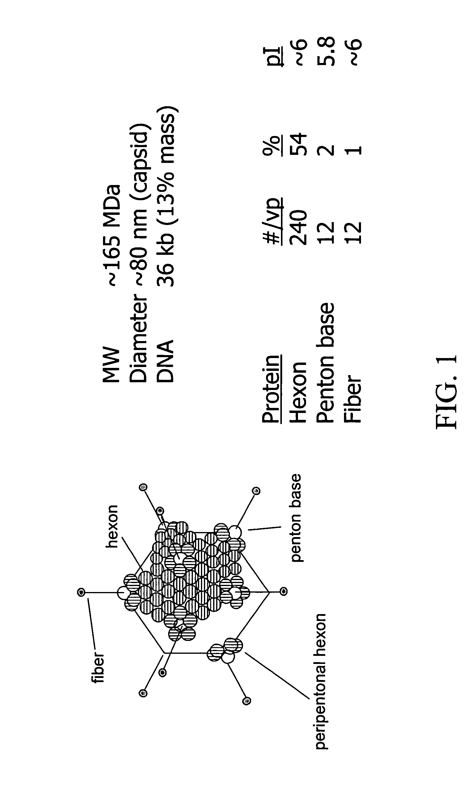
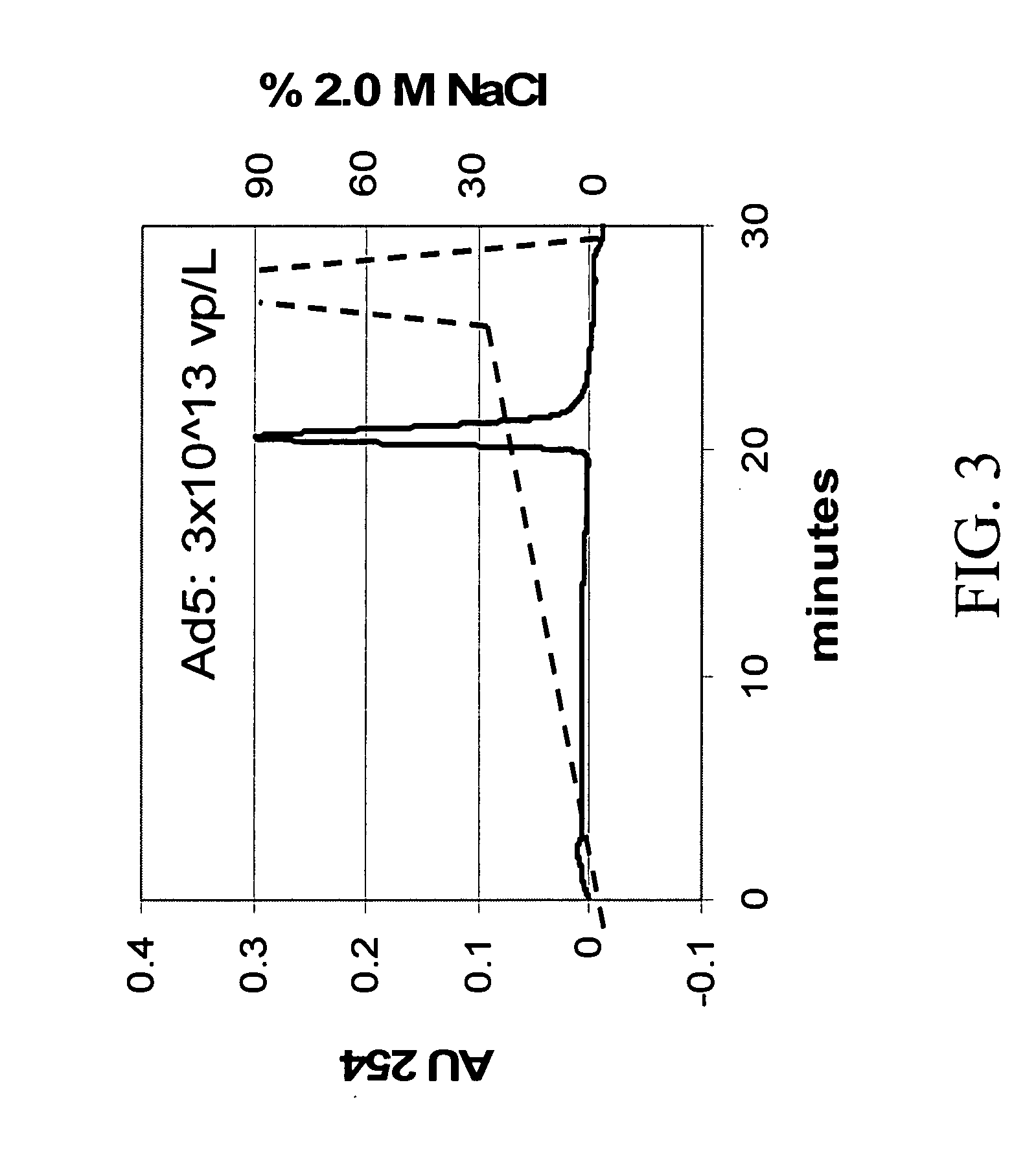
[0079] Ad5 was be purified and analyzed by HPLC with UV detection using Resource™ Q anion exchange resin from Amersham Biosciences (Piscataway N.J., US). The chromatogram is shown in FIG. 3. A quaternary ammonium anion exchange media, Resource Q, distinguished Ad5 from byproducts with a NaCl gradient (dotted line) from 40 to 600 mM at 1 mL / min, using the fact that hexon, penton and fiber proteins have IEP 6. A static capacity of ˜5×10¹¹ virus per mL, a detection limit >1×10⁸ particles per milliliter, and a linear Ad5 virus particle / HPLC area ratio of 20,085 have been reported for this method. [5]


PUM

PropertyMeasurementUnit
refractive indexaaaaaaaaaa
massaaaaaaaaaa
ionic strengthsaaaaaaaaaa

Abstract

Described are methods allowing measurement of adsorption and desorption of analytes with membranes or resins directly using surface plasmon resonance (SPR). Also described are methods for assembling intact resins or membranes on SPR surfaces. Such methods provide estimates of mass-action ion-exchange adsorption and desorption rates accounting for steric (σ) and characteristic charge (ν) effects. The methods further permit accurate estimation of rate constants for uniform adsorption of a homogeneous analyte solution on homogeneous adsorptive sites distributed heterogeneously in space, relative to the planar boundary. Solutions are obtained for locally porous media and solid spheres. The methods are extendible to other media and heterogeneous adsorptive sites. The methods further provide for enhancement of lateral mass transport in such optical measurement instruments through radial hydrodynamic diffusion (radial dispersion) by, for example, incorporating porous media in flow cells of detection devices such as, but not limited to, SPR or TIRF instruments.

Description

CROSS-REFERENCE TO RELATED APPLICATION [0001] This application claims priority under 35 U.S.C. 199(e) to U.S. Provisional Application 60 / 626,566, filed Nov. 9, 2004, the contents of the entirety of which is incorporated herein by reference.TECHNICAL FIELD [0002] The invention relates to the field of biotechnology and optical multi-analyte biosensor technology, more particularly to the use of optical multi-analyte biosensor technology employing the principal of internal reflection of polarized light for use in biological, biochemical and chemical analysis and in particular for detecting interactions or binding events between biological molecules, such as adsorption of viral particles to three-dimensional surfaces allowing measurement of adsorption and desorption of analyte(s) to intact membranes or resins directly. The detection method used in the biosensor system may be based on the evanescent wave phenomenon at total internal reflection, such as surface plasmon resonance (SPR), cri...

Claims


Application Information

Patent Timeline
Patent Type & Authority Applications(United States)
IPC IPC(8): C12Q1/70G01N33/53C12M1/34
CPCB82Y15/00B82Y30/00G01N2333/80G01N2333/075G01N33/54373
Inventor ROPER, D. KEITH
Owner UNIV OF UTAH RES FOUND
Why Eureka
Try Eureka