
Method for effecting the rapid release of a signature chemical from bacterial endospores, and for detection thereof

a technology of signature chemical and endospore, which is applied in the field of rapid release of signature chemical from bacterial endospore, and detection thereof, and can solve the problems of difficult determination of low spore concentration, significant time-consuming due to sample handling and data analysis, and insufficient unique emission spectrum to differentiate it from common biological materials

Active Publication Date: 2006-11-16
REAL TIME ANALYZERS
View PDF0 Cites 20 Cited by
  • Summary
  • Abstract
  • Description
  • Claims
  • Application Information

AI Technical Summary

Benefits of technology

[0016] It is a broad object of the present invention to provide a novel method for rapidly obtaining information to aid in the assessment of health risks associated with exposure to spores, and in particular spores of the genera Bacillus or Clostridium.
[0019] A further specific objective of the invention is to provide a novel method in which one or more weak acid compounds, having a pKa value of 0.1 to 11, is employed to release the signature chemical from the endospores, and in which additional physical means, such as heat or sonication, are not required to ensure release of the signature chemical in a timely manner, e.g., within 10 minutes.
[0025] A SERS-active medium will, in general, consist of a SERS-active metal in the form of particulates or other appropriately sized structures that can support a surface plasmon field. The SERS-active metal will normally be selected from the group consisting of copper, gold, silver, and alloys and mixtures thereof, and the SERS-active medium will generally be of a form selected from metal colloids in solution, metal colloids deposited on surfaces, metal-coated particles on a surface, metal depositions on surface structures, electrochemically generated metal surfaces, and metal-doped porous materials, in particular metal-doped sol-gels. Preferably, such a metal-doped sol-gel will be sufficiently porous to permit the analyte substance to be transported therethrough so as to effectively separate the analyte chemical from other components of the analyte substance, with at least a portion of the analyte chemical being retained in the sol-gel in sufficiently intimate proximity to the dopant metal to enable generation, in the irradiation step, of surface-enhanced Raman spectra of the analyte chemical.
[0027] Such a sol-gel will advantageously be of a chemical composition that is effective to extract the analyte chemical from the analyte substance, to thereby improve sensitivity. For the same reason, the dopant metal of the sol-gel may desirably have an electro-potential that is effective to attract the analyte chemical from the analyte substance, e.g., electropositive silver effective to attract a negatively-charged, deprotonated acidic analyte chemical.
[0029] The ability of weak acids to release CaDPA (or other signature chemicals) from endospores, without requiring physical aids such as high temperatures, ultrasound, or the like, overcomes limitations of prior techniques that do require such methods and apparatus, while also serving to avoid the production or accumulation of associated cell debris that is generated thereby, such debris having been found to interfere with analysis or to limit measurement sensitivity. As used herein, a weak acid is defined as an acid having a dissociation constant, Ka, of between 10−0.8 and 10−11, or, stated otherwise, to have a pKa between 0.1 and 11. Suitable weak acids include all mono-, di-, tri- and tetra-carboxylic acids (C1 through C16), branched or straight-chained, aliphatic, aromatic, or substituted acids, as well as their salts (e.g., sodium acetate) that produce such acids when added to water or another solvent or solvent mixture. In the preferred embodiments, acetic, citric, formic, fumaric, lactic, malic, propionic, sorbic, tetraacetic, trifluoroacetic acid, or a mixture thereof, with or without solvents, would be used.

Problems solved by technology

Although this method provides a relatively high degree of discrimination and sensitivity, it requires significant time due to sample handling and data analysis.
It has been shown that fluorescence can be obtained from CaDPA (Nudelman, R, B V Bronk, S Efrima, “Fluorescence Emission Derived from Dipicolinate Acid, its Sodium, and its Calcium Salts”Appl Spectrosc 54, 445-449, 2000), however the emission spectrum is not sufficiently unique to differentiate it from common biological materials that also fluoresce.
This, coupled with the fact that the Tb3+ cation produces the same luminescence spectrum, makes determinations of low spore concentrations problematic.
Furthermore, the combination of heat and the DDA surfactant severely degrade the spore, generating cell debris (here defined as biochemical fragments of the spore).
However, Raman spectroscopy is inherently an insensitive technique, and these measurements were of milligram, and not the required nanogram, sample amount.
The value of this technique is limited by the extremely low energy conversion of ultraviolet lasers, which require substantial power supplies and thus confine measurements to laboratory settings, and which also require spectral acquisition times as long as one hour.
However, both measurements were of pure DPA in water and no method to extract the DPA from spores was mentioned.
The use of heat or ultrasound had two deleterious consequences, 1) it produced spore debris that likely reduced the sensitivity of the SERS measurement, and 2) it unduly complicated the device by requiring a heat or ultrasound source.
In either case, however, germination takes several hours at room temperature, and consequently none of these chemicals provide sufficient speed for the purpose to satisfy the objects of this invention.
However, only limited studies have involved the analysis of DPA.
Again, however, the inclusion of a physical means, such as heat, pressure, sonication, or their combination (e.g. autoclaving), are required if the DPA release times are to be substantially reduced, and in such cases significant cell debris is generated, thus compromising the sensitivity of DPA detection.

Method used


Image

  • Method for effecting the rapid release of a signature chemical from bacterial endospores, and for detection thereof
  • Method for effecting the rapid release of a signature chemical from bacterial endospores, and for detection thereof
  • Method for effecting the rapid release of a signature chemical from bacterial endospores, and for detection thereof

Examples


example 1

Analysis of a Mail Sorter for Anthrax-Causing Endospores

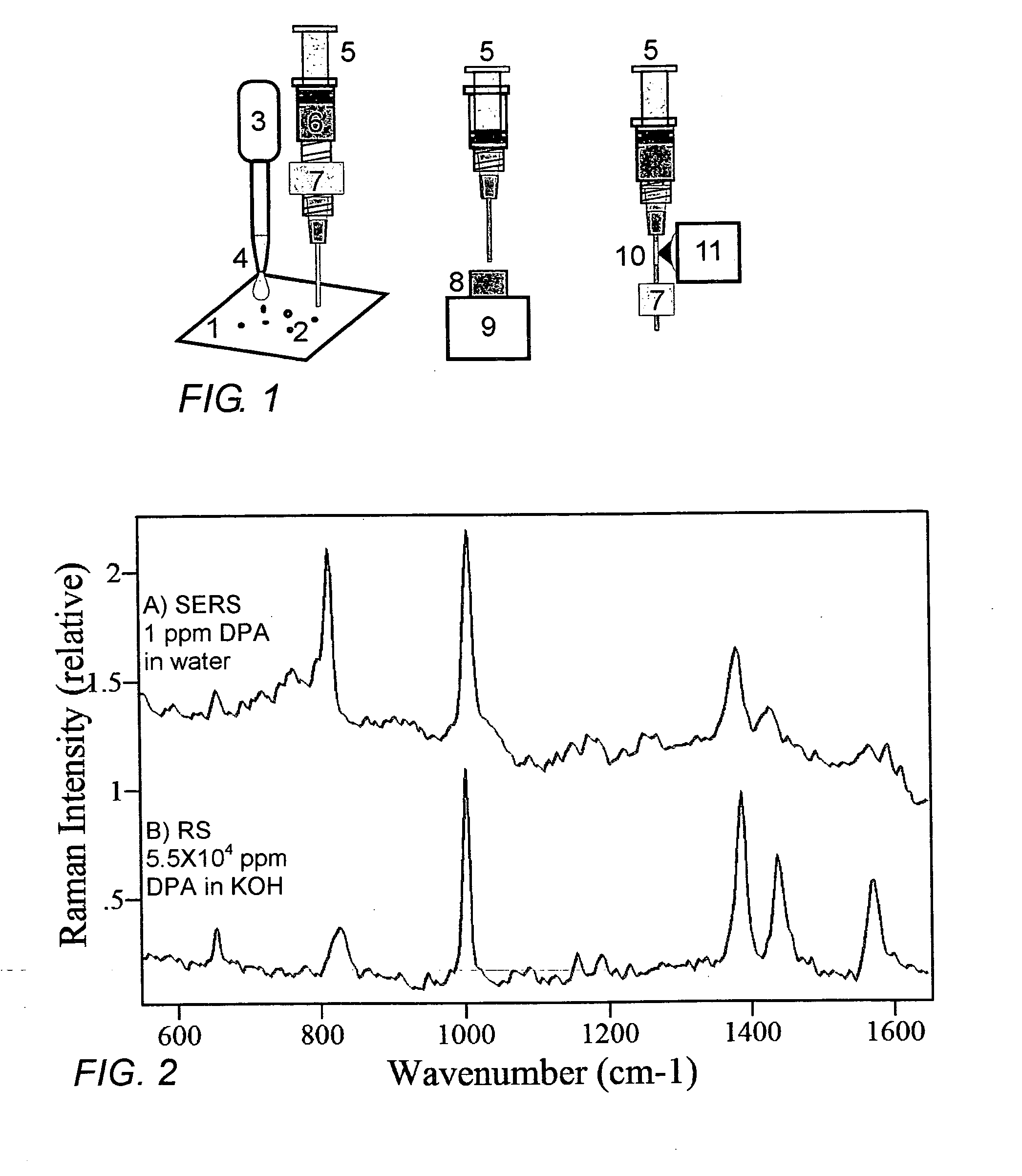
[0092] An envelope containing 1 g of Bacillus anthracis spores is passed through a mail sorting machine, hypothetically leaving behind an average of 100 picograms, or 10 spores per cm2 (invisible to the unaided eye), on the equipment surfaces 1. With reference to FIG. 1, 50 microL of glacial acetic acid is dispensed onto the surface, which spreads over a 100 cm2 circular area of the machine. During the course of 1 minute, the acetic acid effects the release of CaDPA from the estimated 1000 spores, producing from 0.5 to 1.5 nanograms of DPA in the solution. A 1 mL syringe 5 is used to draw 10 microliters of the acetic acid solution 6, containing on average 0.2 ng of DPA, through a PTFE filter 7 into a 1-mm diameter glass capillary containing a SERS-active sol-gel 10. The capillary is then mounted in the sample holder of a Raman spectrometer 11, and 85 mW of 785 nm laser excitation radiation is focused into the silver-doped sol-...

example 2

Continuous Analysis of Air to Detect B. anthracis Endospores

[0093] A container of B. anthracis endospores is opened and left in the lobby of a commercial building, causing the release of 20,000 spores per cubic meter of air. Ambient air is drawn into a return duct of the building ventilation system, and is passed, at the rate of one cubic meter per minute, through a large-particle filter or large-particle cyclone-separator, and (with reference to FIG. 12) into a cyclone unit 35. The cyclone is designed to retain particles of 1-5 micron in collector 37 while passing particulate matter of finer sizes. A thin film of acetic acid is caused to flow from the reservoir 36 over the inner, lower portion of the conical enclosure of the cyclone, thereby collecting 50 percent of the deposited particles; air is passed through the cyclone at the rate of one cubic meter per minute, and acetic acid is collected at the rate of 50 mL per minute. A pump 38, and appropriate valves (not shown), are use...

example 3

Analysis of Hospital Air for Clostridia Endospores

[0095]Clostridia are one of the most common bacterial causes of food-borne illnesses (Clostridium perfengens), including botulism (Clostridium botulinum). They also cause tetanus (Clostridium tetani), pseudomembranous colitis, and anti-biotic associated diarrhea, the latter being the most frequently identified cause of hospital-acquired diarrhea (Clostridium defficile). For example, see McDonald L C, M Owings, D B Jernigan, “Increasing Rates of Clostridium difficile Infection Among Patients Discharged from US Short-Stay Hospitals, 1996-2003”, Emerg Infect Dis, 12, 1-20, 2006.

[0096] Similar to the previous Example, the ambient air is drawn into a return duct of a hospital ventilation system, and is passed, at the rate of one cubic meter per minute, through a multi-stage impactor that captures particles of decreasing size range as it passes through each stage, until finally, particles of 1-5 micron are deposited into numerous vials r...


PUM

PropertyMeasurementUnit
Fractionaaaaaaaaaa
Fractionaaaaaaaaaa
Timeaaaaaaaaaa

Abstract

A weak organic acid is used to effect the release of CaDPA from Bacillus or Clostridium endospores, rapidly and at room temperature, to enable detection and measurement of DPA and thereby the assessment of risk associated with exposure to Bacillus anthracis, Clostridium botulinum, and like spores. The method can be applied to airborne, food-borne, and water-borne spores, as well as to spores collected from surfaces or contained in body fluids, and analysis is advantageously carried out using surface-enhanced Raman spectroscopy.

Description

CROSS REFERENCE TO RELATED APPLICATION [0001] This application claims the benefit of U.S. Provisional Patent Application No. 60 / 654,664, filed Feb. 18, 2005 in the names of the same inventors.STATEMENT REGARDING FEDERALLY SPONSORED RESEARCH AND DEVELOPMENT [0002] The U.S. Government has rights in this invention pursuant to US Army Contract No. DAAD13-02-C-0015, NSF Contract No. DMI-0214280 and NSF Contract No. DMI-0215819.BACKGROUND OF THE INVENTION [0003] Calcium dipicolinate (CaDPA, calcium 2,6-pyridinedicarboxilate) represents 5 to 15% by weight of Bacillus anthracis spores (Murrell, W G, in The Bacterial Spores, Gould, G W, A Hurst, Eds. Academic Press, London, 1969, p. 215), and consequently a number of methods are being developed and used to detect CaDPA or its derivatives as an indicator or biochemical signature for the presence of B. anthracis, a bacterium that has been used in bioterrorism due to its ability to cause anthrax. The appropriateness of measuring CaDPA or its de...

Claims


Application Information

Patent Timeline
IPC IPC(8): C12Q1/68
CPCC12Q1/04G01N2333/33G01N2333/32Y02A50/30
Inventor FARQUHARSON, STUARTINSCORE, FRANK E.GIFT, ALAN D.SHENDE, CHETAN SHRIKANT
Owner REAL TIME ANALYZERS
Learn More