
Method of constructing artificial cell tissue and base material therefor

Inactive Publication Date: 2007-05-31
DAI NIPPON PRINTING CO LTD
View PDF8 Cites 29 Cited by
  • Summary
  • Abstract
  • Description
  • Claims
  • Application Information

AI Technical Summary

Benefits of technology

[0011] The present invention has been completed to address the above problems in conventional technology. Specifically, an object of the present invention is to array and culture cells in a finely patterned state using a convenient method without damaging the cells and thus to promote tissue formation by cultured cells.

Problems solved by technology

Artificial skin or the like containing a synthetic polymer is not preferable for transplantation because it may cause rejection or other problems.
Animal cells are thus unable to survive for a long time period in a floating state ex vivo.
However, such cells generally form a tissue with difficulty, so that it is impossible to obtain original cellular functions.
Chemical modification of a biopolymer or the like to impart such photosensitivity is often difficult.
This leads to a problem such that the selectivity range of a cell adhesive material is extremely narrowed.
Moreover, biomaterials and the like having high ability to culture cells are generally difficult to decompose by plasma.
Thus, patterning using a plasma etching method also has low industrial productivity and thus is impractical.
Thus, such process is problematic in that treatment steps become complicated, the possibility of contamination becomes high, cells are denatured or damaged, original cellular functions may deteriorate, and the like.
However, it is difficult to efficiently form a fine pattern using the method disclosed herein.
Furthermore, the method also requires variation of temperature when cells are removed from the support, so that the steps are complicated.
Moreover, direct patterning on a biomaterial such as an organ is also difficult.
However, in order to prepare many artificial blood vessels by this method, many finely processed cell culture substrates must be prepared and tissue formation requires much time.
Thus, such method has also low industrial productivity.

Method used

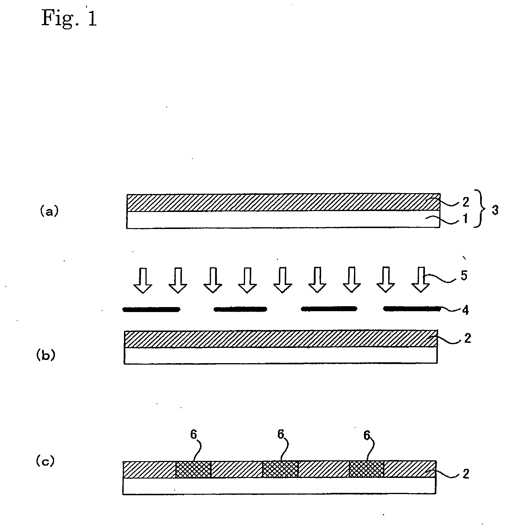
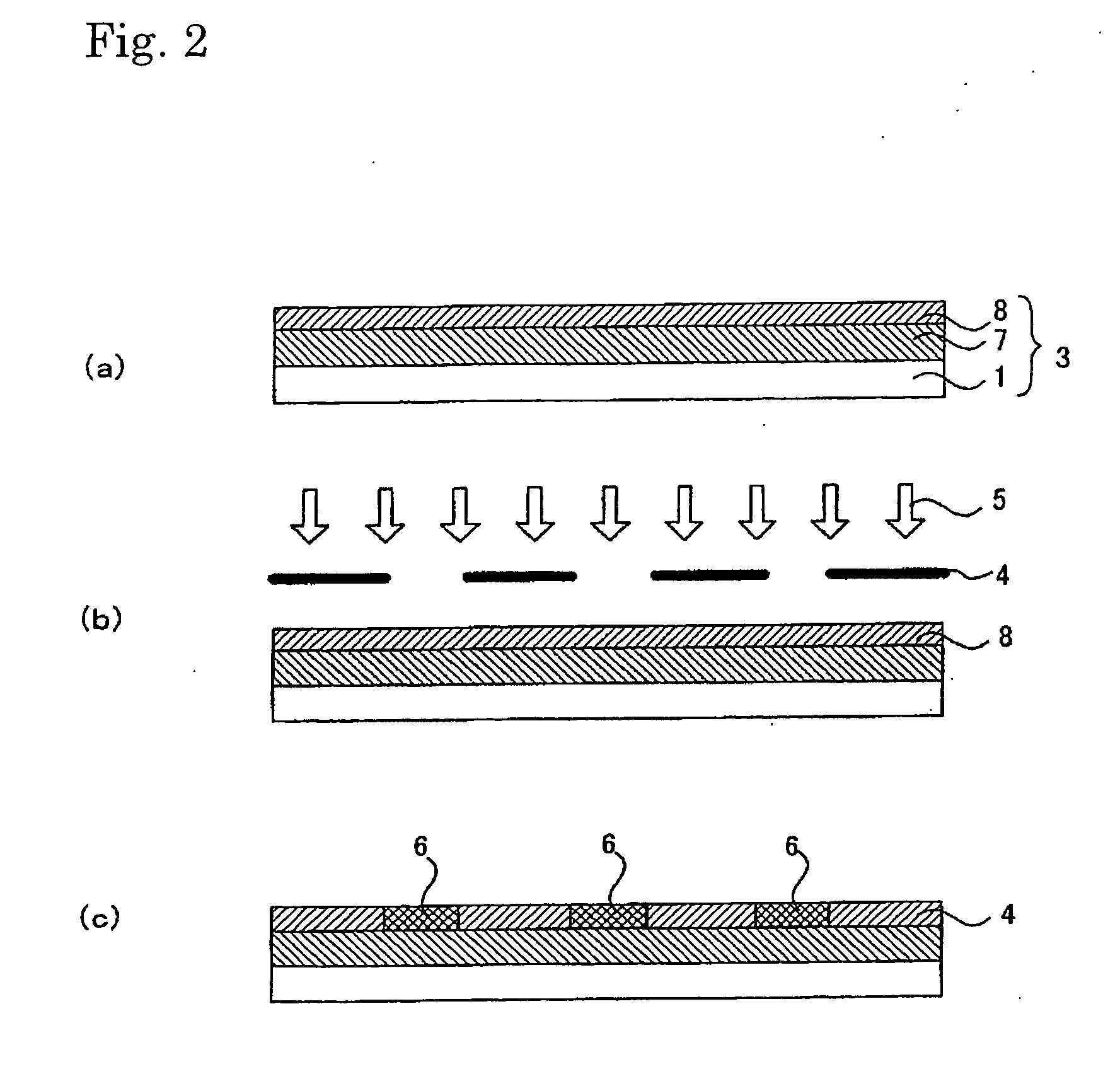
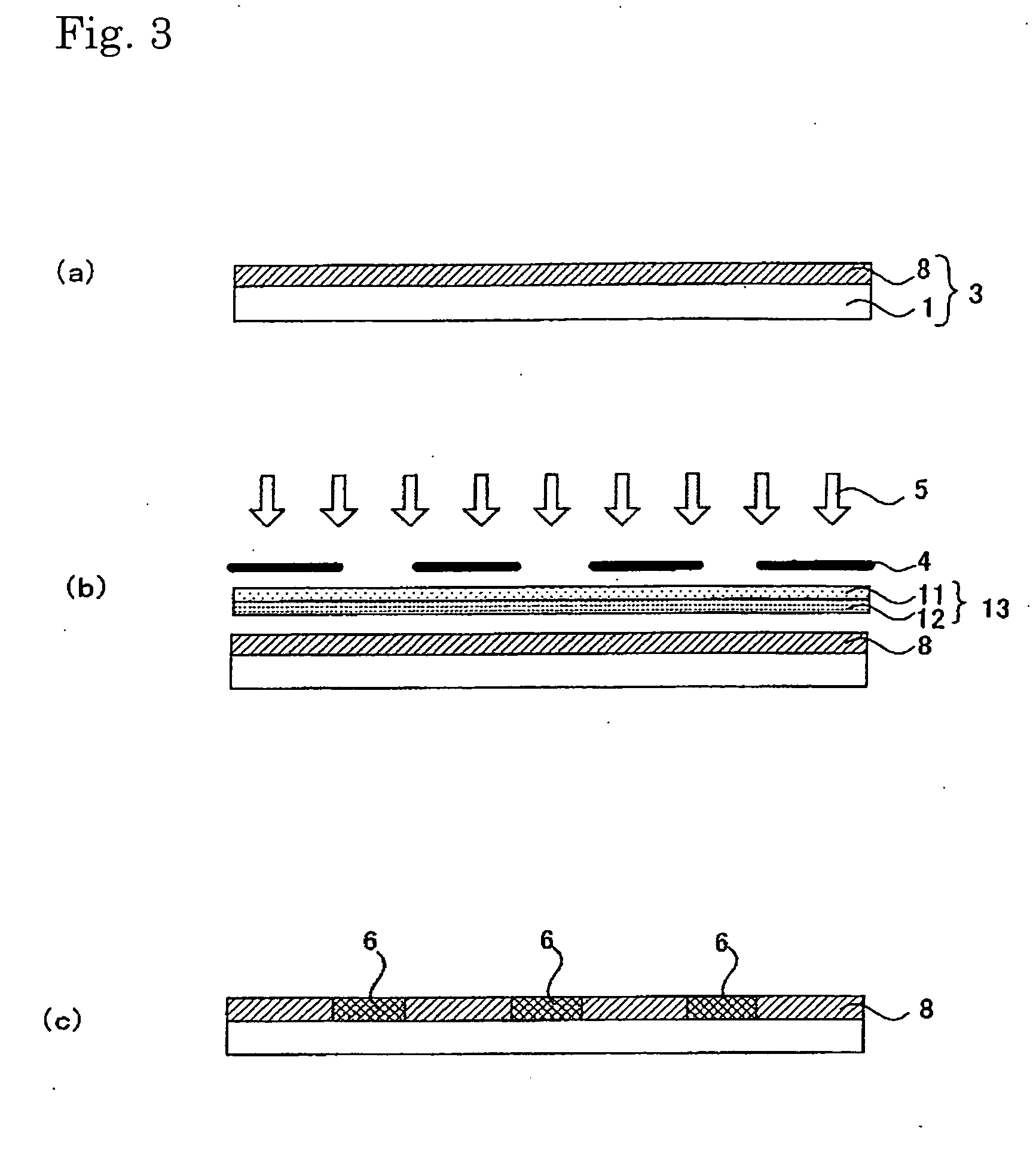

Image

  • Method of constructing artificial cell tissue and base material therefor
  • Method of constructing artificial cell tissue and base material therefor
  • Method of constructing artificial cell tissue and base material therefor

Examples


example 1

[0311] 1.5 g of fluoroalkyl silane TSL8233 (GE Toshiba Silicones), 5.0 g of tetramethoxysilane TSL8114 (GE Toshiba Silicones), and 2.4 g of 5.0×10⁻³N HCl were mixed for 12 hours and then diluted 10-fold with isopropyl alcohol.

[0312] Next, 2.0 g of the solution was applied to a 10 cm×10 cm soda glass substrate using a spin coater at 1000 rpm for 5 seconds. The substrate was dried at 150° C. for 10 minutes.

[0313] Next, 3.0 g a titanium oxide sol solution (ISHIHARA SANGYO KAISHA, LTD. STK-03) diluted 3-fold with isopropyl alcohol was used as a composition for a photocatalyst-comprising layer.

[0314] The above composition for a photocatalyst-comprising layer was applied to the patterned surface (on which line portions each having a width of 60 μm and space portions each having a width of 300 μm had been arranged alternately) of a line & space negative photomask (quartz) using a spin coater at 700 rpm for 3 seconds, followed by 10 minutes of drying treatment at 150° C. Thus, a photomas...

example 2

[0320] As cells to be cultured, bovine carotid-derived vascular endothelial cells (Onodera M, Morita I, Mano Y, Murota S: Differential Effects of Nitric Oxide on the Activity of Prostaglandin Endoperoxide h Synthase-1 and-2 in Vascular Endothelial Cells, Prostag Leukotress 62: 161-167, 2000) of 10th to 17th generations obtained by successive culture were used.

[0321] Bovine carotid-derived vascular endothelial cells that had reached confluence in a 10 cm dish were removed by 0.05% trypsin-EDTA treatment. The number of cells was counted using a Coulter counter™ ZM and then the concentration was adjusted to 106 cells / ml. The cell array substrate (exposure time: 360 seconds) prepared in Example 1 was sterilized with an autoclave. The above endothelial cells were inoculated at 106 cells / 5 ml per well on the culture dish (Heraeus Quadriprem™, 76 mm×26 mm, and 1976 mm2) on which the cell array substrate had been placed. The cells were incubated for 24 hours using a CO2 incubator.

[0322] 0...

example 3

[0324] 10 g of a fluorine coating agent XC98-B2742 (GE Toshiba Silicones) was diluted 10-fold with isopropyl alcohol. 5 g of 1,3-butanediol was further added as a solvent with a high boiling point and then the solution was stirred for 5 minutes.

[0325] A polyester film 150-T60 (Lumilar, Toray Industries, Inc.) having a thickness of 150 μm, which had been cut to A4 size, was spin-coated with the solution. Subsequently, the film was heated in a clean oven at 130° C. for 10 minutes, washed with water, and then dried at 90° C. for 3 minutes.

[0326] In the meantime, on a negative photomask (quartz), line portions (opening) each having a width of 60 μm and space portions (shielding portions) each having a width of 300 μm were arranged alternately. Line portions (openings) each having a width of 60 μm orthogonally crossing the line & space pattern were formed at intervals of 2.5 cm. In a manner similar to that of Example 1, the photomask was coated with a photocatalyst layer. Thus, a photo...


PUM


Abstract

The present invention relates to a method for culturing cells, which comprises the steps of: causing cells to adhere to the surface of a cell array substrate having a cell adhesiveness variation pattern that comprises regions having good cell adhesiveness and regions having inhibited cell adhesiveness patterned on a substrate; transferring the adhered cells to a cell culture substrate in such patterned state; and culturing the transferred cells.

Description

TECHNICAL FIELD [0001] The present invention relates to a method for culturing cells in a patterned state, a cell tissue prepared by the method, and a substrate to which cells have adhered in a patterned state. BACKGROUND ART [0002] Recently, technology for direct transplantation of artificial alternates or cell tissues obtained by culturing cells has been a focus of attention. Typical examples of such technology include artificial skin, artificial blood vessels, and cultured cell tissues. Artificial skin or the like containing a synthetic polymer is not preferable for transplantation because it may cause rejection or other problems. On the other hand, with a cultured cell tissue, there is no concern about rejection, because such tissue is obtained by culturing the cells of a subject into which the tissue will be transplanted and thus it is preferable for transplantation. Such cultured cell tissue is prepared by collecting cells from a subject for transplantation and then culturing ...

Claims


Application Information

Patent Timeline
IPC IPC(8): C12N5/06A61L27/38C12N5/00C12N5/07C12N5/071C12N5/077
CPCA61L27/38C12N5/0068C12N2533/00C12N5/00C12M3/00
Inventor MORITA, IKUONAKAMURA, MAKOTOMIYAKE, HIDEYUKIHATTORI, HIDESHIKOBAYASHI, HIRONORIKURIHARA, MASAAKI
Owner DAI NIPPON PRINTING CO LTD
Learn More