Antibody specific to intact human autotaxin, method of screening the same and method and reagent for examining malignant lymphoma by assaying autotaxin

an autotaxin and autotaxin technology, applied in the field of antibodies, can solve the problems of incomplete analysis of autotaxin detail, and achieve the effect of enabling quantification of human autotaxin in a short period of tim

Inactive Publication Date: 2010-05-13
THE UNIV OF TOKYO +1
View PDF4 Cites 3 Cited by
  • Summary
  • Abstract
  • Description
  • Claims
  • Application Information

AI Technical Summary

Benefits of technology

[0007]We found a technique for efficiently acquiring antibody to naturally-occurring form of human autotaxin, and used the antibody to the naturally-occurring form of human autotaxin acquired with this method to successfully provide a method and reagent enabling accurate quantification of human autotaxin that is not affected by intrinsic substances that cause problems during measurement of activity and does not require pretreatment for denaturing the sample such as by denaturing with reduction treatment or denaturing using a protein denaturant such as guanidine hydrochloride or urea.
[0017]According to the inventions as described above, a monoclonal antibody capable of specifically recognizing and binding naturally-occurring form of human autotaxin can be efficiently acquired, and a measurement method enabling quantification of autotaxin in a human specimen using this antibody can be constructed without requiring pretreatment and the like. Measurement of human serum using a naturally-occurring form of human autotaxin measurement reagent obtained according to the aforementioned inventions enables diagnosis of cancer or diagnosis of chronic liver disease. In addition, according to the aforementioned method, a method and reagent enabling quantification of human autotaxin can be provided that allows quantification to be carried out in a short period of time and without being affected by intrinsic measurement-inhibiting factors or competitive enzymes that conventionally caused problems in the estimation of autotaxin levels based on lysophospholipase D enzyme activity.
[0018]Moreover, the inventors of the present invention also found that human autotaxin contained in blood and the like can be quantified both easily and in a short period of time by using a versatile human autotaxin quantitative measurement system such as ELISA using a monoclonal antibody that specifically reacts with naturally-occurring form of human autotaxin present in a human specimen. As a result of conducting extensive studies using this measurement system, concentrations of autotaxin in serum were clearly determined to be high in patients with lymphomas, and particularly patients with follicular lymphomas. Malignant lymphomas are broadly categorized into two types consisting of Hodgkin's lymphomas and non-Hodgkin's lymphomas, and roughly 90% of the lymphomas in Japan are categorized as non-Hodgkin's lymphomas. Non-Hodgkin's lymphomas are categorized on the basis of such factors as morphological characteristics (pathological classification), cell traits (“B cell-like, T cell-like or NK cell-like) or chromosomal and / or genetic information, and are categorized into an extremely diverse range of types according to WHO classification. Follicular lymphomas, which constitute one of the types thereof, have been reported to account of 10 to 15% of malignant lymphomas in Japan, and that number has demonstrated an increasing trend in recent years. Diagnosis of malignant lymphomas is made by assessing the progress of the disease by lymph node biopsy, chest X-ray, computerized tomography (CT), magnetic resonance imaging (MRI), gallium (Ga) scintigraphy or positron emission tomography (PET) and the like. Although blood tests are also used, such as measurement of lactose dehydrogenase (LDH), C-reactive protein (CRP) or soluble interleukin-2 (IL-2) receptors, none of these are diagnostic markers specific for malignant lymphomas, thereby creating a need for a diagnostic marker specific for malignant lymphomas.
[0019]Although there have been reports indicating that the concentrations at which autotaxin is present in human tissues or body fluids fluctuate due to various diseases, analyses of the causative relationship with various diseases have not been conducted since there has been no method for quantification thereof. According to the present invention, concentrations of autotaxin in serum and other human body fluids are able to be quantified easily, in a short period of time and with high reliability. This measurement method has made it possible to identify the relationships between autotaxin concentrations and various diseases that were heretofore unable to be determined through the use of measurement reagents. Moreover, as a result of further conducting extensive studies using this measurement system, testing for malignant lymphomas, and particularly follicular lymphomas, for which diagnosis was previously both complex and difficult due to the absence of a serum marker, has also become possible.
[0020]The inventors of the present invention were able to accurately quantify human autotaxin by constructing an immunochemical measurement system using antibody to human autotaxin without being affected by intrinsic substances that cause problems during measurement of enzyme activity and without requiring pretreatment of the specimen. As a result of conducting extensive studies using this measurement reagent, it was found that serum autotaxin concentrations exhibit high values in malignant lymphoma patients, and particularly in follicular lymphoma patients, thereby making it possible to provide a reagent capable of detecting or facilitating detection of follicular lymphomas. In addition, this reagent also enables detection or facilitates detection of follicular lymphomas by measurement of the lysophospholipase D enzyme activity of autotaxin, although inferior in terms of complexity and accuracy.
[0022]According to the aforementioned inventions, malignant lymphomas, and particularly follicular lymphomas, can be detected by quantifying the concentration of autotaxin in a human specimen with a quantification reagent using a monoclonal antibody specific to human autotaxin without requiring pretreatment of the autotaxin in the human specimen. The use of this immunoassay reagent makes it possible to provide a reagent enabling quantification of human autotaxin in a short period of time and without being affected by intrinsic measurement-inhibiting factors or competitive enzymes contained in specimens. In addition, although inferior to the aforementioned immunochemical quantification method in terms of complexity and accuracy, the aforementioned inventions also allow assessment of malignant lymphomas and are able to provide a reagent for that purpose even in the case of measurement of lysophospholipase D enzyme activity.

Problems solved by technology

Although there have been reports suggesting that concentrations at which human autotaxin is present in human tissues or body fluids fluctuate due to various diseases, since there has heretofore been no method for quantifying human autotaxin, detailed analyses thereof have not been performed.

Method used


Image

  • Antibody specific to intact human autotaxin, method of screening the same and method and reagent for examining malignant lymphoma by assaying autotaxin
  • Antibody specific to intact human autotaxin, method of screening the same and method and reagent for examining malignant lymphoma by assaying autotaxin
  • Antibody specific to intact human autotaxin, method of screening the same and method and reagent for examining malignant lymphoma by assaying autotaxin

Examples


example 1

Expression of Recombinant Human Autotaxin

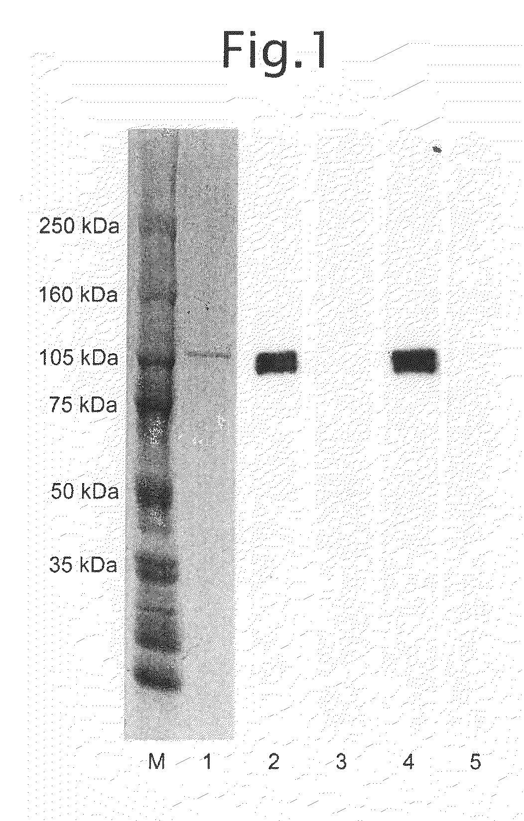
[0058]Nucleotide numbers 1 to 2589 (SEQ ID NO: 1) of Autotaxin-t (GenBank accession number L46720) were cloned from a human liver cDNA library using RT-PCR in accordance with ordinary methods. This cDNA was inserted into a baculovirus transfer vector pFASTBac-1 (Invitrogen), and a baculovirus for expressing full-length human autotaxin was prepared using the Bac-to-Bac System (Invitrogen) in accordance with the protocol. The stop codon (TAA nucleotide sequence 2590 to 2592) was removed from the cloned full-length human autotaxin cDNA followed by the addition of 6 histidine residues to prepare a baculovirus for expressing polyhistidine-tagged human autotaxin having polyhistidine in accordance with the protocol. Culture supernatants containing expressed full-length human autotaxin and polyhistidine-tagged human autotaxin were able to be prepared by infecting into sf9 or sf21 and the like in accordance with ordinary methods using these baculoviru...

example 2

Purification of Polyhistidine-Tagged Human Autotaxin

[0059]One liter of cells of insect cell line sf21 (5×10⁵ cells / mL) were infected with baculovirus for expressing polyhistidine-tagged human autotaxin followed by culturing for 4 days at 28° C. Following completion of culturing, the cells were separated by centrifugal separation (10 minutes at 3000 rpm) followed by removal of cell fragments and other precipitates with a 0.45 μm filter. The recovered culture supernatant was dialyzed with TBS (Tris-buffered saline: 10 mM Tris-HCl, 150 mM NaCl, pH 7.4) and then purified using a metal chelating column packed with BD-TALON Metal Affinity Resin (BD Biosciences, Cat. No. 63501) in accordance with the manual provided. More specifically, the column was packed with 5 mL of resin by volume. 50 mL of a 50 mM CoCl2 solution were added to the column to bind the cobalt. After washing the column with an aqueous solution containing 300 mM NaCl, the column was equilibrated with a pH 7.7 aqueous solut...
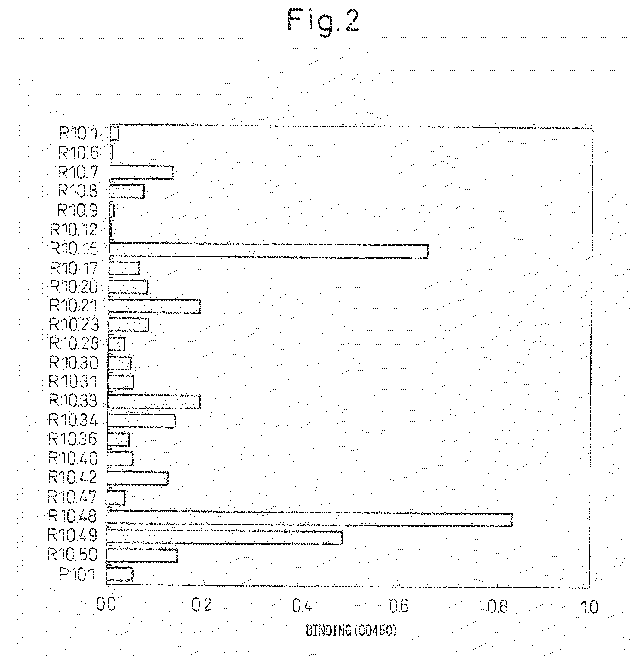
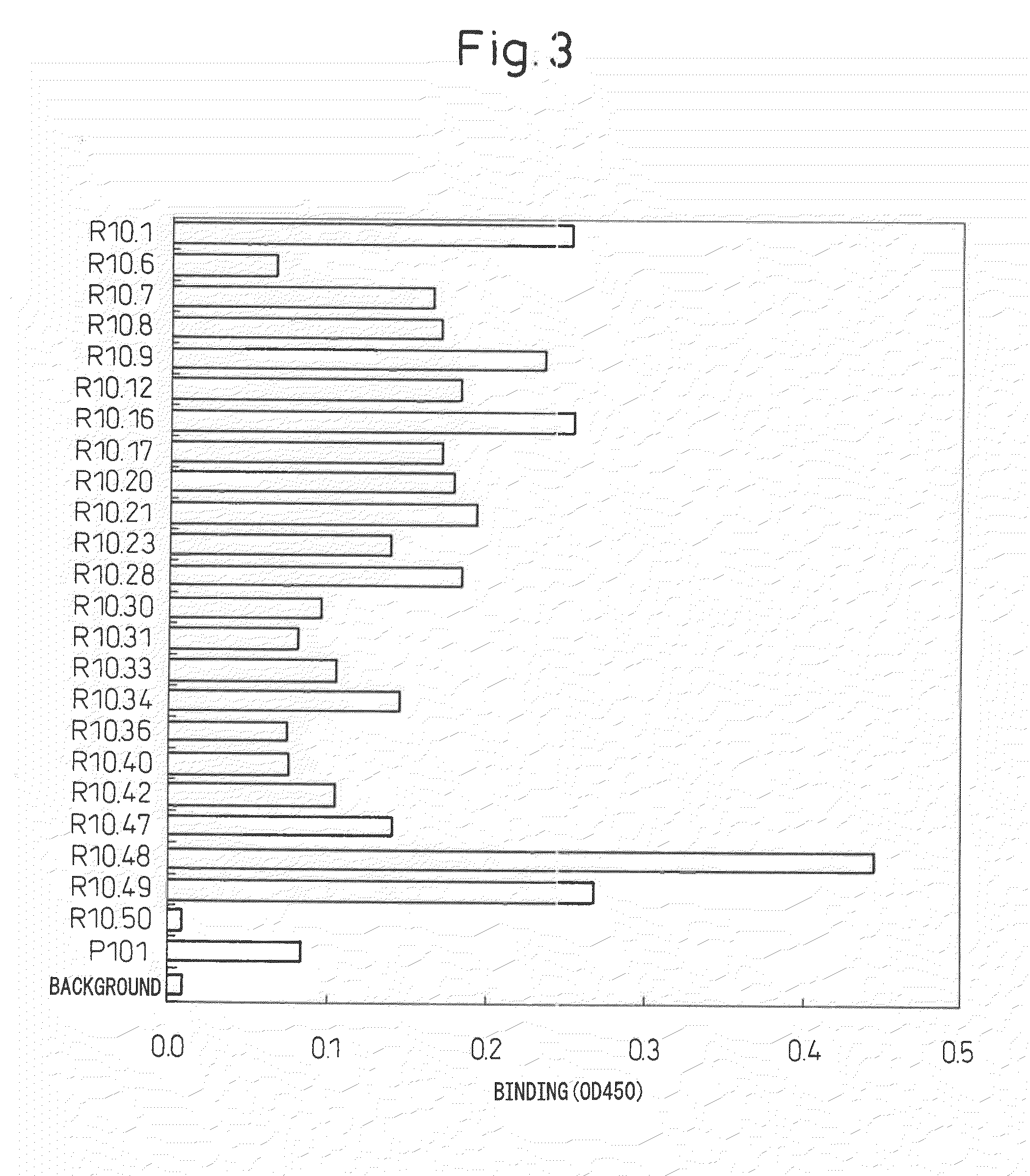
example 3

Monoclonal Antibody Production

[0060]Seven-week-old female Wistar-Lewis rats were immunized with 250 μg of antigen together with Freund's complete adjuvant into a hind limb under ether anesthesia. One month later, the inguinal lymph nodes and iliac lymph nodes were excised from the rats followed by recovery of B cells. Cell fusion was carried out in accordance with conventional methods with mouse myeloma cell line PAI in the presence of polyethylene glycol, selection was carried out for about 10 days using HAT medium, and target antibodies were selected by screening for antibody-producing hybridomas in accordance with Example 4. Cells in those wells that screened positive were established as hybridomas by monocloning using the limiting dilution method. At that time, after culturing for about 10 days using HT medium, culturing was finally continued with hybridoma medium and the supernatant was recovered to recover the antibody. Medium containing 27.5 mL of NCTC-109 medium (Invitrogen)...


PUM

PropertyMeasurementUnit
molecular weightaaaaaaaaaa
concentrationsaaaaaaaaaa

Abstract

A screening method is provided for antibody specifically recognizing naturally-occurring form of human autotaxin in a state in which it is present in the body without being denatured, comprising the steps of: binding a binding factor capable of capturing a candidate antibody of the antibody to a solid phase; binding the candidate antibody of the antibody to the binding factor; allowing naturally-occurring form of human autotaxin to react on a system in which the candidate antibody has reacted; and selecting an antibody that specifically recognizes the naturally-occurring form of human autotaxin by using as an indicator thereof the binding strength of the naturally-occurring form of human autotaxin to the antibody. Moreover, a detecting method for malignant lymphoma is provided comprising: measuring the concentration of autotaxin in a human specimen, and judging to be a malignant lymphoma in the case that value exhibits a significantly higher value than normal values comprised of measured values from normal healthy subjects.

Description

TECHNICAL FIELD[0001]The present invention relates to an antibody that specifically recognizes and binds naturally-occurring form of human autotaxin, a screening method thereof, and a method and reagent for detecting malignant lymphoma based on measurement of autotaxin concentration in a specimen.BACKGROUND ART[0002]Human autotaxin is a glycoprotein having a molecular weight of about 125 kDa first isolated from a culture medium of A2058 human melanoma cells by M. L. Stracke, et al. in 1992 as a substance that induces cell motility (J. Biol. Chem., 256, 2524, 1992). Subsequently, as a result of an analysis of its structure by J. Murata, et al. in 1994 using cDNA cloning, it was determined to have a single transmembrane moiety on the amino terminal, contain two somatomedin B regions, i.e., type I phosphodiesterase activity and an EF hand loop regions in the extracellular domain, and have phosphodiesterase activity (J. Biol. Chem., 269, 30479, 1994). However, there was yet to be any we...

Claims


Application Information

Patent Timeline
Patent Type & Authority Applications(United States)
IPC IPC(8): G01N33/573G01N33/566G01N33/53C07K16/18
CPCC07K16/28C07K16/40C07K2316/96C07K2317/34G01N33/573G01N2333/916G01N33/564C07K2317/76
Inventor AOKI, JUNKENARAI, HIROYUKIYATOMI, YUTAKAIKEDA, HITOSHINAKAMURA, KAZUHIROIGARASHI, KOJIIDE, KAZUFUMI
Owner THE UNIV OF TOKYO
Why Eureka
Try Eureka