Detection of nucleic acids and nucleic acid units

a nucleic acid and nucleic acid technology, applied in the field of nucleic acids and nucleic acid units, can solve the problems of high technical skill, complicated use, and high cost, and achieve the effects of reducing operator time, preventing the risk of pcr product crossover contamination, and high autofluorescen

Inactive Publication Date: 2000-10-03
RENISHAW DIAGNOSTICS
View PDF3 Cites 200 Cited by
  • Summary
  • Abstract
  • Description
  • Claims
  • Application Information

AI Technical Summary

Benefits of technology

A) Genetic disease, acquired or inherited, arises when the DNA sequence of an organism's genome varies from the wild-type sequence at certain crucial points. Analysis of this variation is currently carried out using sequencing, SSCP and DGGE to analyse DNA which has been amplified by the PCR, or other amplification based techniques. The use of ARMS (EP-B-3,332,435) is useful for generating a PCR product in a sequence dependent manner which precludes the need subsequently to analyse the sequence of the amplified fragment. Direct analysis of genetic disease by using ARMS to generate linear extension products from genomic DNA, with subsequent detection of the extension using the method of the present invention, would speed genetic analysis. Avoiding PCR amplification would also prevent the risk of PCR product crossover contamination. The use of ARMS to detect sequence variation is not limited to mutations associated with disease states but could also be applied to detecting single nucleotide polymorphisms (SNPs) which are silent mutations useful in distinguishing individuals genetically.
B) Variable numbered tandem repeat (VNTR) analysis is a means of identifying individuals based on genetic variability from person to person. Genomic DNA is treated with restriction enzymes to generate DNA fragments, some of which contain tandem repeat sequences of variable length. These fragments are separated by electrophoresis and the gel is denatured with alkali. The DNA is then transferred to a membrane and a labelled single stranded oligonucleotide probe complementary to the repeat sequence is hybridised to the DNA. After the probe has been subjected to a stringency wash it is detected via its label. For example, probes labelled with the enzyme alkaline phosphatase might be detected by their generating chemiluminescence from a suitable phosphorylated substrate or radiolabelled probes could be detected by autoradiography. Once the probe has been detected it is stripped off and the next probe is hybridised in order to detect another VNTR. Usually three to five probings are carried out per individual and each probing takes a working day. The probes are not multiplexed because enzyme-linked detection and autoradiography do not lend themselves to such multiplex detection and fluorescent probing is not sensitive enough due to the small amounts of input DNA and the high autofluorescene of hybridisation membranes. It would be an advantage in reducing operator time if all the probings could be carried out simultaneously. The methods of the present invention would allow the multiplex detection of several probes with their unique Raman vibrational "fingerprints" without the problem of autofluorescent backgrounds.
C) Difficulties in multiplexing fluorescence are also observed in the field of cytogenetic analysis by Fluorescence In-Situ Hydridisation (FISH). Due to the broad fluorescence of most organic fluorophores it is difficult to select more than five visible fluorophores whose fluorescence does not overlap so that many cytogenetics laboratories use only two or three fluorophores to label their chromosome probes which limits multiplex detection. One solution is to use varying mixtures of fluorophores to colour different chromosome regions with sequential detection of each colour with a separate filter and computer merging of the resulting images to identify each differently labelled region. This technique requires expensive imaging and analysis equipment and the technique is used by relatively few laboratories. It would be advantageous to use the discrete spectral lines of Raman spectra to allow different chromophores to be "fingerprinted" by hybridisation to different SER(R)S-active probes. A further advantage of such a technique is that Raman signals do not autoquench so that labelling chromophores can be packed more densely onto the chromosome surface than can fluorophores, with a corresponding build-up of signal.

Problems solved by technology

a) fluorescence spectroscopy--this is technically very demanding if high sensitivities are to be achieved. In biological assays, its use tends to be complicated by autofluorescence of the analytes.
b) radiolabelling--this also requires high levels of technical skill but tends to be less sensitive than fluorescence spectroscopy. It also suffers from the obvious hazards involved in handling radioactive materials.
c) chemiluminescence--although this technique can be relatively quick to carry out, and avoids the problem of autofluorescence and the need to handle toxic substances, it is unfortunately relatively insensitive and yet is still technically demanding.
A disadvantage common to many known techniques is their need for large amounts of the target analyte, ie, their relatively low sensitivity.
Often in the situations mentioned above the target is simply not available in sufficiently high concentrations.
Clearly, the need for amplification makes a detection method more complex, costly and time-consuming and introduces greater potential for error and for contamination of the target material.
Normal Raman lines are relatively weak and Raman spectroscopy is therefore too insensitive, relative to other available detection methods, to be of use in chemical analysis.
Raman spectroscopy is also unsuccessful for fluorescent materials, for which the broad fluorescence emission bands (also detected orthogonally to the incident light) tend to swamp the weaker Raman emissions.
This suggests that prior art methods have not been sensitive enough to detect unamplified samples.
However, there is no enabling disclosure of a technique with sufficient sensitivity to be used without prior DNA amplification.
These detection levels, and the reference to the use of PCR in the paper's conclusion, indicate that the technique disclosed would still be unsuitable for detecting unamplified DNA samples.
None has yet demonstrated sensitivities high enough to allow the detection of unamplified nucleic acid samples (ie, the detection of perhaps 1-100 molecules in a sample).
That SERS and SERRS have never been proposed for use in the detection of unamplified nucleic acids is due at least in part to the obvious difficulties in achieving the appropriate sensitivities.
These difficulties are partly due to problems specific to nucleic acids, problems which are therefore not addressed in the more general SER(R)S literature.
The skilled person seeking to detect nucleic acids or nucleic acid units would thus consider SER(R)S spectroscopy to lack the necessary sensitivity or robustness, certainly without target amplification.

Method used


Image

  • Detection of nucleic acids and nucleic acid units
  • Detection of nucleic acids and nucleic acid units
  • Detection of nucleic acids and nucleic acid units

Examples


example 2

Preparation of benzotriazole azo dyes - Specific examples

A) 4-(5'-azobenzotriazolyl)-phenylamine Aniline (1 eq) was dissolved in sodium acetate buffer (1.0M, 5 ml, pH 6.0) and acetone (5 ml). Diazotised aminobenzotriazole was added to this solution dropwise at 0.degree. C. with stirring over 1 hour. The solid produced was isolated by filtration and washed with saturated KCl (3.times.50 ml) to leave a dark yellow residue (0.892 g, 3.74 mmol, 84%), R.sub.f [dichloromethane / methanol (A) 9 / 1] 0.37; .delta..sub.H (DMSO-d6) 4.10 (1H, s, NH) 7.14-8.25 (7H, m, ar) 12.73 (2H, br s, NH.sub.2) ; .lambda..sub.max (MeOH) 360 nm.

B) 3-methoxy-4-(5'-azobenzotriazolyl)-phenylamine Anisidine (1 eq) was dissolved in sodium acetate buffer (1.0M, 5 ml, pH 6.0) and acetone (5 ml). Diazotised aminobenzotriazole was added to this solution dropwise at 0.degree. C. with stirring over 1 hour. The solid produced was isolated by filtration and washed with saturated KCl (3.times.50 ml). The residue was recrystal...

example 3

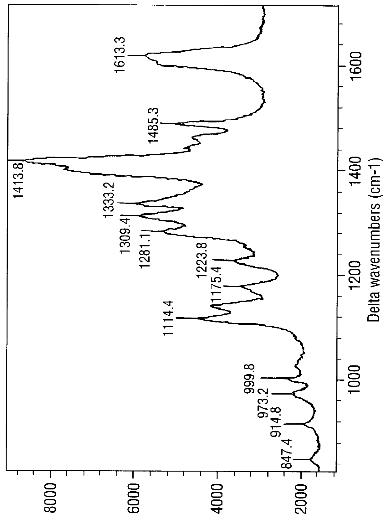
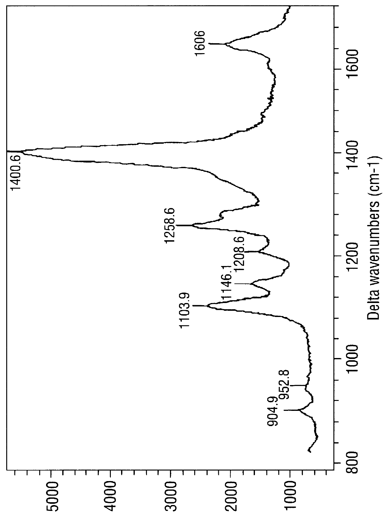
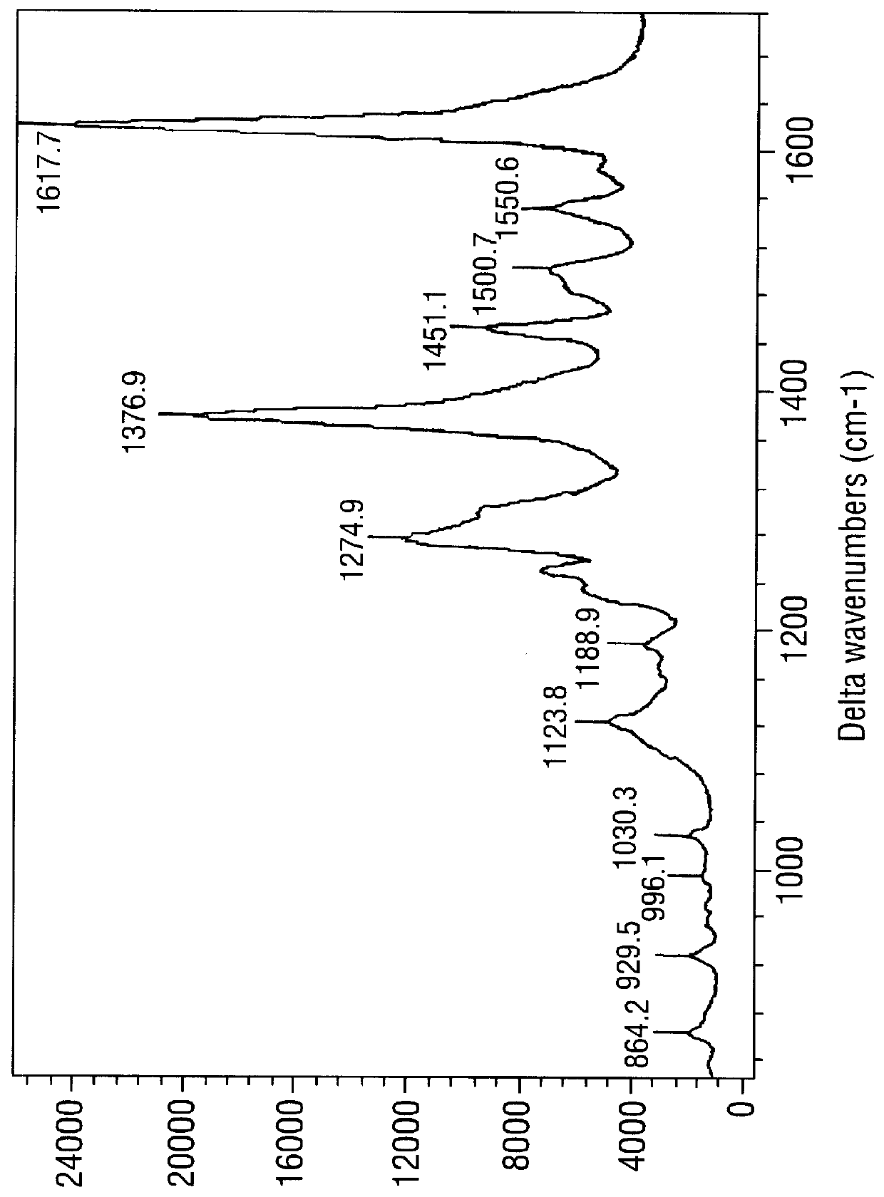
SERRS spectra for modified (benzotriazole) azo dyes

FIGS. 3-6 are SERRS spectra obtained for the modified azo dyes A-D respectively prepared in Example 2.

These are dyes containing the chemi-adsorptive benzotriazole group which tends to "seek out" a SER(R)S-active silver colloidal surface. Such dyes are of use as labels in the invention.

The conditions for obtaining the spectra were as follows. The SERRS-active surface was a citrate-reduced silver colloid prepared as in Example 5 below, in the form of a colloid / water mixture (1:1, 1 ml). To this was added a methanol solution of the dye in question (10 .mu.l, approximately 10.sup.-5 M) followed by spermine (20 .mu.l, 8.times.10.sup.-4 M). Spectra were obtained using apparatus also described in Example 5. .lambda..sub.max for the laser was 514.5 nm; .lambda..sub.max values for the dyes were .about.394 nm (FIG. 3), 409 nm (FIG. 4), 444 nm (FIG. 5) and 468 nm (FIG. 6).

example 4

Attachment of benzotriazole monoazo dyes to a nucleic acid

General method

After synthesis of the benzotriazole monoazo dye the primary amine is protected by the dimethylformamide group thus allowing selective protection of the benzotriazole secondary amine by the monomethoxytrityl group. After removal of the formamide group by treatment with base, the free amine is coupled with succinic anhydride to produce the carboxylic acid. The acid is then coupled with a methylene linker bearing a primary amine and a protected alcohol. After selective removal of the protecting group, the alcohol is phosphitylated thus producing a monomer capable of undergoing routing solid phase synthesis.

Alternatively the acid produced in the above scheme may be converted into a suitable active ester (N-hydroxysuccinimide, pentafluorophenol) and coupled with a nucleophilic primary amine at the 5'-terminus of a nucleic acid. (See J Goodchild, supra.)


PUM

PropertyMeasurementUnit
concentrationaaaaaaaaaa
diameteraaaaaaaaaa
diameteraaaaaaaaaa

Abstract

PCT No. PCT/GB96/01830 Sec. 371 Date Apr. 21, 1998 Sec. 102(e) Date Apr. 21, 1998 PCT Filed Jul. 25, 1996 PCT Pub. No. WO97/05280 PCT Pub. Date Feb. 13, 1997The invention relates to the detection of target nucleic acids or nucleic acid units in a sample, by obtaining a SER(R)S spectrum for a SER(R)S-active complex containing, or derived directly from, the target. The complex includes at least a SER(R)S-active label, and optionally a target binding species containing a nucleic acid or nucleic acid unit. In this detection method, the concentration of the target present in the SER(R)S-active complex, or of the nucleic acid or unit contained in the target binding species in the SER(R)S-active complex, is no higher than 10-10 moles per liter. Additionally or alternatively, one or more of the following features may be used with the method: i) the introduction of a polyamine; ii) modification of the target, and/or of the nucleic acid or nucleic acid unit contained in the target binding species, in a manner that promotes or facilitates its chemi-sorption onto a SER(R)S-active surface; iii) inclusion of a chemi-sorptive functional group in the SER(R)S-active label. The invention also provides SER(R)S-active complexes for use in such a method, a kit for use in carrying out the method or preparing the complexes and a method for sequencing a nucleic acid which comprises the use of the detection method to detect at least one target nucleotide or sequence of nucleotides within the acid.

Description

This invention relates to methods for detecting the presence or absence of, and analysing the sequence of, target nucleic acids in a sample. Such methods may also be applied to target nucleic acid units such as nucleotides and nucleosides and their analogues. The invention also relates to chemical complexes for use in such methods, to a kit of reagents for use in carrying out the methods and to certain novel compounds of use in the methods.BACKGROUND TO THE INVENTIONDetection of Nucleic AcidsThere are many situations in which it is necessary to detect, either qualitatively or quantitatively, the presence of nucleic acids such as DNA and RNA or their constituent nucleotides. Examples of such situations include medical diagnosis (eg, the detection of infectious agents like bacteria and viruses, the diagnosis of inherited and acquired genetic diseases and the establishment of tissue type), forensic tests in criminal investigations and paternity disputes and of course the more general a...

Claims


Application Information

Patent Timeline
Patent Type & Authority Patents(United States)
IPC IPC(8): C12Q1/68G01N21/65G01N21/63G01N33/543C07D249/18C12N15/09C12Q1/6816G01N33/566
CPCC12Q1/6816G01N21/658Y10T436/24C12Q2565/632
Inventor GRAHAM, DUNCANLINACRE, ADRIAN MATTHEW THORNTONMUNRO, CALLUM HUGHSMITH, WILLIAM EWANWATSON, NIGEL DEANWHITE, PETER CYRIL
Owner RENISHAW DIAGNOSTICS
Why Eureka
Try Eureka