Probe for mass spectrometry

a mass spectrometry and probe technology, applied in the direction of material analysis by resonance, particle separator tube details, peptides, etc., can solve the problems of disfavorable radiolabels, limited current methods for analysing protein microarrays, and limitations in both methods, so as to achieve more accurate measurement

Inactive Publication Date: 2006-06-06
SENSE PROTEOMIC LTD
View PDF7 Cites 31 Cited by
  • Summary
  • Abstract
  • Description
  • Claims
  • Application Information

AI Technical Summary

Benefits of technology

[0029]Another significant development enabling the “miniaturisation” of a protein array formed on a MALDI target derives from the application of the Applicant's COVET technology described in WO 01 / 57198. Briefly, using this technology they are able to create from cDNA libraries expressed proteins, which carry a “sequence tag” that can be used to capture the proteins with a high affinity and in a specific orientation on the microarray surface. This firstly enables proteins e.g. a protein library to be stably immobilized such that leaching of protein from the surface is avoided and secondly the oriented immobilisation of the fusion protein onto the surface ensure maximum biological activity.
[0033]In this way the Applicant has been able to introduce the protein capture moieties not only in a homogeneous, spatial defined arrangement but also in a manner which enables high affinity binding in a specific manner. The resulting surface combines selectivity for the capture of biological macromolecules on the probe with reduced non specific binding of the type commonly observed on underivatised glass or metal surfaces and additionally results in a homogeneous distribution and orientation of the captured biological macromolecules.
[0035]In a preferred embodiment the probe has as it's protein capture moieties either a biotin binder e.g. neutravidin, avidin or streptavidin or a bleomycin resistant protein binder e.g. bleomycin. The proteins are bound to the probe to create a protein microarray by printing a plurality of bacterial, yeast, sf9 or mammalian cell lysates containing fusion proteins in which a high affinity tag e.g. biotin or zeocin resistant protein (ZRP) is expressed onto the capture surface. Proteins are derived from the expression of a cDNA library and each individual clone is tagged at the C-terminus and / or on the N-terminus with a consensus sequence, which will enable high affinity recognition of the protein even in the presence of the otherwise protein repellent molecules. Only the recombinant, tagged protein can recognise the capture surface and other proteins from the lysate can be washed away as they do not bind to the protein repellent surface and do not have a high affinity to the protein binding moieties present in the layer.
[0068]In contrast to the prior art in which matrix and analyte are co crystalised in an aqueous solvent, the Applicant uses two distinct steps in which first the protein is deposited in an aqueous solvent and then the energy absorbing molecules are deposited such that they crystallise out from the non aqueous solvent on the probe. This has the advantage that the protein is deposited in its biological form. However, using a non aqueous solvent to deliver the energy absorbing molecules allows the formation of a homogenous layer of microcrystals. This has two benefits. First the formation of a homogenous layer means it is not necessary to search for a sweet spot as the homogenous layer guarantees protein in the presence of energy absorbing molecules and secondly it results in more accurate measurement due to the even nature of the layer.

Problems solved by technology

However, both methods suffer limitations due to their bias towards highly expressed proteins and the destructive method of separation.
The current methods of analysing protein microarrays are therefore restricted by the availability of appropriate labelled ligands.
However, for drug-like small molecules, which often have molecular weights of less than 1000 Da, neither radio- or fluorescent labels are desirable; radiolabels are disfavoured for health and safety reasons, whilst the introduction of a fluorophore into the small molecule could significantly perturb the structure activity profile in an unpredictable manner.
However, the development of a MALDI MS-compatible protein microarray is complex since existing methods for forming protein microarrays do not transfer readily onto to a MALDI target.
There are a number of reasons why this is the case, inter alia the specialised nature of the probe surfaces and the potential for salts present in reaction buffers to interfere with the detection method.
As a consequence it is often observed that individual crystals contain insufficient analyte for analysis by MALDI.
However, all these methods suffer from one or more of the following limitations:a) Partial or total loss of biological activity because of amine-based coupling of the analyte or the bait onto the probe;b) Low specificity between the analyte and the surface which can lead to the non-specific binding of several analytes to the surface (e.g. ion-exchange surfaces);c) Low affinity of the analyte to the surface which can lead to leaching of the analyte from the surface during any wash procedures (e.g. ion-exchange and nitriloacetic acid surfaces);d) The affinity capture surface lacks non specific protein resistance, which can lead to high levels of non-specific protein binding which would interfere with the analysis of a protein microarray;e) The availability of only a limited number of affinity capture proteins.
Thus existing methods do not enable the immobilisation of large numbers of different, purified proteins in the form of a MALDI MS-compatible microarray suitable for functional analysis of the muicroarrayed proteins.

Method used


Image

  • Probe for mass spectrometry
  • Probe for mass spectrometry
  • Probe for mass spectrometry

Examples


example 1

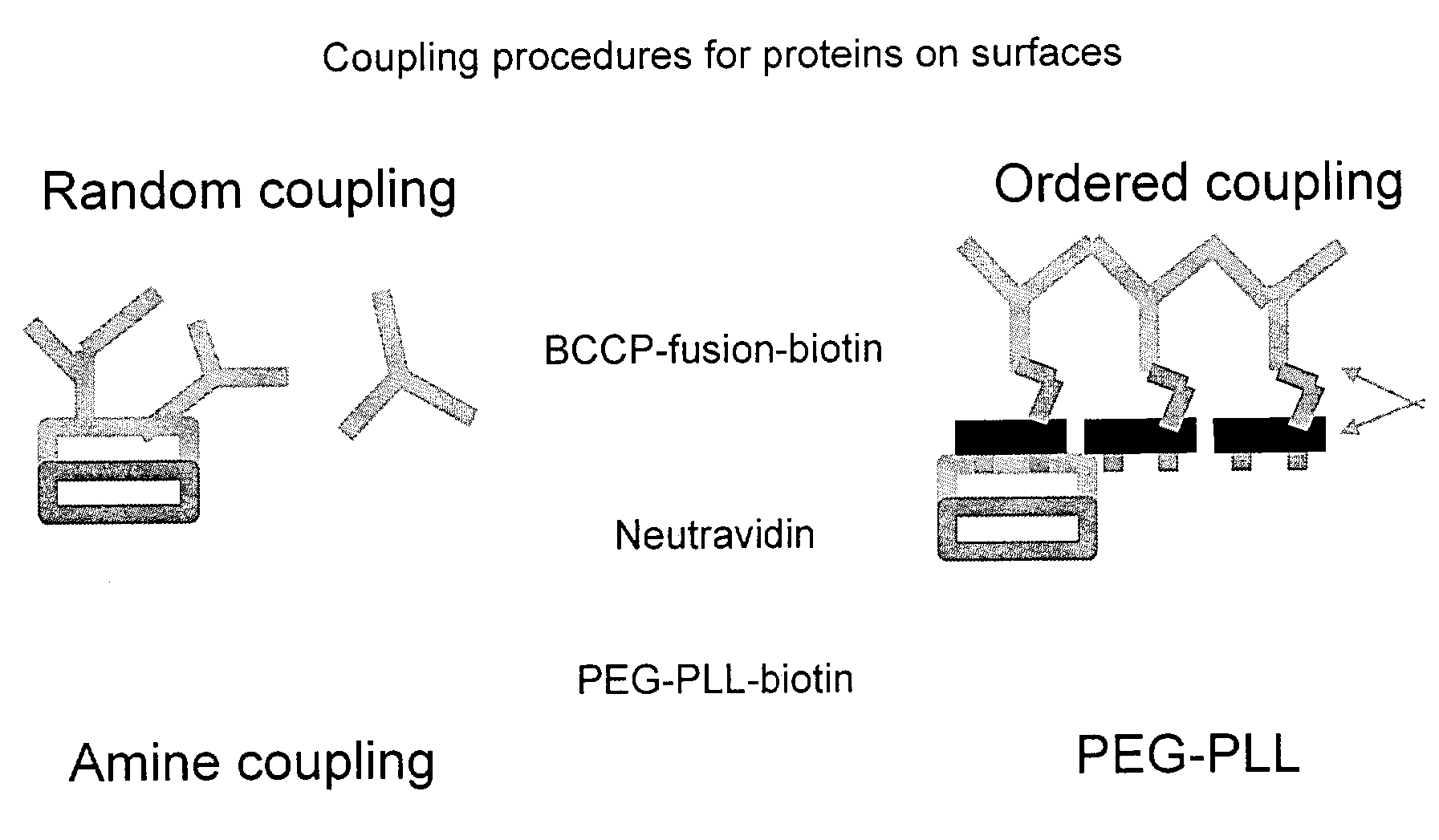
[0119]Affinity capture of a variety of tagged proteins can be demonstrated using for example PEG-PLL-biotin or PEG-PLL-Bleomycin B6 as the protein capturing moieties.

[0120]FIGS. 2, a and b show the mass spectra acquired from a protein microarray demonstrating respectively the capture of 1500 and 15 femtogram of biotin tagged insulin. The biotin tagged insulin was arrayed onto an affinity capture surface on a gold coated microscope glass slide in a 3 nanoliter volume using 300 micrometer pins (Q-Array, Genetix, New Milton, UK). The gold coated PEG-PLL-Biotin Neutravidin surface, was washed three times with 1 mM Tris-HCl pH 7.5, dried under a stream of nitrogen and overlaid with 3 nanolitre of α-cyano-4-hydroxy-cinnamic acid dissolved in 99% acetone v / v, 1% glycerol resulting in a spot with an radius of approximately 200 micrometer. The probe was analysed with a mass spectrometer MALDI TOF mass spectrometer. Several biotin tagged insulin peaks are visible due to the different degree o...

example 2

[0122]A PLL-PEG-biotin neutravidin surface on a MALDI target is overlaid with 500 nanoliters of a biotinylated protein mixture derived from an E. coli lysate expressing a human recombinant protein in conjunction with a sequence tag in this case Biotin carboxyl carrier protein (BCCP) from E. coli. The protein was captured for a period of 2 hours on the surface, washed twice with washing buffer followed by two washes with desalting buffer, and overlaid with 300 nanoliters of an energy absorbing matrix, namely saturated α-cyano-4-hydroxycinnamic acid in acetone. The mass spectrum acquired in linear mode using the delayed extraction technique at low laser power is illustrated in FIG. 5. The advantage of this method is that the sample can be applied as a complex mixture of proteins and after washing only the molecules of interest remain. Secondly the analyte is captured in a spatially defined position before it is released from the affinity capture surface by the addition of matrix.

4.3 C...

example 3

[0123]FIG. 6 shows the peptide fingerprint analysis of Glutathione-S-transferase-Biotin Carboxyl Carrier Protein (GST-BCCP). A bacterial crude lysate containing the fusion protein and bacterial proteins was placed on the MALDI target previously coated with PEG-PLL-biotin and neutravidin. The BCCP fusion partner of GST contained a biotinylation consensus sequence such that it becomes biotinylated when expressed in E. coli. allowing the fusion protein to bind specifically to the PEG-PLL-biotin neutravidin surface, whilst allowing the bacterial proteins to be washed away with buffer. For identification purpose the surface captured protein was digested by overlaying it with trypsin and analysed by MALDI MS. A protein fingerprint analysis revealed 12 peptides belonged to GST from Schistosoma mansoni, 4 peptides belonging to Neutravidin and 3 to trypsin (see table 1), but no bacterial protein was identified using the remaining un-matched peptides. This experiment demonstrates that PEG-PLL...


PUM

PropertyMeasurementUnit
areaaaaaaaaaaa
areaaaaaaaaaaa
areaaaaaaaaaaa

Abstract

The present invention relates to a probe for the analysis of one or more analytes, particularly proteins or compounds capable of binding or otherwise interacting therewith, by laser desorption / ionization mass spectrometry, more particularly MALDI MS. It also relates to a protein microarray, a method of producing a protein microarray and a method of analyzing a protein microarray. The probe comprises a support having an electroconductive target surface thereon characterized in that the target surface comprises a micro array having a plurality of discrete target areas presenting one or more analyte capture moieties. Each discrete target area has an area of less than 1000 μm2, more preferably still less than 500 μm2, and more preferably still less than 100 μm2.

Description

[0001]The present invention relates to a probe for the analysis of one or more analytes, particularly proteins or compounds capable of binding or otherwise interacting therewith, by laser desorption / ionisation mass spectrometry, more particularly MALDI MS; It also relates to a protein microarray, a method of producing a protein microarray and a method of analysing a protein microarray.[0002]Such a mass spectrometry probe, upon which a microarray has been fabricated, enables interrogation of protein—small molecule interactions in a label-free manner by desorption and ionisation of analytes (e.g. protein, drug or drug candidate, carbohydrate, DNA, RNA or other test molecule). The probe and methods are particularly useful in the drug discovery process, for example in hit series evaluation, lead optimisation, predictive toxicogenomics and metabolite profiling.[0003]Analysis of disease processes and drug effects have traditionally focussed on genomics, whereas proteomics, the study of th...

Claims


Application Information

Patent Timeline
Patent Type & Authority Patents(United States)
IPC IPC(8): B01D59/44G01N27/64A61K47/48C07K14/00G01N27/62G01N33/483G01N33/53G01N33/94G01N37/00H01J49/04H01J49/16
CPCA61K47/48407B82Y30/00C07K14/00H01J49/0418G01N33/9446Y10T436/24G01N2415/00A61K47/6809
Inventor KOOPMAN, JENS-OLIVERBLACKBURN, JONATHAN M.
Owner SENSE PROTEOMIC LTD
Why Eureka
Try Eureka