Antioxidant enhancement of therapy for hyperproliferative conditions

a technology of anti-proliferative conditions and anti-oxidant enhancement, which is applied in the field of medicinal chemistry, can solve the problems of limited clinical utility of fluoropyrimidines, tumors lacking functional p53 are frequently refractory, and not all intolerant patients showed reduced dihydrouracil dehydrogenase activity, etc., to reduce pp2a enzymatic dephosphorylation, prevent dephosphorylation, and reduce methylcarboxylation

Inactive Publication Date: 2006-07-04
SALUTRIA PHARMACEUTICALS LLC
View PDF66 Cites 14 Cited by
  • Summary
  • Abstract
  • Description
  • Claims
  • Application Information

AI Technical Summary

Benefits of technology

[0015]It has been discovered that antioxidants, including those specifically disclosed herein, induce cell cycle arrest (G1, G2, S and M type), and thus are useful to enhance the efficacy of antineoplastic drugs for the treatment of abnormal cell proliferation. In one embodiment, therefore, the invention is a method to enhance the cytotoxic activity of an antineoplastic drug comprising administering an effective amount of the antineoplastic drug to a host exhibiting abnormal cell proliferation in combination with an effective cytotoxicity-increasing amount of an antioxidant.
[0017]Importantly, it has also been discovered that not only do antioxidants increase the cytotoxicity of antineoplastic agents to abnormally proliferating cells, they also decrease the toxicity of antineoplastic agents to normal cells. Therefore, antioxidants both increase the effectiveness and decrease the toxicity of antineoplastic agents. The palliative effect on normal cells is pronounced in epithelial cells. It has, in particular, been discovered that antioxidants exhibit this effect on cells other than white blood cells, as previously reported by Borch in U.S. Pat. Nos. 5,035,878 and 5,294,430.
[0018]Therefore, the invention includes a method to decrease the toxicity of an antineoplastic agent administered for the treatment of a solid growth of abnormally proliferating cells, comprising administering an antioxidant prior to, with, or following the antineoplastic treatment, as well as a method to increase the therapeutic index of an antineoplastic agent administered for the treatment of a solid growth of abnormally proliferating cells, comprising administering an antioxidant prior to, with, or following the antineoplastic treatment.
[0019]At least in certain cell lines, it has been discovered that antioxidants increase the cytotoxicity of antineoplastic drugs by affecting a post translational modification of C / EBPβ (CCAAT / Enhancer Binding Protein (C / EBP) β), also known as NF_IL6, AGP / EBP, LAP, IL-6DBP, rNF_IL6, and CRP2(5–11)), which is a member of a diverse group of nuclear transcription factors that contain a leucine zipper motif required for dimer formation and a basic DNA binding domain which facilitates the interactions between these factors and the regulatory domains of promoters and / or enhancers of target genes. C / EBPβ activates several acute-phase protein genes through the NF_IL6 responsive elements, implying that it has a nuclear target(s). C / EBPβ also has been shown to be responsible for the regulation of genes encoding albumin, c-fos and several adipocyte-specific proteins. Furthermore, C / EBPβ has been implicated in the activation of various genes involved in inflammatory and immune responses, including the interleukin-1 (IL-1) and interleukin-8 (IL-8), granulocyte macrophage / colony-stimulating factor, and immunoglobulin genes. Thus, C / EBPβ is a pleiotropic transactivator involved in a myriad of signal transduction and cell differentiation events.
[0020]In one pathway, which may not be exclusive, it has been discovered that antioxidants increase the cytotoxicity of antineoplastic drugs through a cascade of events that include: (I) increasing the level of cAMP, which causes the activation of protein kinase A, an enzyme that phosphorylates C / EBPβ, which on phosphorylation is then translocated from the cytosol to the nucleus of the cell wherein it mediates the induction of p21, which causes an arrest of cell growth; and (II) preventing the dephosphorylation of C / EBPβ in the nucleus (and thus deactivation and delocalization) through the inhibition of protein phosphatase 2A (PP2A). The inhibition of PP2A activity is caused by a decrease in methyltransferase activity, an enzyme which carboxymethylates the catalytic subunit of PP2A, which is involved in maintaining PP2A in an active form. Decreased methylcarboxylation results in decreased PP2A enzymatic dephosphorylation of C / EBPβ as a substrate. By simultaneously inducing the phosphorylation of C / EBPβ and inhibiting the dephosphorylation of C / EBPβ, antioxidants maintain C / EBPβ in an active state in the nucleus of the cell, which induces the continued expression of a cyclin-dependent kinase inhibitor, p21WAF1 / CIP1 and subsequent cell cycle arrest.
[0022]As one nonlimiting example, the present invention demonstrates that compounds exhibiting antioxidant properties (for example, pyrrolidinedithiocarbamate, “PDTC,” and the vitamin E analogue, Trolox®) decrease DNA replication in human colorectal cancer cells by the induction of G1 cell cycle arrest and / or apoptosis. However, antioxidant compounds have no effect on normal human colonocytes, keratinocytes or mammary epithelial cells (see Table 1). Cell cycle perturbations were more pronounced in colorectal cancer cells expressing mutant p53 compared to wild type p53. Induction of cell cycle arrest and apoptosis correlated with sustained induction of the cyclin-dependent kinase inhibitor, p21WAF1CIP1. Treatment with antioxidants in combination with 5-FU significantly reduced anchorage-independent colorectal cancer cell growth. Furthermore, antioxidants alone significantly reduced growth of established colorectal cancer tumors in athymic mice, and the combination of 5-FU and antioxidant either arrested tumor growth (Trolox®) or caused tumor regression (pyrrolidinedithiocarbamate).

Problems solved by technology

The clinical utility of the fluoropyrimidines is limited by the host-toxicity induced by the administration of these compounds.
However, it appeared that not all intolerant patients showed reduced dihydrouracil dehydrogenase activities.
Thus, tumors lacking functional p53 are frequently refractory to such therapies (S. C. Righetti et al., Cancer Res.

Method used


Image

  • Antioxidant enhancement of therapy for hyperproliferative conditions
  • Antioxidant enhancement of therapy for hyperproliferative conditions
  • Antioxidant enhancement of therapy for hyperproliferative conditions

Examples


example 1

[0164]HCT 116 and HCT 15 human CRC cells were obtained from the American type Culture Collection. p21WAF1 / CIP1− / − cancer cells generated from HCT 116 cells by T. Waldman were provided by J. Pietenpol (Vanderbilt University, Tenn.) and HPV E6-transfected HCT 116 by W. S. E1-Deiry (University of Pennsylvania, Pa.) [W. S. El-Deiry et al., Cell 75, 817 (1993)]. All cancer cell lines used in these studies were grown in Dulbecco's modified Eagle's medium (DMEM) (GIBCO BRL) with high glucose and supplemented with 10% heat-activated fetal bovine serum (FBS), non-essential amino acids, L-glutamine, and penicillin G Sodium (100 U / ml) and streptomycin sulfate (100 mg / ml) at 37° C. in 5% CO2 in air.

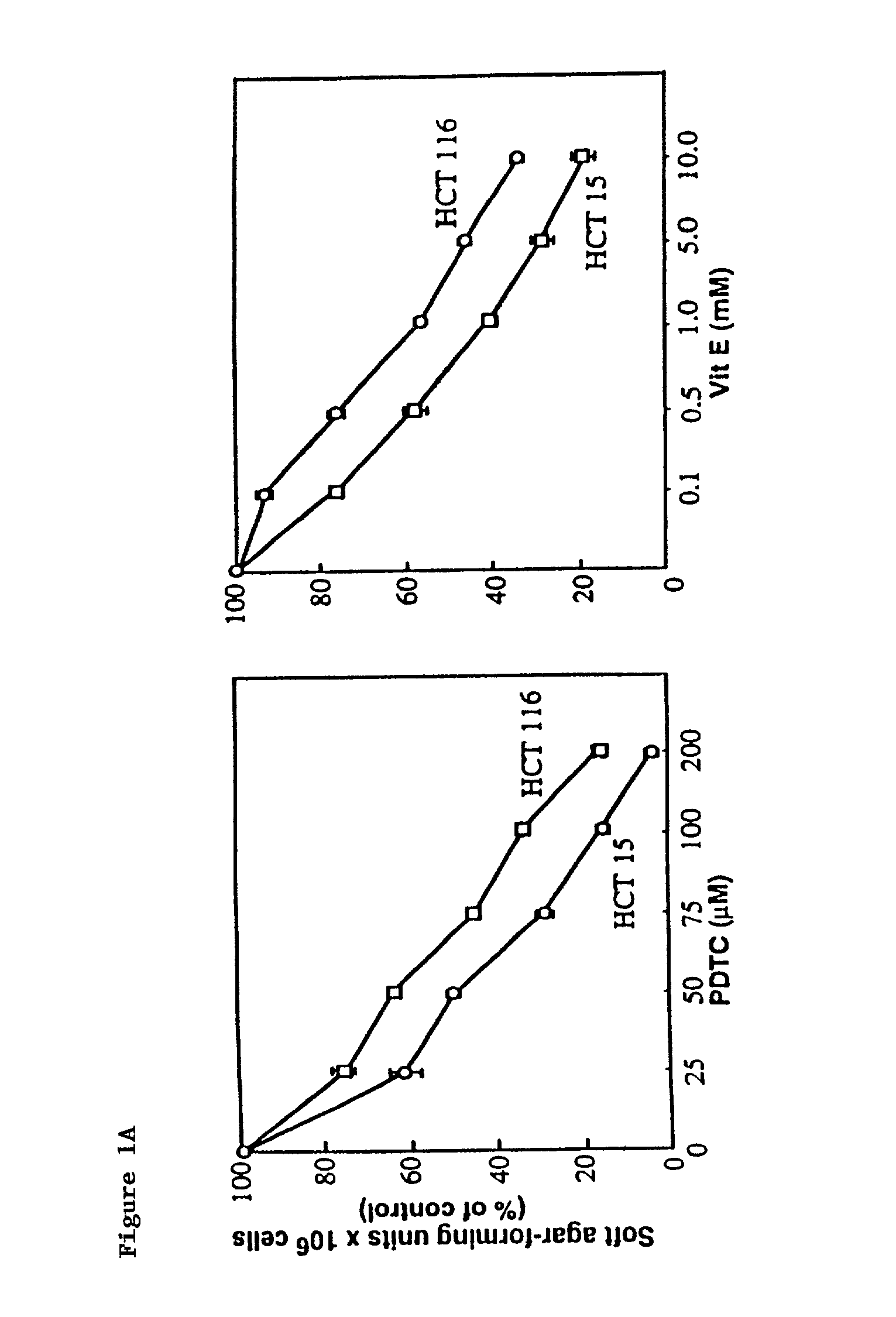
[0165]To determine the effect of pyrrolidinedithiocarbamate (Sigma Chemical Co., Ma.), a vitamin E analogue (6-hydroxy-2.5,7,8-tetramethylchroman-2-carboxylic acid: vit (E) (Aldrich), 5-FU (Hoffmann-LaRoche Inc. Nutley, N.J.) or doxorubicin (Sigma) on anchorage-independent growth, HCT 15 and HCT 116 ...

example 2

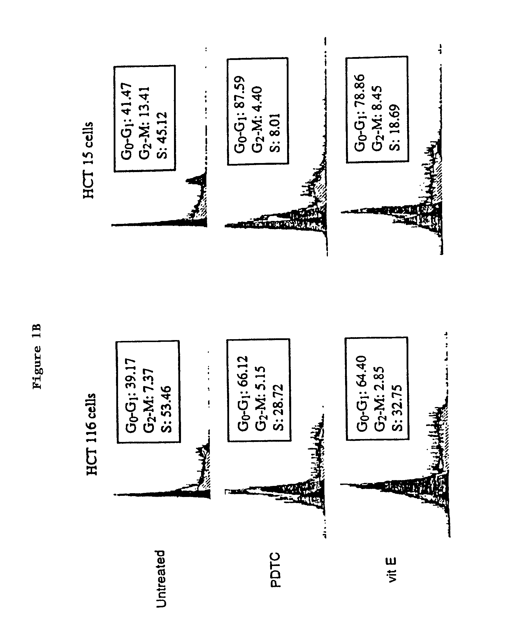
[0166]DNA Content of nuclei was determined as described [I. G. Nicoletti et al., J Immunol. Methods. 139, 271 (1991)] by lysing plasma membranes, staining nuclear DNA with propidium iodide (50 mg / ml), and quantitating the relative DNA content of nuclei using the Becton Dickinson FACSORT fluorescence-activated cell sorter. The proportion of nuclei in each phase of the cell cycle was determined using MODFIT-DNA analysis software. Detection of apoptotic cells either by fluorescence microscopy or by flow cytometry was performed using the ApopTag Plus In Situ Apoptosis Detection Kit (Oncor, Gaithersburg, Md.) as described in the manufacturer's protocol. Briefly, gigoxygenin-labeled nucleotides were added to free 3′OH groups of DNA produced by DNA fragmentation during apoptosis by terminal deoxynucleotidyl transferase (TdT). Digoxygenin was detected by a FTC-conjugated anti-digoxygenin antibody. Analysis was carried out using the fluorescence-activated cell sorter and FITC staining visual...

example 3

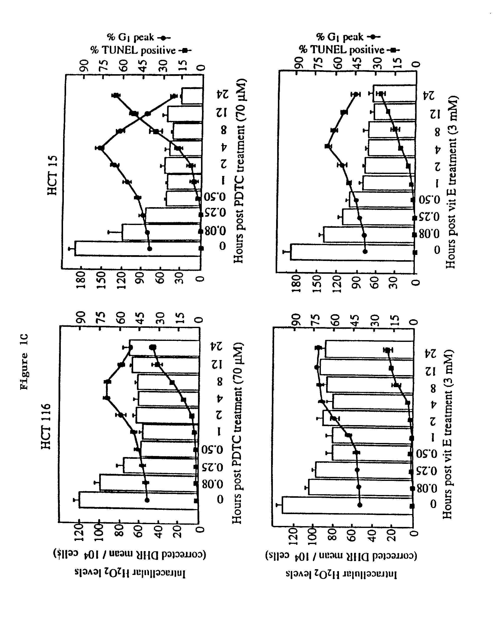
[0167]Intracellular H2O2 levels were analyzed by flow cytometry using dihydrorhodamine 1234 (DHR) as a specific fluorescent dye probe [G. Rothe, A. Emmendorffer, A. Oser, J. Roesler, G. Valet, J Immun. Methods 138, 133 (1991); J. A. Royall, H. Ischiropoulos, Arch. Biochem. Biophysics 302, 348 (1993)]. CRC cells were grown in DMEM containing 1 mM DHR and pyrrolidinedithiocarbamate (70 μM) or vit E (3 mM) for up to 24 hours. Following trypinization, trypsin activity was quenched with 2% FBS in phosphate buffered saline and cells fixed in 1% paraformaldahyde (Sigma). Cellular rhodamine 123 fluorescence intensity of 1×10⁴ cells was measured for each sample using a Becton-Dickenson FACS Vantage flow cytometer with the excitation source at 488 nm and emission wave length of 580 nm. Histograms were analyzed with the software program PC-Lysis (Becton Dickenson). Background fluorescence from blank wells was subtracted from each reading.


PUM

PropertyMeasurementUnit
pHaaaaaaaaaa
timeaaaaaaaaaa
temperatureaaaaaaaaaa

Abstract

A method to enhance the cytotoxic activity of an antineoplastic drug comprising administering an effective amount of the antineoplastic drug to a host exhibiting abnormal cell proliferation in combination with an effective cytotoxicity-increasing amount of an antioxidant. The invention also includes a method to decrease the toxicity to an antineoplastic agent or increase the therapeutic index of an antineoplastic agent administered for the treatment of a solid growth of abnormally proliferating cells, comprising administering an antioxidant prior to, with, or following the antineoplastic treatment.

Description

[0001]This application is a continuation application of U.S. Ser. No. 09 / 108,609 filed on Jul. 1, 1998 now abandoned, which is a continuation application of U.S. Ser. No. 08 / 967,492, entitled “Antioxidant Enhancement of Anti-Neoplastic Therapy for Hyperproliferative Conditions,” filed on Nov. 11, 1997 now abandoned, by Rebecca Chinery, R. Daniel Beauchamp, Robert J. Coffey, Russell M. Medford and Brian Wadsinski, and converted to a provisional application by a petition filed on Jun. 30, 1998; which is a continuation-in-part application of U.S. Ser. No. 08 / 886,653, entitled “Antioxidant Enhancement of Anti-Neoplastic Therapy,” filed on Jul. 1, 1997 now abandoned, by Rebecca Chinery, R. Daniel Beauchamp, and Robert J. Coffey, and converted to a provisional application by a petition filed on Jun. 20, 1998.[0002]This invention was developed in part using funds from the federal government under National Institute of Health grant Nos. CA4613 and GM53319 and CA-69457. The United States gov...

Claims


Application Information

Patent Timeline
Patent Type & Authority Patents(United States)
IPC IPC(8): A61K31/00A61K31/50A61K47/20A61K31/138A61K31/198A61K31/336A61K31/337A61K31/34A61K31/357A61K31/365A61K31/381A61K31/395A61K31/396A61K31/40A61K31/407A61K31/409A61K31/416A61K31/4164A61K31/4166A61K31/4168A61K31/4178A61K31/4188A61K31/4196A61K31/4353A61K31/437A61K31/4375A61K31/4468A61K31/451A61K31/47A61K31/4704A61K31/473A61K31/495A61K31/5025A61K31/506A61K31/513A61K31/519A61K31/52A61K31/53A61K31/566A61K31/585A61K31/67A61K31/675A61K31/70A61K31/7008A61K31/704A61K31/7042A61K31/706A61K31/7064A61K31/7072A61K31/7076A61K31/7135A61K38/43A61K38/45A61K38/55A61K45/00A61K45/06A61P9/00A61P17/06A61P35/00A61P43/00
CPCA61K31/00A61P9/00A61P17/06A61P35/00A61P43/00A61K45/06
Inventor CHINERY, REBECCABEAUCHAMP, R. DANIELCOFFEY, ROBERT J.MEDFORD, RUSSELL M.WADZINSKI, BRIAN E.
Owner SALUTRIA PHARMACEUTICALS LLC
Why Eureka
Try Eureka