Quantitative, high-throughput screening method for protein stability

a protein stability and quantitative analysis technology, applied in the field of protein stability screening, can solve the problems of not being able to disclose the quantitative analysis of native-like proteins, unable to meet the needs of stability measurement, and currently necessitate time-consuming experiments with pure protein samples, etc., and achieve the effect of changing the stability of the test protein

Inactive Publication Date: 2006-12-12
DUKE UNIV
View PDF5 Cites 2 Cited by
  • Summary
  • Abstract
  • Description
  • Claims
  • Application Information

AI Technical Summary

Benefits of technology

[0018]Additionally, a method of quantitatively determining a change in the stability of a test protein imparted by the association of a test ligand with the test protein. In a preferred embodiment, the method comprises: (a) providing a test protein; (b) providing a test ligand; (c) contacting the test ligand with the test protein to form a test mixture; (d) contacting the test mixture with an exchange buffer comprising a denaturant and deuterium, the exchange buffer having a denaturant concentration; (e) contacting the test mixture with a mass spectrometry matrix medium; (f) determining a change in mass of the test protein by mass spectrometry; (g) varying the denaturant concentration of the exchange buffer; (h) repeating steps (a)–(g) a desired number of times; (i) calculating the stability of the test protein in the presence of the test ligand; and (j) quantitatively comparing the stability of the test protein in the presence of the test ligand with the stability of the protein in the absence of ligand to thereby generate a change in the stability of a test protein imparted by the association of a test ligand with the test protein.
[0019]Furthermore, a method of detecting an improperly folded mutant protein is disclosed. In a preferred embodiment, the method comprises: (a) contacting a mutant test protein with an exchange buffer comprising a denaturant and deuterium, the exchange buffer having a denaturant concentration; (b) contacting a mutant test protein with a mass spectrometry matrix medium; (c) determining a change in mass of the mutant test protein by mass spectrometry; (d) varying the denaturant concentration of the exchange buffer; (e) repeating steps (a)–(e) a desired number of times; (f) quantitatively determining protein stability based on the change in mass of the mutant test protein as a function of denaturant concentration; and (g) comparing the stability of the mutant test protein with the stability of a control, non-mutated test protein, a difference in the stabilities being indicative of an improperly folded mutant protein.
[0020]A method of detecting a disease characterized by protein misfolding is also disclosed. In a preferred embodiment, the method comprises: (a) providing a test protein suspected of being misfolded; (b) contacting the protein with an exchange buffer comprising a denaturant and deuterium, the exchange buffer having a denaturant concentration; (c) contacting the test protein with a mass spectrometry matrix medium; (d) determining a change in mass of the test protein by mass spectrometry; (e) varying the denaturant concentration of the exchange buffer; (f) repeating steps (a)–(e) a desired number of times; and (g) analyzing the change in mass of the test protein to determine a stability of the test protein; and (h) comparing the stability of the test protein suspected of being misfolded with a known stability of the test protein, wherein a change in stability of the test protein when compared with the known stability of the test protein is indicative of a disease characterized by protein misfolding.
[0021]Moreover, a method of identifying a protein that unfolds through one or more stable intermediates is disclosed. The method comprises: (a) providing a test protein suspected of unfolding through one or more intermediates; (b) contacting the test protein with an exchange buffer comprising a denaturant and deuterium, the exchange buffer having a denaturant concentration; (c) contacting the test protein with a mass spectrometry matrix medium; (d) determining a change in mass of the test protein by mass spectrometry; (e) varying the denaturant concentration of the exchange buffer, (f) repeating steps (a)–(e) a desired number of times; and (g) plotting the change in mass of the test protein as a function of denaturant concentration to generate an unfolding curve; (h) evaluating the unfolding curve, to thereby identify one or more stable intermediates.
[0022]In the above embodiments it is generally preferable that a test protein is disposed in a crude cell lysate or in vivo, which can comprise whole cells. A reference protein is preferably provided with the test protein. It is also preferable that the test protein has a mass of less than or equal to about 1,000,000 daltons. The test protein can be a multimeric protein. Additionally, it is preferable that the test protein, or a plurality of test proteins are disposed on a microtiter plate and the steps of the preferred embodiments disclosed are repeated for each test protein disposed on the microtiter plate. Finally, it is preferable that the denaturant is selected from the group consisting of detergents, guanidinium chloride and urea, that the mass spectrometry matrix material is a MALDI mass spectrometry matrix material, and that the mass spectrometry is MALDI mass spectrometry. Optionally, the MALDI mass spectrometry matrix material is selected from the group consisting of sinapinic acid, α-cyano-4-hydroxycinnamic acid, 2,5-dihdroxybenzoic acid, 2,5-dihydroxyacetophenone and 3-amino-4-hydroxybenzoic acid. Additionally, analysis can be performed using a computer program.

Problems solved by technology

Despite their utility, stability measurements currently necessitate time-consuming experiments with pure protein samples.
17: 121–27), where a large number of polypeptides often need to be analyzed, stability measurements are not practical.
However, these studies did not disclose the quantitative analysis of native-like proteins.
Conventional denaturation methods used for the analysis of protein stability have at least the following identified experimental limitations: 1) stability can only be measured under conditions where the protein is partially unfolded; 2) the energetics of localized fluctuations can not be easily measured; 3) many proteins cannot be analyzed because they aggregate during the course of denaturation; 4) stability measurements cannot be obtained in complex mixtures, extracts or living cells; 5) relatively large amounts of protein are required for analysis; and 6) denaturation experiments are time-consuming and not amenable to high-throughput analysis.
These constraints drastically limit the number of biological problems that can be addressed through stability measurements.
This method, however, does not take advantage of the high sensitivity available from an analytical technique such as MALDI mass spectrometry, and cannot be employed at the low protein levels that MALDI mass spectrometry can detect.
Moreover, proteolytic methods can require additional steps to isolate and analyze proteolytic fragments and cannot be performed in an in vivo setting.
Finally, this method cannot be employed to generate quantitative measurements of protein stability.

Method used


Image

  • Quantitative, high-throughput screening method for protein stability
  • Quantitative, high-throughput screening method for protein stability
  • Quantitative, high-throughput screening method for protein stability

Examples


example 1

Laboratory Example 1

Quantitative, High-Throughout Screen for Protein Stability

[0293]1.A. Sample Preparation

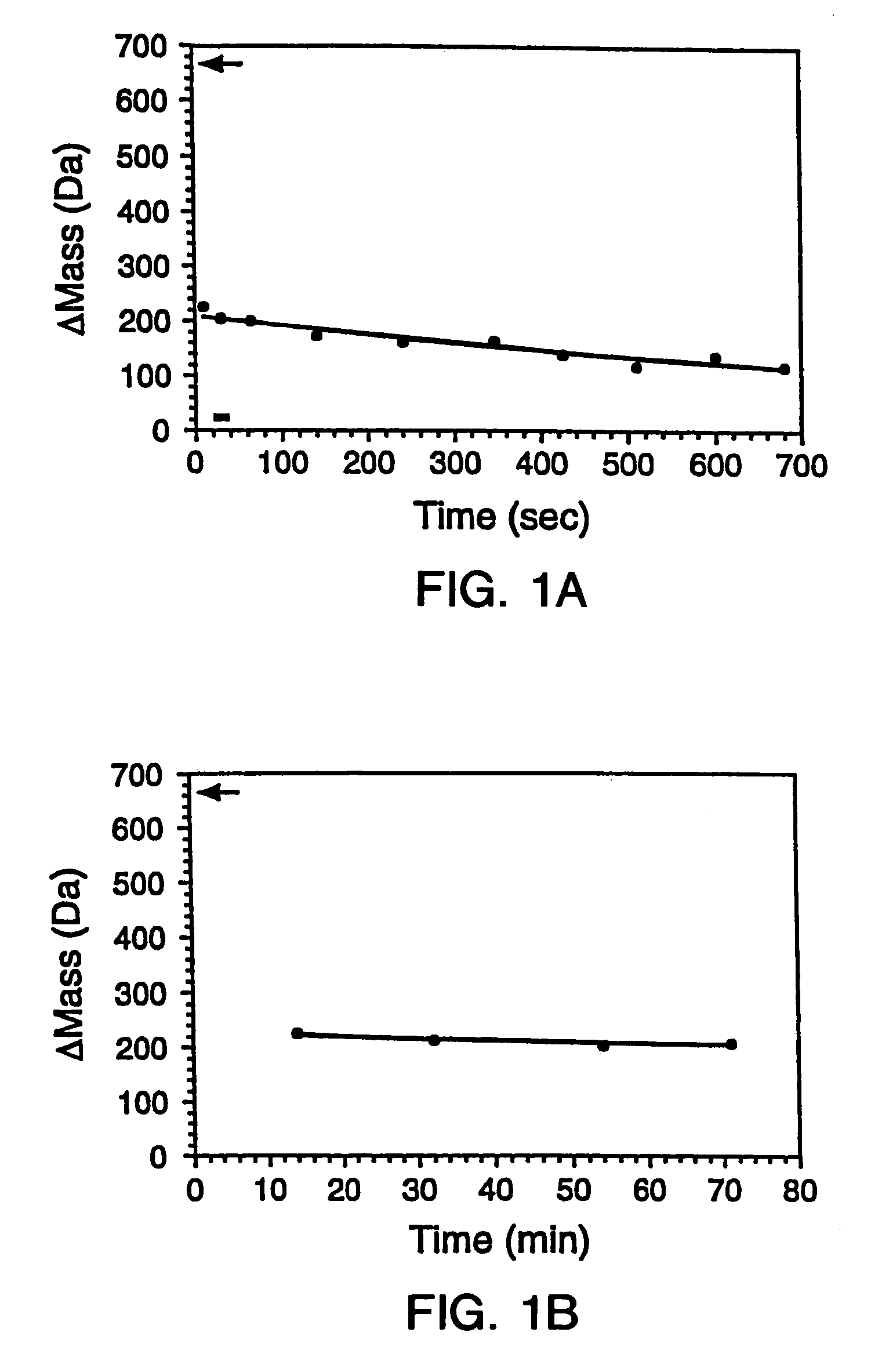
[0294]The gene encoding maltose binding protein or λ6-85 was cloned into a T7 expression vector and expressed in E. Coli strain BL21-(DE3). 200 μL LB cultures of the recombinant E. Coli were grown in 96 well plates and induced by the addition of 0.4 mM isopropylthio-β-D-galactosidase (IPTG). The cells were subsequently pelleted and lysed by suspension in 10 mL BUG BUSTER™ solution (Novagen, Inc., Madison, Wis.). The lysates were centrifuged and the supernatant was used for hydrogen exchange experiments without any further manipulation. SDS-PAGE indicated that the crude samples comprised ˜10%–50% expressed protein in a background of cellular impurities. From the gel, the concentrations of the proteins were estimated to range between 50 to 500 μM.

[0295]1.B. Hydrogen Exchange

[0296]Hydrogen exchange was initiated by adding 10-fold excess deuterated exchange buffet (20 mM sodium pho...

example 2

Laboratory Example 2

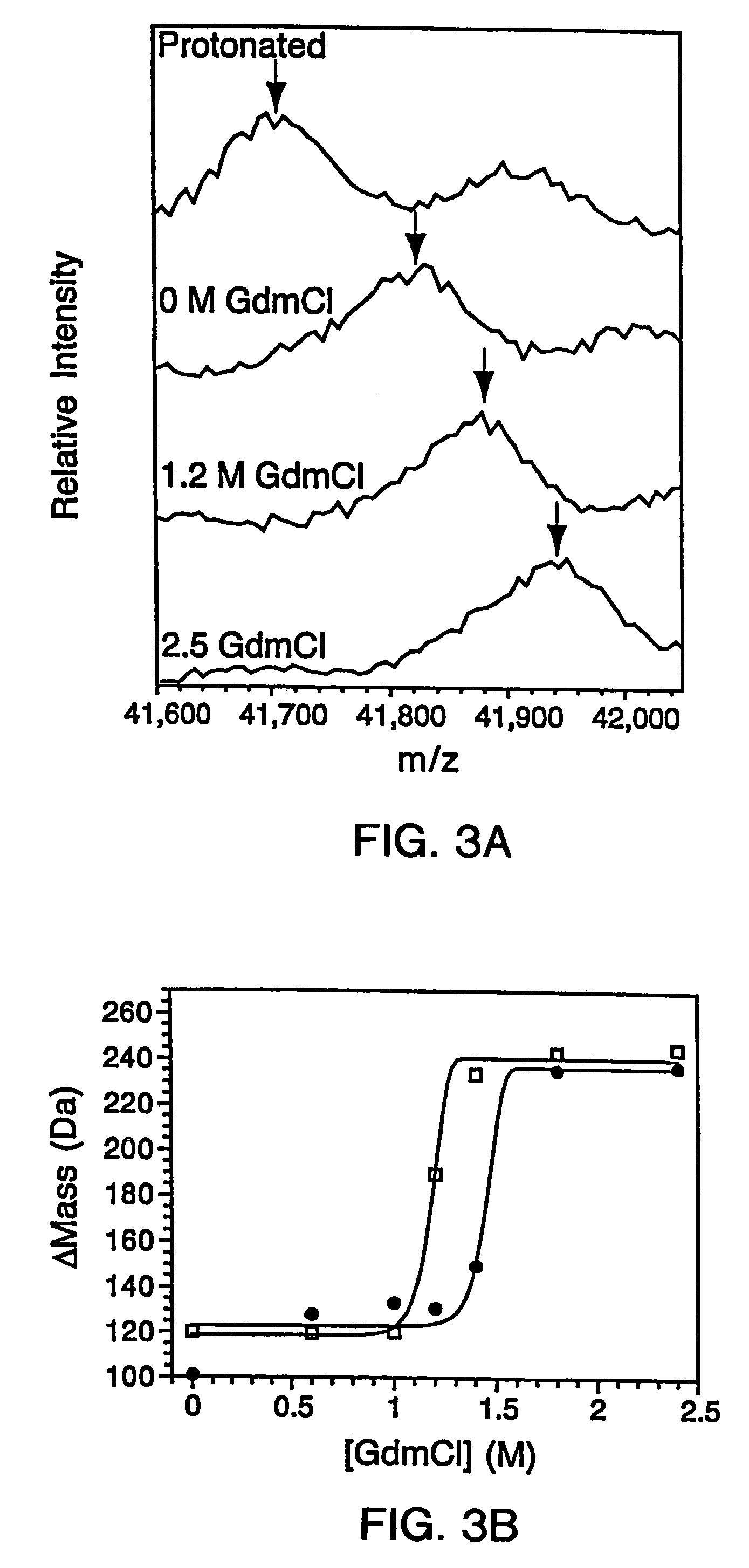
SUPREX as a Tool for Measuring Protein-Protein Binding

[0306]2.A. Mutagenesis and Protein Expression

[0307]The genes for the B1 domain (Q32C) and its mutants were constructed as previously described (Sloan & Hellinga, (1999) Protein Sci. 8: 1643–48) and cloned in the pKK223 expression vector (Pharmacia, Peapack, N.J.). Mutations were constructed in the Q32C (WT*) background. The plasmids were transformed into XL1-Blue E coli and grown in 10 mL cultures of LB media containing 100 μg / mL ampicillin. Expression was induced by the addition of 0.4 mM isopropylthio-β-D-galactosidase (IPTG). The cells were subsequently pelleted and lysed by suspension in 500 μL of BUG BUSTER™ solution (Novagen, Inc., Madison, Wis.). The lysates were centrifuged and the supernatant was used for hydrogen exchange experiments without any further manipulation. SDS-PAGE indicated that the crude samples consisted of −30%–40% expressed protein in a background of cellular impurities. The exact pro...

example 3

Laboratory Example 3

SUPREX as a Tool for Measuring Protein Stabilities In Vivo

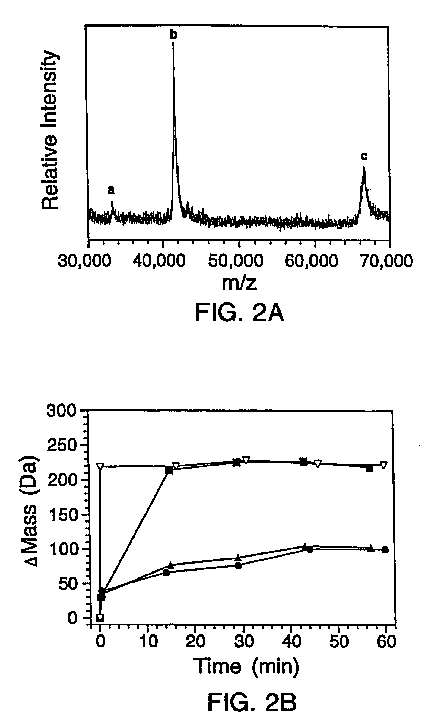
[0317]The G46A / G48A thermostable variant of monomeric lambda repressor (λ6-85) (Burton et al., (1996) J. Mol. Biol. 263: 311–22) was overexpressed using a T7 vector in E. coli in H2O-based media. To initiate hydrogen exchange, the bacteria were transferred to a deuterated media. Time-course studies indicated that under these conditions the D2O freely diffuses across the cell membrane and the solvent deuterons exchange with labile protons of the protein within the cytoplasm of the cell. Chloramphenicol was added to the deuterated media in order to inhibit further protein synthesis during the course of the exchange reaction, thereby preventing deuteration during synthesis. The E. coli cells were then lysed and the extent of exchange determined using MALDI mass spectrometry to measure the increase in mass, as generally depicted in the schematic of FIG. 8.

[0318]3.A. Sample Preparation

[0319]In vivo experiments ...


PUM

PropertyMeasurementUnit
massaaaaaaaaaa
mass gainaaaaaaaaaa
pHaaaaaaaaaa

Abstract

In proteomic research, it is often necessary to screen a large number of polypeptides for the presence of stable structure. Described herein are methods (referred to as MALDI MS-HX and SUPREX) for measuring the stability of proteins in a rapid, high-throughput fashion. The method employs hydrogen exchange to estimate the stability of quantities of unpurified protein extracts, using matrix-assisted laser desorption/ionization (MALDI) mass spectrometry. A method of quantitatively determining the stability of a test protein under native conditions is disclosed. The method includes the steps (a) providing a test protein; (b) contacting the protein with an exchange buffer comprising a denaturant and deuterium, the exchange buffer having a denaturant concentration; (c) contacting the test protein with a mass spectrometry matrix medium; (d) determining a change in mass of the test protein by mass spectrometry; (e) varying the denaturant concentration of the exchange buffer; (f) repeating steps (a)–(e) a desired number of times; and (g) quantitatively determining protein stability based on the change in mass of the test protein as a function of denaturant concentration, whereby the stability of a test protein under native conditions is quantitatively determined.

Description

CROSS REFERENCE TO RELATED APPLICATIONS[0001]This application is a divisional of U.S. patent application Ser. No. 09 / 845,363, filed Apr. 30, 2001 now U.S. Pat. No 6,734,023, herein incorporated by reference in its entirety, which is based on and claims priority to U.S. Provisional Application Ser. No. 60 / 200,311, filed Apr. 28, 2000, herein incorporated by reference in its entirety.GRANT STATEMENT[0002]This work was supported by NIH grant GM45322. Thus, the U.S. Government has certain rights in the invention.TECHNICAL FIELD[0003]The present invention relates generally to a method of screening for protein stability. More particularly, the present invention relates to a quantitative method of screening for protein stability that can be employed in a high throughput setting using relatively small amounts of pure and impure protein samples.[0004]Table of AbbreviationsBSAbovine serum albuminCDcircular dichroismDSCdifferential scanning calorimetryESIelectrospray ionizationFEfraction excha...

Claims


Application Information

Patent Timeline
Patent Type & Authority Patents(United States)
IPC IPC(8): G01N1/00G01N33/68
CPCG01N33/6803G01N33/6848G01N33/6851G01N33/726Y10T436/24G01N2458/15G01N2800/00Y10S436/811G01N2333/805
Inventor OAS, TERENCE G.GHAEMMAGHAMI, SINAPOWELL, KENDALL D.FITZGERALD, MICHAEL C.
Owner DUKE UNIV
Why Eureka
Try Eureka