Eureka AIR delivers breakthrough ideas for toughest innovation challenges, trusted by R&D personnel around the world.

Method and apparatus for detecting vulnerable atherosclerotic plaque

a technology of atherosclerotic plaque and detection method, which is applied in the field of methods and apparatus for detecting vulnerable atherosclerotic plaque, can solve the problems of hammering the prognosis and treatment of patients suffering from cardiovascular disease, plaques with inflamed surfaces or a high density of activated macrophages, and a thin overlying cap at risk of tirombosis, so as to reduce the risk of restenosis, reduce the risk of vasculopathy

Inactive Publication Date: 2008-09-16
BOARD OF RGT THE UNIV OF TEXAS SYST
View PDF138 Cites 118 Cited by
  • Summary
  • Abstract
  • Description
  • Claims
  • Application Information

AI Technical Summary

Benefits of technology

[0031]The present invention overcomes many of the failures of the prior art by providing a sensitive method of identifying vulnerable or at-risk atherosclerotic plaque in a vessel. Moreover, the methods of the invention are able to distinguish such vulnerable plaques from the relatively stable plaques by detecting regions of elevated temperature along the vessel wall as an indicator of populations or clusters of activated inflammatory cells. An important advantage of the present methods is that they assist the physician in diagnosing plaques at imminent risk of rupturing or occluding so that appropriate interventional steps may be taken to avert a possibly fatal cardiovascular event. Certain embodiments of the invention also permit the physician to monitor the condition of grafts and transplants so that the influx of inflammatory cells and the development of transplant vasculopathy can be detected and treated before the occurrence of a critical event.
[0034]The devices and methods of the present invention also are capable of effectively identifying patients who have arterial wall areas of unusually low temperature and which represent previously undetected arterial at-risk areas. Just as excess heat can identify regions at risk due to inflammation, sub-normal heat (i.e., areas cooler than the rest of a vessel) indicates a lack of actively metabolizing healthy cells, since heat in the body results from actively metabolizing cells. Non-cellular areas are typically regions of hemorrhage, thrombosis, cholesterol pools, or calcium—all indicators of high risk plaques. The devices and methods of the invention achieve these ends by identifying and analyzing thermal discrepancies in the wall temperature of blood vessels. In some embodiments of the invention, devices are provided that are also capable of providing a thermal image of the vessel wall and of applying therapeutic gentle heat treatment.
[0056]The invention's methods and devices will have a number of utilities, including reduction of morbidity and mortality from coronary and carotid artery atherosclerosis, reduction of restenosis and thus the need for repeated angioplasties or atherectomies, and also including reduction of vasculopathy in organ-transplant patients. In turn, these improved outcomes will produce the benefits of better health care, improved public health, and reduced health care costs. These and other objects, features and advantages of the present invention will become apparent with reference to the following description and drawing.

Problems solved by technology

Myocardial infarction is not predictable by presently available clinical means, which greatly hampers prognosis and treatment of patients suffering from cardiovascular disease (Fuster et al.
The rupture, fissure, or ulcer causes a large thrombus or blood clot to form on the inside of the artery, which may completely occlude the flow of blood through the artery, thereby injuring the heart or brain.
It has been observed that plaques with inflamed surfaces or a high density of activated macrophages and a thin overlying cap are at risk of tirombosis.
Modalities such as intravascular ultrasound, which might identify plaque vulnerability on the basis of cap thinness has been proven to be incapable of identifying which plaques are at risk of rupturing (de Feytia et al.
One problem with currently-available infrared optical fibers is that they are not made of biocompatible material, and are therefore unsuitable for directly contacting tissues and fluids inside a human blood vessel.
Also, the somewhat brittle nature of conventional infrared optical fibers makes them incapable of bending sufficiently to be aimed directly into a plaque (i.e., perpendicular to the linear axis of the vessel).
Very few of these intravascular heat sensing devices are actually capable of, or practical for, discerning slight to moderate temperature elevations (i.e., 0.01° C. or more) along the luminal wall of a vessel.
Most of the prior art methods and catheter-based heat sensing devices are unable to differentiate plaque from normal vessel wall, much less distinguish between unstable plaques and the more stable ones that are not at imminent risk of rupturing or occluding.
Even when the presence or absence of histological features such as calcification in plaque can be assessed by one of the existing devices, it is not adequately predictive of whether the plaque is unstable or whether it will tend to rapidly progress in severity of vascular stenosis.
Infrared thermography did not have the sensitivity of x-ray mammography for deep masses, however, because the temperature of many such masses does not directly reach the skin.
Despite this work, infrared is little used clinically today, and workable IR catheters have not been developed, due at least in part to the large size, brittleness and expense of commercially available IR optical fibers.
There are a number of commercially available catheters that monitor blood temperature in order to measure cardiac output (e.g. American Edwards / Baxter Swan Ganz catheter) by continuous thermo-dilution, but these catheters cannot measure the temperature of the vessel wall itself, or of specific plaque regions on the vessel wall.
Of the existing catheter-based and non-invasive methods and devices, none have proved to be adequate or practical for clinical use to identify unstable atherosclerotic plaques.

Method used


Image

  • Method and apparatus for detecting vulnerable atherosclerotic plaque
  • Method and apparatus for detecting vulnerable atherosclerotic plaque
  • Method and apparatus for detecting vulnerable atherosclerotic plaque

Examples


Embodiment Construction

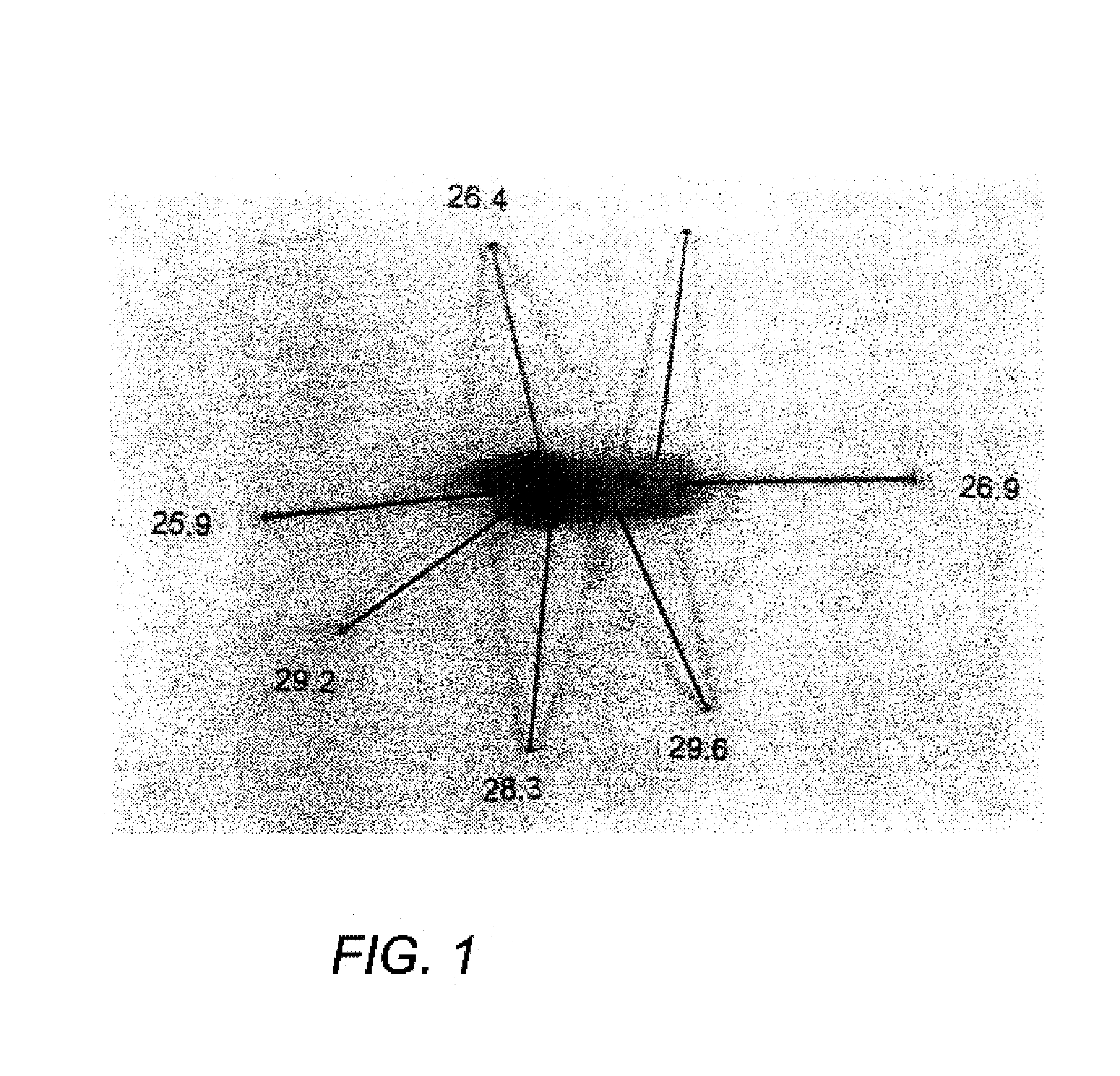
[0133]In the description which follows, the inventors confirm and extend their earlier observations that approximately one-third of human atherosclerotic plaque specimens have temperature heterogeneity ranging from about 0.2° to 4.0° C. as measured in live, freshly excised carotid endaiterectomy specimens. FIG. 1 is a photomicrograph of one such specimen, with pins marking the temperatures of various regions of the plaque. The term “temperature heterogeneity” means that the surface temperature of a plaque is not uniform along its length and width. The term also refers to the temperature differences between certain plaque regions and other vessel sites or an average or ambient vessel wall temperature. The fact that human atherosclerotic plaque with measurable temperature heterogeneity has the morphological characteristics of plaque that is likely to ulcerate provides a new and sensitive way to detect and treat these dangerous plaques before myocardial infarction and its consequences ...


PUM


Abstract

Methods and devices are disclosed for detecting vulnerable atherosclerotic plaque, or plaque at risk of reducing blood flow in a vessel, by identifying a region of elevated temperature along a living vessel wall. The disclosure that human atherosclerotic plaque with measurable temperature heterogeneity has the morphological characteristics of plaque that is likely to ulcerate provides a new and sensitive technique for detecting and treating these dangerous plaques before myocardial infarction and its consequences occur. The disclosed methods are advantageous over conventional plaque detection techniques because they are capable of differentiating between those plaques that are at great risk of rupture, fissure, or ulceration, and consequent thrombosis and occlusion of the artery, and those that are not presently at risk. Infrared heat-sensing catheters useful for identifying potentially fatal arterial plaques in patients with disease of the coronary or other arteries are also described. In some embodiments a coherent infrared fiber optic bundle is employed to radially and longitudinally explore a luminal wall to identify inflamed, heat-producing, atherosclerotic plaque. Certain other methods and devices are disclosed which are particularly suited for non-invasively identifying and then monitoring the progression or amelioration of an inflamed plaque in a patient, and for monitoring for onset of inflammation in an implanted arteriovenous graft. Also disclosed are thermocouple basket catheters and thermistor basket catheters which are also capable of detecting temperature heterogeneity along a vessel wall.

Description

CROSS-REFERENCE TO RELATED APPLICATION[0001]The present application is a continuation application of Ser. No. 09 / 340,089, filed Jun. 25,1999, now U.S. Pat. No. 6,615,071 which is incorporated by reference herein.STATEMENT REGARDING FEDERALLY SPONSORED RESEARCH OR DEVELOPMENT[0002]Not applicableBACKGROUND OF THE INVENTION[0003]1. Field of the Invention[0004]The present invention relates generally to methods of identifying in a living vessel an atherosclerotic plaque at risk of rupture or thrombosis. More particularly, the invention relates to such methods that include detecting sites of inflammation in a vessel wall exhibiting about 0.2°-5° C. temperature elevation above adjacent or ambient vessel wall temperature. The invention also relates to intravascular and non-invasive devices for measuring vessel wall temperatures and detecting about 0.2-5° C. temperature differences between regions of living vessel wall.[0005]2. Description of the Related Art[0006]Despite the declining age-sp...

Claims


Application Information

Patent Timeline
IPC IPC(8): A61B1/00A61B1/04A61B6/00A61B5/00
CPCA61B5/0071A61B5/0073A61B5/0075A61B5/0084A61B5/0086A61B5/015A61B5/02007A61B5/415A61B5/418A61B5/6853
Inventor CASSCELLS, III, S. WARDWILLERSON, JAMES T.NAGHAVI, MORTEZAGUO, BUJIN
Owner BOARD OF RGT THE UNIV OF TEXAS SYST
Why Eureka
Eureka Blog
Learn More