Use of high wavenumber Raman spectroscopy for measuring tissue
a raman spectroscopy and tissue technology, applied in the field of instruments, can solve the problems of limiting the applicability of raman spectroscopy, strong signal background, and inability to enable detailed in vivo analysis of the molecular composition of plaques
- Summary
- Abstract
- Description
- Claims
- Application Information
AI Technical Summary
Benefits of technology
Problems solved by technology
Method used
Image
Examples
example 1
Raman Mapping of Atherosclerotic Artery
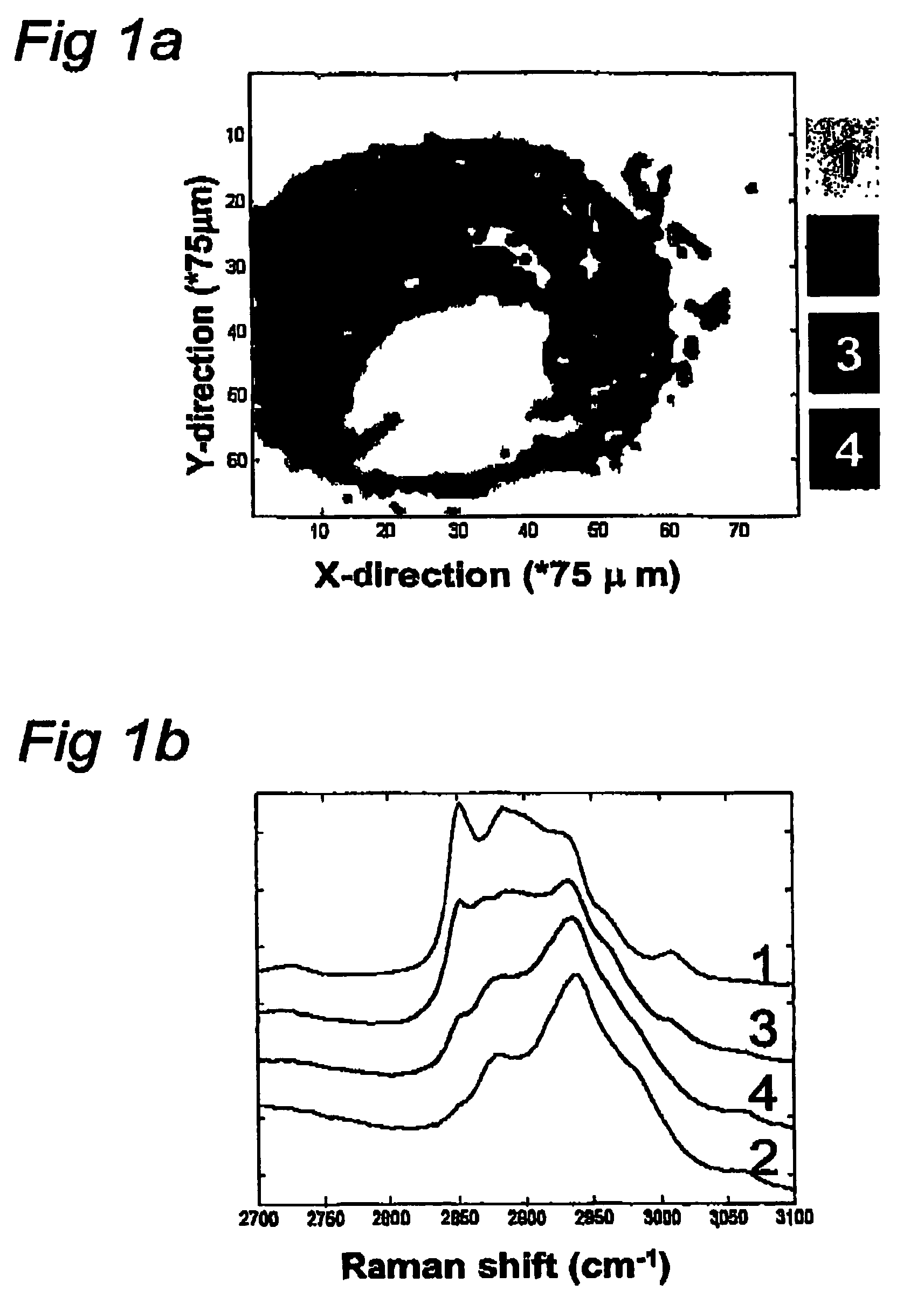
[0109]This experiment describes the possibilities of Raman spectroscopy in the spectral region of the invention for studying artherosclerotic plaque.
[0110]The human coronary artery sample used to create the Raman map shown in FIG. 1 was obtained at the time of autopsy (less than 24 hour post mortem). It was snap frozen in liquid nitrogen and stored at −80° C. until use. For the Raman measurements a 20 μm thick cryosection was placed on a calcium fluoride (CaF2) window (Crystan UK) and passively warmed to reach room temperature. After Raman measurements it was stained with a standard hematoxylin and eosin staining procedure.
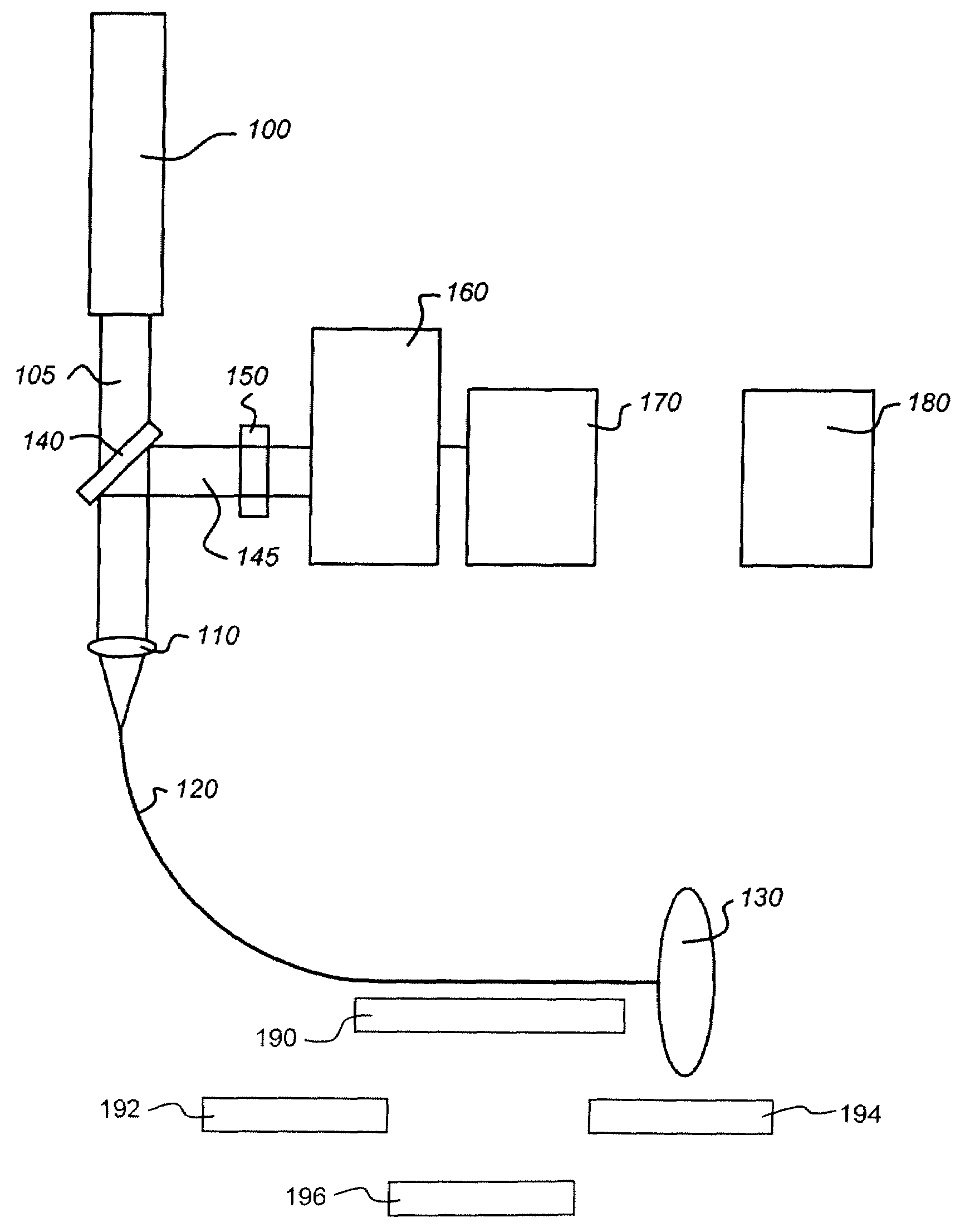
[0111]To collect Raman specta, 719 nm laser light from an argon-ion pumped Titanium: Sapphire laser system (Spectra Physics, Mountain View, Calif.) was used. The Raman microspectrometer system that was used has been described in detail in Van de Poll S W E, Bak Schut T C, Van der Laarse A, Puppels G J “In Situ Investigation o...
example 2
Raman Mapping of Cancerous Tissue
[0125]This experiment describes the possibilities of Raman spectroscopy in the spectral region of the invention for studying cancerous tissue.
[0126]The high wavenumber region can also be used advantageously in various clinical oncology applications. For instance, FIG. 4A shows a Raman map obtained of a thin tissue section of human dura infiltrated by meningioma in a way similar to the map of an atherosclerotic lesion in FIG. 1A. Currently no good intra-operative assessment of excision margins is possible. However, it is known that meningioma tissue that is left behind may lead to recurrence of the tumor. FIG. 4B shows a picture of an adjacent tissue section after staining with hematoxylin and eosin (H&E stained). Surprisingly, the histopathological evaluation of this section and its comparison with the Raman map show that the light gray areas in the Raman map correspond to dura, while the dark areas correspond to meningioma (MG).
[0127]This experiment...
example 3
Raman Mapping of Cancerous Tissue
[0128]This experiments describes the possibilities of Raman spectroscopy in the spectral region of the invention for studying cancerous tissue.
[0129]FIG. 5A shows a Raman map of a thin section of human glioblastoma with both vital tumor areas and areas with necrotic tissue. Surprisingly, comparison of the Raman map with the H&E stained adjacent section evaluated by a neuropathologist, shows that the light gray area corresponds to vital tumor tissue while the dark gray area in the Raman map corresponds to necrosis.
[0130]This experiment shows that Raman measurements in the spectral region of the invention give valuable information on cancerous tissue of the brain, enabling Raman spectroscopy for discriminating between vital tumor tissue and necrosis.
PUM
| Property | Measurement | Unit |
|---|---|---|
| wavenumber | aaaaa | aaaaa |
| wavenumber | aaaaa | aaaaa |
| wavenumber | aaaaa | aaaaa |
Abstract
Description
Claims
Application Information
 - R&D
- Intellectual Property
- Life Sciences
- Materials
- Tech Scout
- Unparalleled Data Quality
- Higher Quality Content
- 60% Fewer Hallucinations
Browse by: Latest US Patents, China's latest patents, Technical Efficacy Thesaurus, Application Domain, Technology Topic, Popular Technical Reports.
© 2025 PatSnap. All rights reserved.Legal|Privacy policy|Modern Slavery Act Transparency Statement|Sitemap|About US| Contact US: help@patsnap.com


