

Keratinocyte-fibrocyte concomitant grafting for wound healing
a keratinocyte and fibrocyte technology, applied in the field of wound healing, can solve the problems of large cost and time, difficult to achieve the effect of cell growth and integration, and easy clinical us
- Summary
- Abstract
- Description
- Claims
- Application Information
AI Technical Summary
Benefits of technology
Problems solved by technology
Method used
Image

Examples
example 1
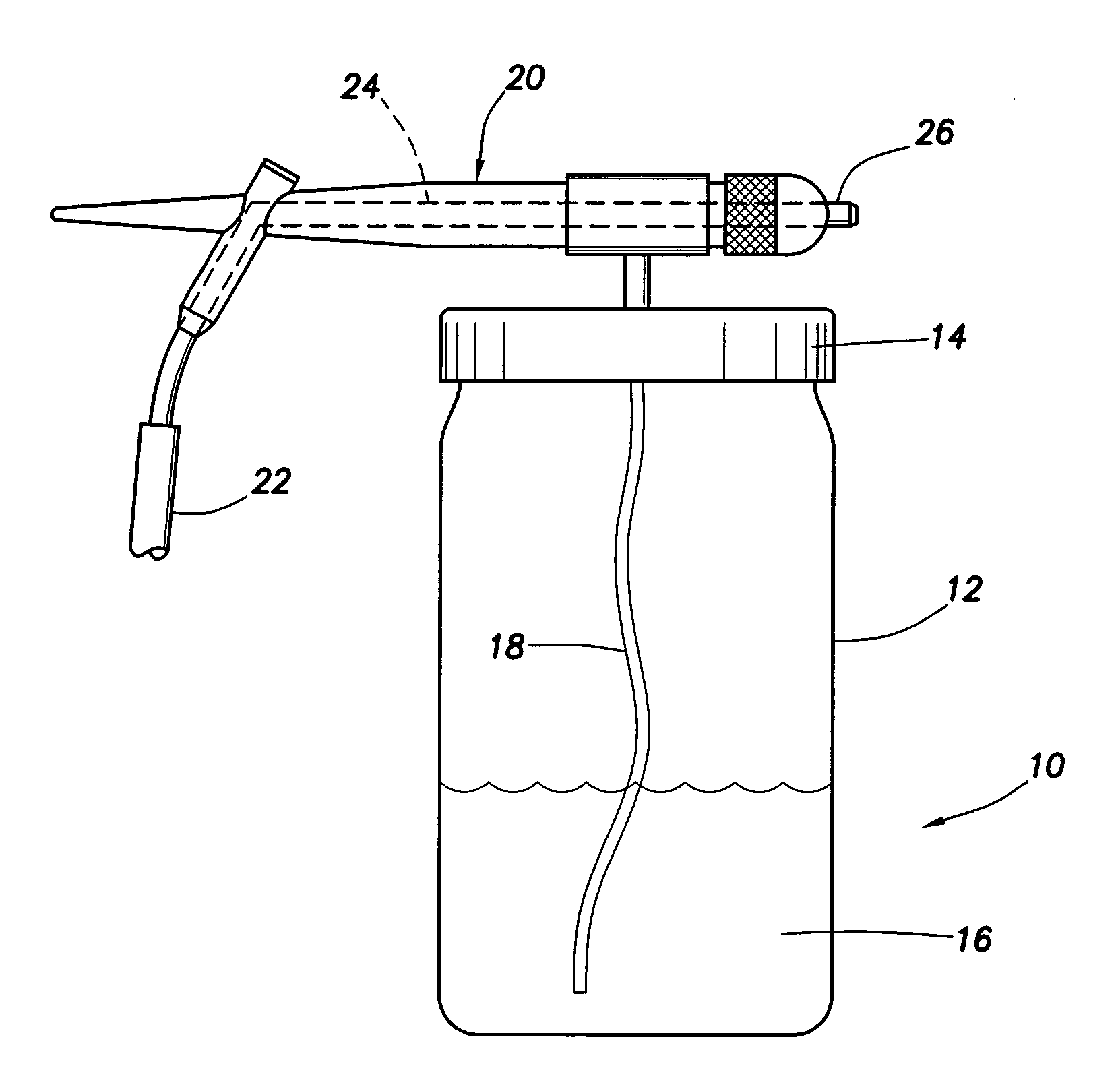
Air-Jet Cell External Seeding (ACES)
[0037]The invention may include a technique, termed Air-Jet Cell External Seeding (ACES), in which cells and / or adhesion proteins or growth factors may be seeded onto a dermal substrate by a hand held air-jet sprayer. In brief: (a) a commercially available hand held air-jet sprayer may be used to deposit cells which are suspended in autologous serum or other specified media; (b) the cells are seeded onto a commercially available dermal substrate (allograft) obtained from human cadaver skin; and (c) the construction of the resulting full thickness graft material can occur in vitro or in vivo.
[0038]This technology may utilize some techniques employed in a process known as cytoscribing which utilizes: (a) an ink cartridge of an inkjet printer filled with either fibronectin or other adhesion molecule; and (b) the deposition of the molecule on a substratum by a computer controlled printer. Subsequently, the cells only bind at sites where the adhesion m...
example 2
Harvesting and Culturing of Autologous Skin Graft Material in vitro
[0039]Engineered tissues of the present invention may contain keratinocytes and fibrocytes harvested from the same wound patient or from other sources. Specifically, keratinocytes may be obtained from a keratinized stratified epithelial surface which is unaffected by trauma or wounding, while fibrocytes may be obtained by liposuction. Keratinocytes may also be cultured from human neonatal foreskins. Fibrocytes may be obtained from adipose and dermal tissue via a tulip syringe during liposuction. Fibrocytes and undifferentiated adipocytes may be separated from the extracted adipose tissue along with collagen by enzymatic digestion and gentle agitation. Fibrocytes may also be obtained from human postpartum umbilical cord or neonatal foreskin dermis. The resultant fibrocytes or keratinocytes may be rinsed and concentrated prior to resuspension in autologous serum (or other media) and subsequent dispersion on the allogra...
example 3
Construction of in vitro Full Thickness Grafts
[0040]Dermal allograft material may be obtained from human cadavers. This dermal substitute consists primarily of collagen and may be used to construct an in vitro wound model analog by acting as an attachment matrix and in the orientation of a three-dimensional architecture for cell growth. This facilitates the in vitro study and development of clinical techniques in the construction of immediate autologous graft tissues. Autologous constructed tissues may be used in both transplantation studies and investigations into the cell biology of tissue engineering and wound healing.
[0041]ACES may be used to disperse and culture cells on the allograft substratum. Purified cell adhesion proteins and / or growth factors may be pre-incorporated into the allograft dermal matrix. Cell adhesion proteins (fibronectin, vitronectin, laminin, tenascin, fibrin) as well as growth factors (IGF, PDGF, TGF-β) may be used in tissue engineered autologous grafts.
PUM
| Property | Measurement | Unit |
|---|---|---|
| pore size | aaaaa | aaaaa |
| thickness | aaaaa | aaaaa |
| pore sizes | aaaaa | aaaaa |
Abstract
Description
Claims
Application Information