
Parstatin peptides

a technology of parstatin and peptides, which is applied in the direction of peptide/protein ingredients, drug compositions, angiogenin, etc., to achieve the effect of preventing or inhibiting endothelial cell growth

Inactive Publication Date: 2015-11-10
TSOPANOGLOU NIKOS +2
View PDF3 Cites 2 Cited by
  • Summary
  • Abstract
  • Description
  • Claims
  • Application Information

AI Technical Summary

Benefits of technology

The invention is about a group of peptides and their uses in medicine and biotechnology. These peptides have been found to have various effects on the kidneys, heart, and blood vessels. They can be used to treat kidney disorders, heart-related diseases, and angiogenesis (the formation of new blood vessels). The invention also includes methods for using these peptides to prevent or treat these disorders, as well as methods for inducing cell death and inhibiting cell growth. The invention also includes nucleic acid sequences and antibodies that target these peptides. Overall, the invention provides new tools for research and development of new treatments for kidney and heart-related diseases.

Problems solved by technology

In age-related macular degeneration, the associated visual problems are caused by an ingrowth of choroidal capillaries through defects in Bruch's membrane, with proliferation of fibrovascular tissue beneath the retinal pigment epithelium.
Diabetic retinopathy is a leading cause of blindness in adults of working age.
Age-related macular degeneration is a leading cause of vision loss in people over 65 years old.
Multiple theories exist but, the exact etiology and pathogenesis of AMD are still not well understood.
The condition is associated with severe visual impairment and is a high risk factor for graft rejection after allograft corneal transplantation.
In addition, corneal neovascularization and subsequent opacification remain the most frequent causes of blindness after severe alkali burn trauma.
To date, there are no pharmacological or surgical treatment options for the inhibition of corneal neovascularization that have been proven to be both safe and effective.
Despite the routine use of topical steroids, the inflammatory response can lead to oedema, lipid deposition and corneal scarring that may not only significantly alter visual acuity, but also worsen the prognosis of subsequent penetrating keratoplasty.
In addition, longer-term use of these drugs can lead to various adverse side effects such as cataracts, glaucoma, infection, and delay corneal epithelial healing.
This may be largely due to the fact that FGF binds very tightly to charged components of the extracellular matrix and may not be readily available in a freely diffusible form that would be detected by standard assays of intraocular fluids.
Viable and approved current treatments for diseases related to ocular neovascularization are limited.
Laser photocoagulation alone or photodynamic therapy with VISUDYNE® are therapies that involve laser-induced occlusion of the affected vasculature, which can result in localized damage to the retina.
Unfortunately, laser photocoagulation is a cytodestructive procedure and the visual field of the treated eye is irreversibly compromised.
The results in a larger patient population may be less robust.
In addition, adverse effects on neurons and vessels have been observed in primates after a single administration of the humanized anti-VEGF antibody (Bevacizumab) (Peters et al., 2007, Am J Ophthalmol, 91: 827-831) and sporadic case reports of complications of anti-VEGF therapy related to regression of blood vessels, increased risk for stroke and myocardial infarction, and local side effects due to the intravitreal application mode have appeared (Fraunfelder et al., 2005, Drugs Today, 41: 703-709; Tobin et al., 2006, Insight, 31: 11-14; Rosenfeld et al., 2006, Ophthalmol Clin NA, 19: 361-372; Baffert et al., 2006, Am J Physiol Heart Circ Physiol, 290: H547-H559; Hurwitz et al., 2004, Clin Colorectal Cancer, 4(suppl): 2: S62-S68).
The limited efficacy and potential adverse effects of currently implemented therapies emphasize the need for alternative therapeutic strategies.
The white cells bound to the endothelium eventually migrate into the cardiac tissue causing significant tissue destruction.
Damage of the cell membrane may in turn causes release of more free radicals signaling apoptosis.
Leukocytes may also build up in small capillaries, obstructing them and leading to more ischemia.
Coronary artery disease can lead to prolonged or irreversible episodes of cardiac ischemia that result in myocardial infarction (MI) which is associated with a high rate of mortality.
During myocardial infarct, stoppage of the heart or damage occurs which reduces the flow of blood to myocardium, and ischemia results.
Cardiac tissue itself is also subjected to ischemic damage.
During various surgeries, reduction of blood flow, clots or air bubbles generated can lead to significant ischemic damage of the myocardium.
Even with the successful treatment of occluded vessels, a significant risk of additional tissue injury after reperfusion may still occur.
If the ischemic episode has been of sufficient severity and / or duration to cause significant changes in the metabolism and the structural integrity of heart muscle, reperfusion may paradoxically result in a worsening of heart function, out of proportion to the amount of dysfunction expected simply as a result of the duration of blocked flow.
ARF secondary to a renal tubular cell injury, including an ischemic injury or a nephrotoxic injury remains a common and potentially devastating problem in clinical medicine and nephrology, with a persistently high rate of mortality and morbidity despite significant advances in supportive care (Chatterjee and Thiemermann, 2003, Expert Opin Emerg Drugs, 8: 389-435).
While these studies have suggested possible therapeutic approaches in animal models, translational research efforts in humans have yielded disappointing results.
The reasons for this may include the multifaceted response of the kidney to ischemic injury and nephrotoxins, and a paucity of early biomarkers for ARF with a resultant delay in initiating therapy.
The notion that only severe renal failure impacts on long-term morbidity is dispelled by the fact that even modest degrees of renal insufficiency significantly increase the risk of death for critically ill patients
The white cells bind to the endothelium and eventually migrate into the kidneys causing significant tissue destruction.
Damage of the cell membrane may in turn promotes release of more free radicals signaling apoptosis.
Leukocytes may also built up in small capillaries obstructing them and leading to more ischemia.
Even with the successful treatment of occluded vessels, a significant risk of additional tissue injury after reperfusion may still occur.
If the ischemic episode has been of sufficient severity and / or duration to cause significant changes in the metabolism and the structural integrity of renal tissue, reperfusion may paradoxically result in a worsening of renal function, out of proportion to the amount of dysfunction expected-simply as a result of the duration of blocked flow.
Renal ischemia, whether caused by shock or during surgery or transplantation, is a major cause of acute kidney injury.
Although reperfusion is essential for the survival of ischemic tissue, there is strong evidence that reperfusion itself caused additional cellular injury and together ischemia-reperfusion of the kidney leads to ischemic ARF.
CIN is an important problem in clinical practice and contrast-induced morbidity has become a significant cause of hospital morbidity and mortality with the increasing use of iodinated contrast media in diagnostic imaging and interventional procedures such as angiography.
Despite increasing ability to support vital organs and resuscitate patients, the incidence and mortality of septic ARF remain high.
Despite the wealth of information relating to the role of thrombin and PAR1 in physiology and diseases states, the information regarding the biological role of cleaved peptides upon activation of PAR1 is very limited.

Method used


Image

  • Parstatin peptides
  • Parstatin peptides
  • Parstatin peptides

Examples


example 1

Parstatin Peptides Synthesis and Compositions

[0301]Parstatin peptides used in our assays were synthesized in the core peptide facility of Peptide Specialty Laboratories GmbH (Heidelberg, Germany) or Bio-Synthesis Inc., (Lewisville, Tex.) or at the Protein and Nucleic Acid Shared Facility at the Medical College of Wisconsin. Synthesized peptides were purified by HPLC technology, characterized by mass spectrometry technology and sequenced. The synthesized peptides were as follows:

[0302]1. Human parstatin, parstatin (1-41), which corresponds to 1-41-amino acids cleaved N-terminal fragment of human PAR1.

[0303]

Sequence:SEQ ID NO: 1MGPRRLLLVAACFSLCGPLLSARTRARRPESKATNATLDPR.(molecular weight of 4468 Da).

[0304]2. Mouse parstatin, which corresponds to 1-41-amino acids cleaved N-terminal fragment of mouse PAR-1.

[0305]

Sequence:SEQ ID NO: 2MGPRRLLIVALGLSLCGPLLSSRVPMSQPESERTDATVNPR.(molecular weight of 4420 Da).

[0306]3. Scrambled human parstatin, which contains to randomly rearranging the amino ...

example 2

Parstatin (1-41) Inhibits Angiogenesis In Vivo

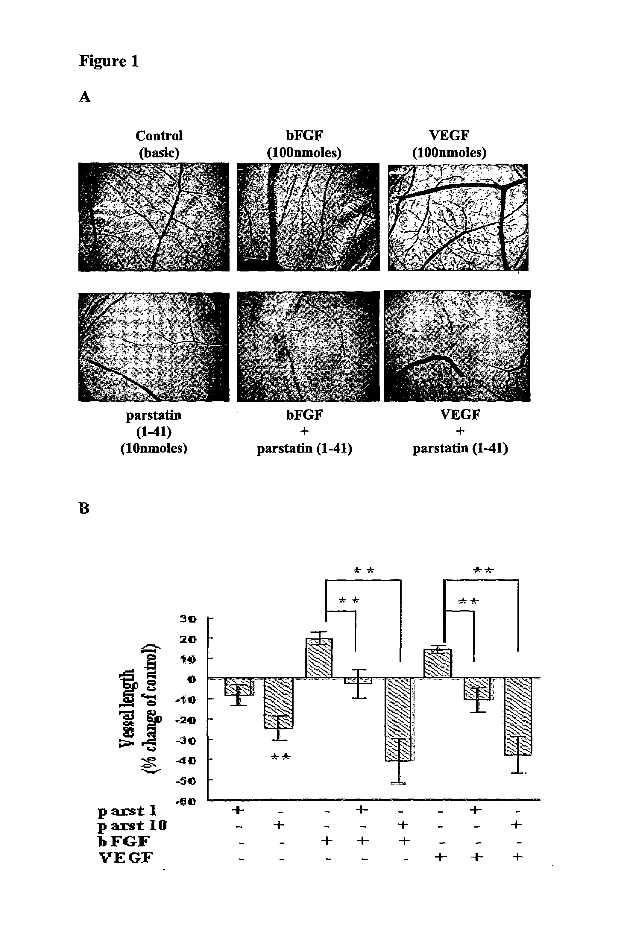
[0320]The in vivo chick chorioallontoic membrane (CAM) angiogenesis model was used to evaluate the effect of parstatin (1-41) in angiogenesis. On incubation day 9 of fertilized chicken eggs, an O-ring (1 cm2) was placed on the surface of the CAM and the vehicle or the indicated substances were placed inside this restricted area. After 48 h, CAMs were fixed in saline-buffered formalin, photographed, and analyzed using the Scion Image software (Scion Image Release Beta 4.0.2 software; Scion Corporation, Frederick, Md.). Image analysis was performed on at least 18 eggs for each group. Vessel number and length were evaluated by pixel counting, and the results expressed as mean percentage of control ±SE. Statistical analyses were performed using a Student's t test.

[0321]Parstatin (1-41) was very potent anti-angiogenic substance (FIG. 1A). The application of human parstatin on CAM of chick embryo, at concentration of 10 nmoles, resulted in a s...

example 3

Parstatin (1-41) Inhibits Angiogenesis in Rat Aortic Ring Assay

[0322]The recognition that angiogenesis in vivo involves not only endothelial cells but also their surrounding cells, has led to development of angiogenic assays using organ culture methods. Of these, the rat aortic ring assay has become the most widely used.

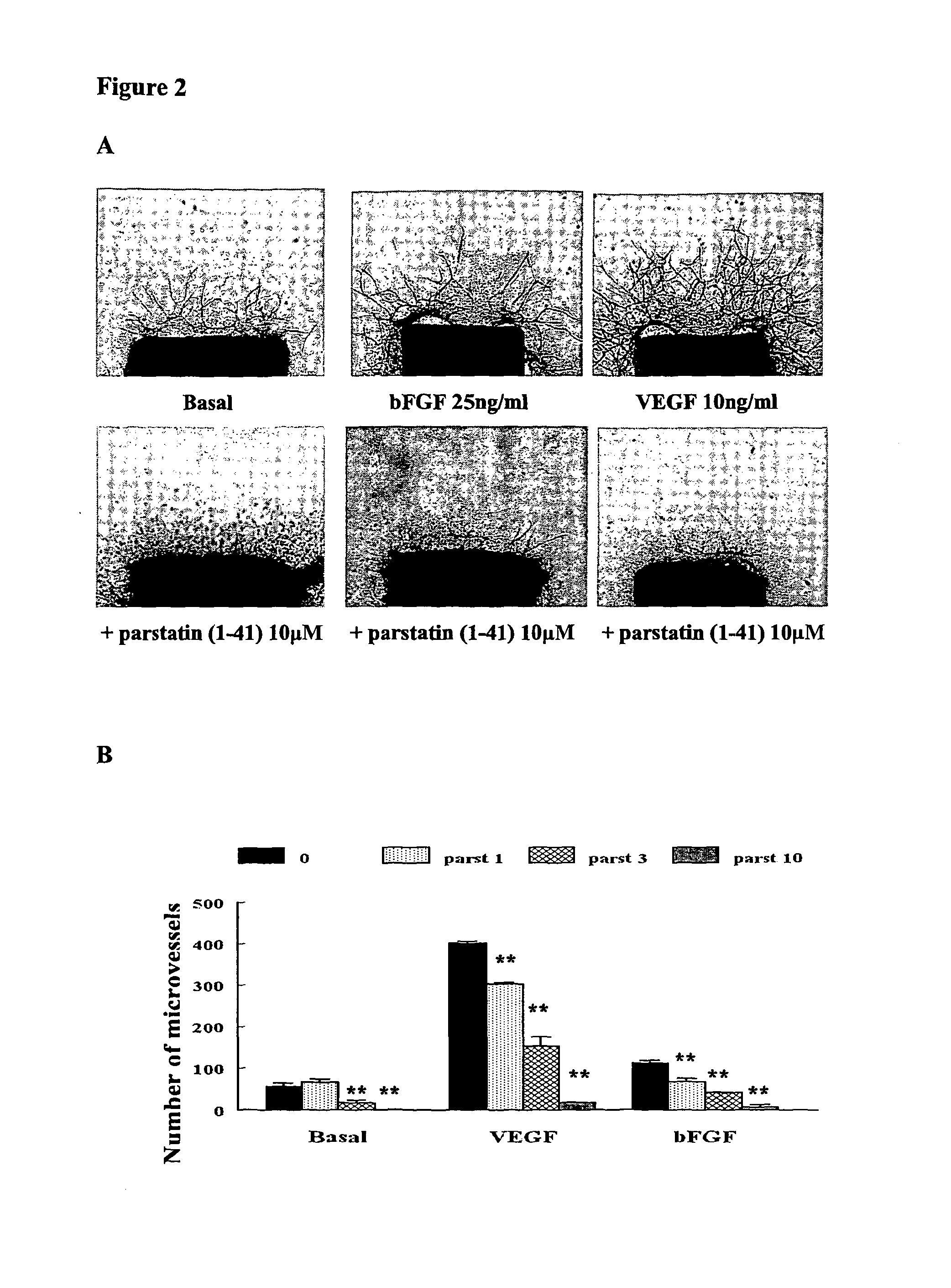
[0323]Freshly cut aortic rings obtained from 5- to 10-week-old Fischer 344 male rats were embedded in collagen gels and transferred to 16-mm wells (4-well NUNC dishes) each containing 115 ml serum-free endothelial basal medium (EBM, Clonetics Corporation) alone or supplemented with VEGF or bFGF. The angiogenic response of aortic cultures was measured in the live cultures by counting the number of neovessels over time using art accepted methods. Mean number of microvessels ±SE was determined. Statistical analysis was performed using unpaired two-tailed t-test.

[0324]Parstatin (1-41) inhibited microvessel formation (FIG. 2A). This inhibitory effect was in a dose depende...


PUM


Abstract

The present invention relates to parstatin peptides, compositions comprising parstatin peptides and their use in the treatment of various disorders, including angiogenesis-related diseases, ocular neovascularization and related disease states, ischemia-reperfusion injury and myocardial-related disease states, and renal disorders.

Description

RELATED APPLICATIONS[0001]The present application is a National Stage entry of the PCT Application No. PCT / IB2010 / 002142 filed Aug. 5, 2010, which is a continuation of U.S. Utility patent application Ser. No. 12 / 572,018, filed Oct. 1, 2009, which claims the benefit of U.S. Provisional Patent Application No. 61 / 247,611, filed Oct. 1, 2009, and PCT Application No. PCT / IB2010 / 002142 is a continuation of U.S. Utility patent application Ser. No. 12 / 645,991, filed Dec. 23, 2009. The entire contents of each of the above documents are incorporated herein by reference.FIELD OF THE INVENTION[0002]The present invention relates to parstatin peptides, compositions comprising parstatin peptides and their use in the treatment of various disorders, including angiogenesis-related diseases, ocular neovascularisation and related disease states, ischemia-reperfusion injury, myocardial-related disease states and renal disorders.BACKGROUND OF THE INVENTION[0003]A. Angiogenesis and Neovascular Ocular Dise...

Claims


Application Information

Patent Timeline
Patent Type & Authority Patents(United States)
IPC IPC(8): A61K38/00A61K38/17A61K38/16C07K7/08A61K38/10C07K14/705C12N5/071
CPCA61K38/177A61K38/10A61K38/16C07K7/08C07K14/705C12N5/069C12N2501/48C12N2501/998
Inventor TSOPANOGLOU, NIKOSMARAGOUDAKIS, MICHAELSIABLIS, DIMITRIOS
Owner TSOPANOGLOU NIKOS
Learn More