Preparation method of aliphatic polyester-chitosan composite fiber tissue repair bracket

An aliphatic polyester and composite fiber technology, used in stents, medical science, prostheses, etc., can solve the problems of uneven properties at the microscopic level of materials, and achieve good cell adhesion, good mechanical properties, and high porosity. Effect

Inactive Publication Date: 2009-04-29
JILIN UNIV
View PDF0 Cites 15 Cited by
  • Summary
  • Abstract
  • Description
  • Claims
  • Application Information

AI Technical Summary

Problems solved by technology

Duan et al [B.Duan, L.L.Wu, X.Y.Yuan, Z.Hu, X.L.Li, Y.Zhang, K.Yao, M.Wang, Hybridnanofibrous membranes of PLGA/chitosan fabricated via an electrospinning array, Journal of Biomedical Materials Research Part A, 83A, 868-878, 2007.] Using electrospinning, with two sets of syringes, a mixed film composed of

Method used


Image

  • Preparation method of aliphatic polyester-chitosan composite fiber tissue repair bracket
  • Preparation method of aliphatic polyester-chitosan composite fiber tissue repair bracket
  • Preparation method of aliphatic polyester-chitosan composite fiber tissue repair bracket

Examples


preparation example Construction

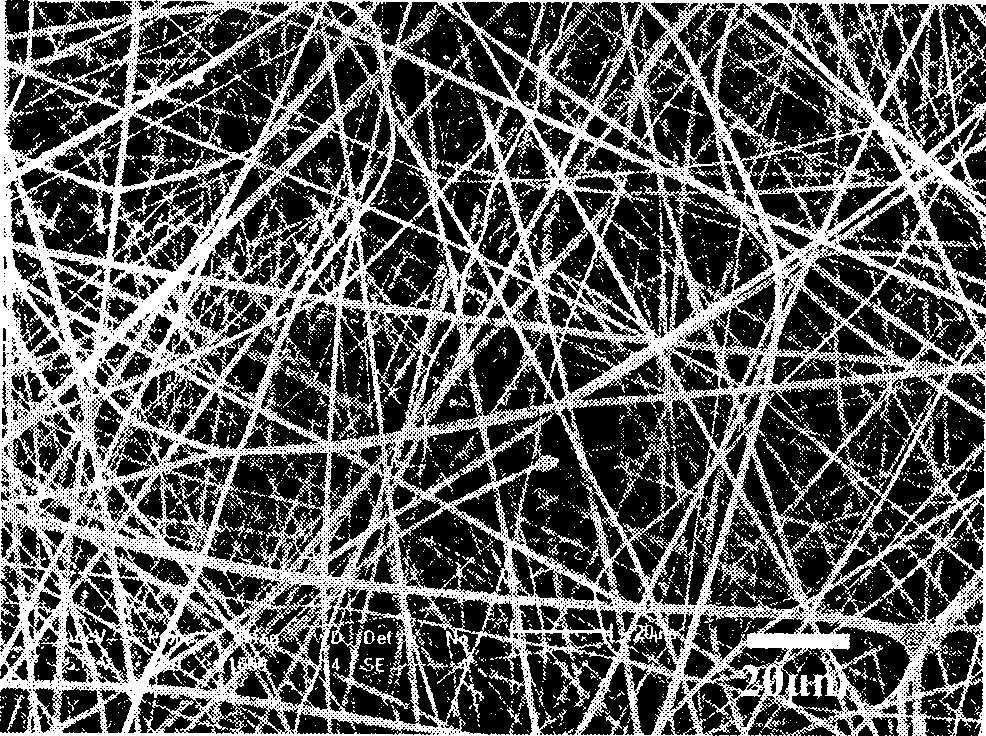
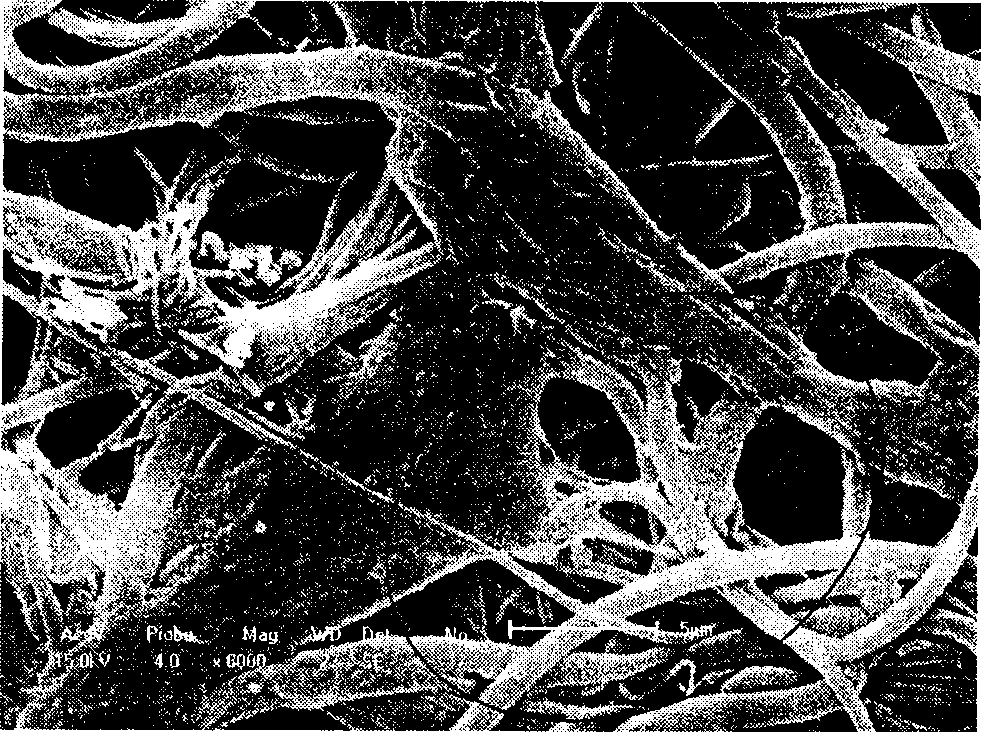
[0018] The preparation method of aliphatic polyester-chitosan composite fiber support comprises the following sequence and steps:

[0019] a. Aliphatic polyester and chitosan are dissolved in co-solvent, stirred evenly to obtain spinning solution, the total mass volume percentage of solute in spinning solution is 4%-25%, the mass fraction of chitosan in solute 9.1%-90%;

[0020] b. Put the spinning solution prepared in step a into a syringe with a needle inner diameter of about 1mm, and the needle of the syringe is connected to the positive pole of the power supply;

[0021] c. Use aluminum foil as the charging device, and ground it, place it opposite the needle, perpendicular to the needle of the glass syringe placed horizontally, and the horizontal distance between the two is 12-30cm;

[0022] d. Adjust the voltage to 11-25KV. Under the high-voltage electric field, the polymer solution will be ejected from the needle and fly to the receiver. The solvent will volatilize duri...

Embodiment 1

[0026] 0.02g polylactic acid and 0.18g crab chitosan were added to a mixed solution (9:1 volume ratio) consisting of 4.5ml trifluoroacetic acid and 0.5ml methylene chloride, and stirred at room temperature for 24 hours. In the obtained spinning solution, the mass ratio of polylactic acid and crab chitosan is 1:9, and the total mass volume percentage of solute is 4%. The above-mentioned polymer solution is pumped into a glass syringe, and the inner diameter of the needle is about 1mm . Connect the needle of this syringe to the positive pole of the power supply providing a voltage of 14kV, and place it horizontally. Use aluminum foil as the receiver and place it at a horizontal distance of 12cm opposite the needle, perpendicular to the syringe, and grounded. Under the high-voltage electric field, the polymer solution will be ejected from the needle and fly to the receiver. The solvent will volatilize during the flight to form micro / nano fibers, and the fiber membrane scaffold w...

Embodiment 2

[0028] 0.18g of polycaprolactone and 0.18g of shrimp chitosan were added to 6.51ml of trifluoroacetic acid and stirred at room temperature for 24 hours. In the prepared spinning solution, the mass ratio of polylactic acid and shrimp chitosan is 1:1, and the total mass volume percentage of solute is 5.5%. The above polymer solution is pumped into a glass syringe, and the inner diameter of the needle is about 1mm. Connect the needle of this syringe to the positive pole of the power supply providing a voltage of 17kV, and place it horizontally. Use aluminum foil as the receiver and place it at a horizontal distance of 18cm opposite the needle, perpendicular to the syringe, and grounded. Under the high-voltage electric field, the polymer solution will be ejected from the needle and fly to the receiver. The solvent will volatilize during the flight to form micro / nano fibers, and the fiber membrane scaffold with an average diameter of 840nm will be received on the aluminum foil.


PUM

PropertyMeasurementUnit
The average diameteraaaaaaaaaa

Abstract

The invention discloses a preparation method of an aliphatic polyester-chitosan composite fibroustissue restoration material, comprising the steps as follows: a. mixture solution of aliphatic polyester and chitosan is prepared; and b. electrostatic spinning is carried out on the mixture solution of aliphatic polyester and chitosan. Compared with the prior art, the preparation method has simple operation and low price, the aliphatic polyester and the chitosan in composite fiber are mixed molecularly, thus avoiding phase separation between the aliphatic polyester and chitosan; the bracket consisting of aliphatic polyester-chitosan composite micro-nano fiber has good fineness degree, good cell adhesion, good mechanic performance, higher porosity and large specific surface area, can be degraded in vivo simultaneously, and is an ideal tissue restoration bracket which has extremely high porosity and similar nano-structure of the extracellular matrix of the natural cell, can promote migration and proliferation of cells, and can be used for the restoration of tissues such as cornea, skin, cartilage and the like.

Description

technical field [0001] The invention belongs to the field of tissue engineering medical materials, in particular to a method for preparing an electrospinning aliphatic polyester-chitosan composite fiber tissue repair bracket. Background technique [0002] Electrospinning, as a processing technique for preparing micro / nano-sized polymer fibers, has attracted more and more attention. Micro / nanofiber fabrics prepared by electrospinning have many special properties, such as high specific surface area and porosity, so they are widely used in biomedical fields, such as wound dressings, drug release, tissue engineering scaffolds, etc. Wait. For tissue engineering scaffolds, microstructures with high porosity and high specific surface area are required to facilitate the growth and attachment of tissue cells. The surface topology of electrospun scaffolds is very similar to natural extracellular matrix (ECM), which can meet the requirements of tissue engineering scaffolds. Chitosan...

Claims


Application Information

Patent Timeline
IPC IPC(8): A61L27/40A61L27/20A61L27/18A61L27/50A61F2/82
Inventor 王洪艳李俊峰徐佳王萍王心蕊贾卉
Owner JILIN UNIV
Why Eureka
Try Eureka