
A fluorescent mesoporous silicon oxide nanomaterial and its preparation method and application

A technology of mesoporous silicon oxide and nanomaterials, applied in the field of mesoporous nanomaterials, can solve the problems of difficulty in controlling the shape and size of porous silicon nanostructures, easy detachment of organic fluorescent dye molecules, affecting production and application, etc., and achieve a uniform spherical shape The effect of morphology and nanometer size, simple preparation process, and good biological safety

Inactive Publication Date: 2011-12-28
SHANGHAI INST OF CERAMIC CHEM & TECH CHINESE ACAD OF SCI
View PDF3 Cites 14 Cited by
  • Summary
  • Abstract
  • Description
  • Claims
  • Application Information

AI Technical Summary

Problems solved by technology

Recent research progress shows that: (1) Organic rare earth complexes or organic fluorescent dye molecules can be monodispersely covalently linked to the inner wall of mesoporous silica mesoporous channels, avoiding fluorescence quenching and improving fluorescence efficiency [ordered Mesoporous silica-based fluorescent nanomaterial and its preparation method CN101525533; J.Mater.Chem.2009, 19, 3395-3403; J.Phys.Chem.C 2010, 114, 12505-12510.], however, organic fluorescent dyes Molecules are easy to fall off, which limits its application to a certain extent; (2) Fluorescent quantum dots or nanoparticles can also be integrated with mesoporous silica by wrapping or connecting [Adv.Funct.Mater.2010, 20, 1166~1172; J.Phys.Chem.B 2003, 107, 11575~11578.], but for the common fluorescent quantum dots containing heavy metal ions, there are biological safety problems caused by the leakage of heavy metal ions [Nano Lett.2004, 4 , 11~18; Toxicol.Appl.Pharmacol.2009, 238, 280~288.]; (3) CN101864308A discloses a mesoporous silicon oxide luminescent material doped with rare earth ions and its preparation method, and CN1583948A discloses a A mesoporous silica / manganese zinc silicate composite phosphor was prepared by mixed sintering
However, only micron-sized powders can be obtained by the above-mentioned preparation method, which limits its application in the field of biomedicine; (4) In addition, porous silicon nanomaterials are considered to be a kind of biosafety, capable of drug delivery and optical However, the preparation of porous silicon nanostructures generally adopts a complex process method of electrochemical corrosion first, and then ultrasonic stripping or mechanical crushing [Nature Mater.2009, 8, 331-336; Nature Nanotechnol .2008, 3, 151~157.]
The biggest defect of this method is that the shape and size of the prepared porous silicon nanostructures are difficult to control, and the yield is relatively low, which seriously affects its actual production and application.

Method used


Image

  • A fluorescent mesoporous silicon oxide nanomaterial and its preparation method and application
  • A fluorescent mesoporous silicon oxide nanomaterial and its preparation method and application
  • A fluorescent mesoporous silicon oxide nanomaterial and its preparation method and application

Examples


Embodiment 1

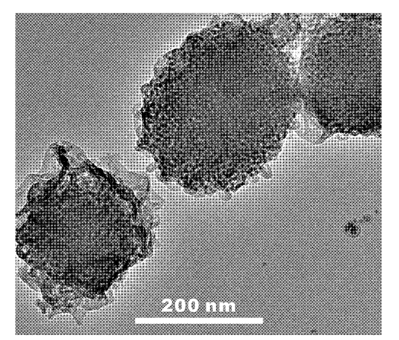
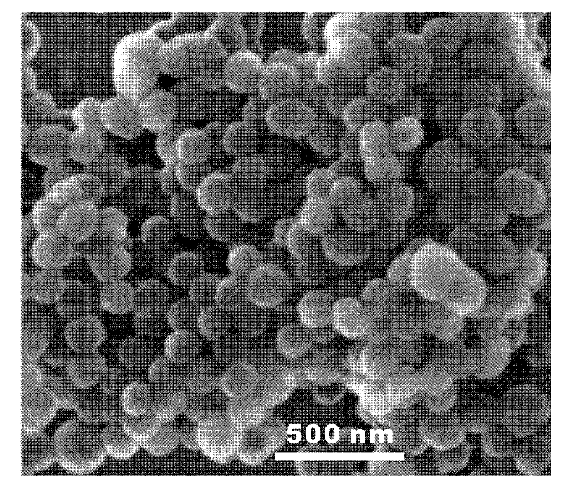
[0037]Under the protection of argon, add 0.17mmol P123 and 50mmol sodium chloride together into 80mL aqueous hydrochloric acid (2mol / L), stir at 25°C to dissolve completely; add 10mmol triethoxysilane, continue stirring for 1 hour Centrifuge, ultrasonically wash three times with ethanol, and then disperse in water and freeze-dry. The obtained freeze-dried powder sample is labeled as "SIC-1". The SIC-1 freeze-dried powder was divided into four parts and placed in a nitrogen atmosphere furnace, and calcined at 500°C, 600°C, 700°C and 900°C for 2 hours respectively, and the four samples prepared were respectively marked as "SIC-500", "SIC-600", "SIC-700", and "SIC-900".

[0038] figure 2 It is the transmission electron micrograph of the "SIC-600" sample prepared in this embodiment, image 3 It is a scanning electron micrograph of the "SIC-600" sample prepared in this example. Depend on figure 2 and image 3 It can be seen that the silicon oxide material prepared by the met...

Embodiment 2

[0041] Take 20 mg of the "SIC-600" sample prepared in Example 1, dry it under vacuum at 120° C. for 12 hours, and then cool it to room temperature under vacuum.

[0042] Add 5 mL of 10 mg / mL doxorubicin ethanol solution, immerse at room temperature for 1 hour, centrifuge, and vacuum dry to prepare a fluorescent nanomaterial loaded with doxorubicin, which is labeled "DOXSIC-600".

[0043] The above-mentioned DOXSIC-600 nanomaterials were co-cultured with MCF-7 breast cancer cells, then the cells were washed with phosphate saline, and finally the cells were observed and photographed under a fluorescent confocal microscope. The photographic results show that the nanomaterials can be taken up by MCF-7 breast cancer cells in large quantities, and release doxorubicin in the cells, and can be fluorescently imaged (such as Figure 7 shown).

[0044] Use the CCK-8 kit (commercially available) to detect the viability of cells after cultivating for three days, and the results show that ...


PUM


Abstract

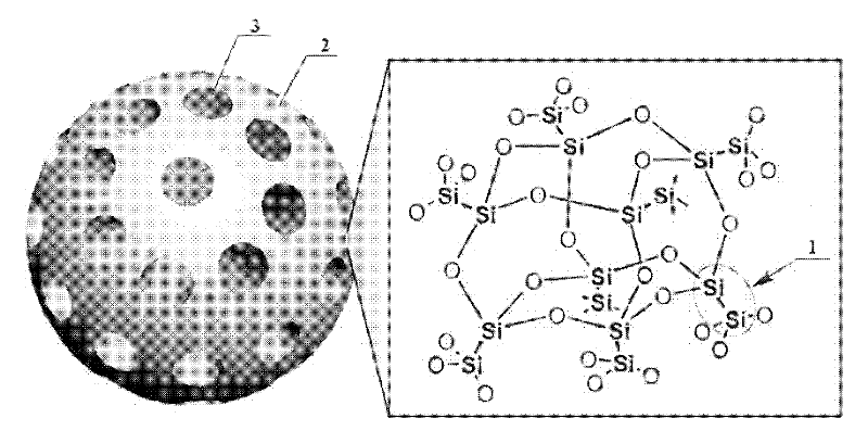
The invention discloses a fluorescent mesoporous silicon oxide nanomaterial and a preparation method and an application thereof. The material is characterized in that a silicon-rich microdomain formed by silicon clusters or silicon quantum dots is embedded in a framework of mesoporous silicon oxide. The preparation method of the material comprises the following steps: adding a water-soluble surfactant and a hydrolyst in water, agitating at 4-60 DEG C and dissolving fully; adding hypoxic silicate ester in the system, continuing agitating for 1-48 h, centrifuging, washing and drying; and calcining in inert atmosphere or vacuum condition to obtain a precursor material. As the fluorescent mesoporous silicon oxide nanomaterial prepared by the preparation method disclosed by the invention has good dispersibility, uniform nanometer size and adjustable aperture and adjustable luminescent performance, the fluorescent mesoporous silicon oxide nanomaterial integrates porous structure and fluorescence function, has double functions of drug loading and conveying and synchronous fluorescence imaging, and has potential application value in synchronous diagnosis and treatment of diseases.

Description

technical field [0001] The invention relates to a fluorescent mesoporous silicon oxide nanomaterial, a preparation method and application thereof, and belongs to the technical field of mesoporous nanomaterials. Background technique [0002] Cancer and cardiovascular disease are two of the most serious diseases that threaten human life and health. The prevention and treatment of major diseases such as cardiovascular disease and cancer have become the strategic focus of public health work in my country, and their early detection, diagnosis and treatment are of great significance. With the help of nanotechnology, the construction of a multifunctional nanostructured material integrating imaging diagnosis, drug delivery and treatment has opened up a new way for the simultaneous diagnosis and treatment of major diseases such as cardiovascular disease and cancer. [0003] Bifunctional nanostructures that couple the drug delivery function of mesoporous silica and the optical imagin...

Claims


Application Information

Patent Timeline
IPC IPC(8): C09K11/59A61K47/04A61K49/00
Inventor 何前军施剑林
Owner SHANGHAI INST OF CERAMIC CHEM & TECH CHINESE ACAD OF SCI
Learn More