Method of reducing disulfide bond in photo-excitation protein to obtain free sulfydryl

A protein and disulfide bond technology, applied in the field of protein fixation and self-assembly, can solve the problems of destroying protein structure and activity, limiting protein application, complicated steps, etc., and achieving the effect of convenient specific fixation, convenient operation and simple operation

Inactive Publication Date: 2013-05-08
NANJING UNIV OF POSTS & TELECOMM
View PDF0 Cites 6 Cited by
  • Summary
  • Abstract
  • Description
  • Claims
  • Application Information

AI Technical Summary

Problems solved by technology

The steps of these methods are relatively cumbersome, and they have relatively high requirements on the solvent environment such as temperature and pH. At the same time, the structure and activity of the protein may be reduced or destroyed. Moreover, the preparation and purification of the modified protein takes a long time and the consumables are relatively expensive, which limits the Application of proteins in biosensors and biomaterials

Method used


Image

  • Method of reducing disulfide bond in photo-excitation protein to obtain free sulfydryl
  • Method of reducing disulfide bond in photo-excitation protein to obtain free sulfydryl
  • Method of reducing disulfide bond in photo-excitation protein to obtain free sulfydryl

Examples


Embodiment 1

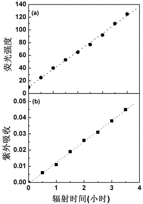
[0022] Photocutinase generates free sulfhydryl groups: Cutinase is a lipolytic enzyme capable of degrading cutin, similar to lipase, which has important industrial uses and can be used to remove oil stains. Prepare 0.1mM cutinase solution, pH 7.5, temperature 25°C, take 2ml and inhale into a quartz cuvette. Use 280-300nm ultraviolet light to irradiate the sample cell of the quartz cuvette, and control the distance of the light so that the light intensity is 0.1W / m 2 , every 30min, detect the strength of the fluorescent signal. Cutinase is a globulin composed of 214 amino acids, of which amino acid No. 69 is tryptophan (Trp69). Such as figure 2(a), In the native state, the fluorescence signal of cutinase tryptophan is quenched. After a certain period of ultraviolet radiation, the fluorescence signal is continuously enhanced. The main reason is that there is a pair of disulfide bonds (Cys31-Cys109) near tryptophan. In the natural state, there is electron and energy tran...

Embodiment 2

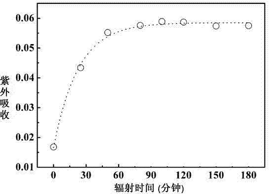
[0024] Illuminating lysozyme to generate free sulfhydryl groups: lysozyme is a glycoside hydrolase that can dissolve the cell wall of Gram-positive bacteria, has a strong killing effect on bacteria, and can also be used to prevent food deterioration. Chemicals, baby food and scientific research have a wide range of applications. Prepare 0.01mM lysozyme solution with TrisHCl buffer (pH 7.8, temperature 25°C), take 2ml and inhale into a quartz cuvette. Use 270-300nm ultraviolet light to irradiate the sample cell of the quartz cuvette, control the light intensity, and after a certain period of time, detect whether there is sulfhydryl group generation by Ellman reagent. Such as image 3 As shown, it can be seen that lysozyme has many pairs of disulfide bonds and tryptophan close to each other. After ultraviolet light irradiation, the protein reacts with DTNB, and the product has ultraviolet absorption at 412nm, indicating that there is free sulfhydryl group formation, and with th...

Embodiment 3

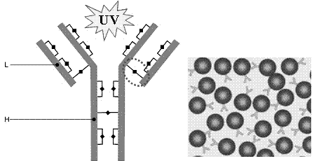
[0026] Combination of immunoglobulin G and gold nanoparticles under illumination: Immunoglobulin G is an important antibody protein, which is often combined with gold nanoparticles for precise positioning of cell surface and intracellular biomacromolecules. It is widely used in immunology, histology, fields of pathology. Such as Figure 4 As shown, the structure of immunoglobulin is a "Y" shape, and the upper "V" is used to recognize the antigen and determine the specificity of the antibody, which is called the Fab segment, and the rest of the carboxyl terminal is the Fc segment. There are more than a dozen pairs of disulfide bonds on immunoglobulins, all of which are close to aromatic amino acids, but most of them are buried inside the structure. Among them, the pair of disulfide bonds marked by a circle has a relatively large solvent exposure, and is prone to reduction when irradiated by ultraviolet light , generating free sulfhydryl groups. At room temperature of 25°C, pr...


PUM


Abstract

The invention discloses a method of reducing disulfide bond in photo-excitation protein to obtain free sulfydryl. The method comprises the following steps: (1) preparing the protein solution, wherein the concentration of the protein is smaller than 1mM, the pH is 7-8, and the temperature of the protein solution is 10-400 DEG C; and (2) conducting ultraviolet irradiation to the protein solution for 10-1000 minutes to obtain free sulfydryl-containing protein, wherein the irradiation intensity is smaller than 30W / m<2> and the irradiation time is 10-1000min. The method is simple to operate, efficient and fast, and has strong specificity and least damage to the structure of the protein. Therefore, the method of obtaining the free sulfydryl through photo-exciting the protein can be used for high coverage rate of the protein in an interface, and is orderly in orientation and constant in specificity; and the technology has significant application prospects in the fields of biosensors, biomaterials, bio-energy sources and the like, thereby bringing tremendous economic benefits and social benefits for the development of industries of medical, chemical and the like.

Description

technical field [0001] The invention relates to a method for obtaining free sulfhydryl groups by reducing disulfide bonds in proteins by light excitation, which is mainly used for protein immobilization and self-assembly in biomaterials and biosensors. Background technique [0002] Adsorption and immobilization of proteins at interfaces are involved in applications such as biomaterials, biosensors, and biofuel cells. In order to improve the orientation distribution of proteins on the interface and better exert their biological functions, it is necessary to modify and process proteins. The usual method is to introduce active groups such as amino groups, carboxyl groups, and sulfhydryl groups, and immobilize the carrier protein on the interface through covalent interaction, so as to realize the specificity of immobilization of biomolecules and the order of molecular orientation, which directly determines the biosensor. Sensitivity and specificity of biomaterials [Rusmini F, e...

Claims


Application Information

Patent Timeline
IPC IPC(8): C07K14/76C07K14/435C07K1/00C07K17/00C12N11/00C12N13/00C12N9/36C12N9/18C12N9/20C12N9/14C12N9/16
Inventor 武灵芝胡栋
Owner NANJING UNIV OF POSTS & TELECOMM
Why Eureka
Try Eureka