
Nanometer structure electrochemical cell sensor preparation method, produced nanometer structure electrochemical cell sensor and use thereof

A technology of cell sensors and nanostructures, applied in the direction of material electrochemical variables, etc.

Inactive Publication Date: 2014-04-09
NANJING UNIV
View PDF4 Cites 11 Cited by
  • Summary
  • Abstract
  • Description
  • Claims
  • Application Information

AI Technical Summary

Problems solved by technology

However, no Fe-based 3 o 4 Report on the Selective Quantitative Analysis of the Expression of Leukemia Cells and Their Surface Death Receptors Using an Electrochemical Cell Sensor Based on Au / Au Composite Nanostructures

Method used


Image

  • Nanometer structure electrochemical cell sensor preparation method, produced nanometer structure electrochemical cell sensor and use thereof
  • Nanometer structure electrochemical cell sensor preparation method, produced nanometer structure electrochemical cell sensor and use thereof
  • Nanometer structure electrochemical cell sensor preparation method, produced nanometer structure electrochemical cell sensor and use thereof

Examples


Embodiment 1

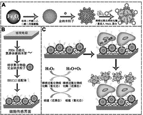
[0035] Example 1. Preparation of multifunctional hybrid nanoprobes for cell sensors

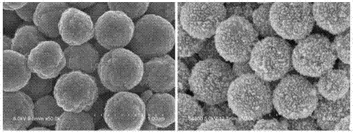
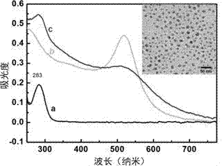
[0036] Weigh 1.35 g of ferric chloride hexahydrate, 3.2 g of anhydrous sodium acetate and 0.5 mL of polyacrylic acid (number average molecular weight: 3000) and add it to 38 mL of ethylene glycol for ultrasonic mixing, then add the mixture into a polytetrafluoroethylene reactor 473 After hydrothermal reaction at K for 6 hours, magnetic separation was carried out, washed several times with ethanol and deionized water respectively, and black magnetic Fe was obtained after drying. 3 o 4 Nanospheres with a particle size of 500 nm ± 20 nm. Take Fe 3 o 4 Disperse 10 mg of nanospheres in 1 mL of deionized water, add 1% PDDA and 1 mL of ultrasonic reaction for 30 minutes, then separate the product under a magnetic field, and wash three times with deionized water to obtain Fe 3 o 4 / PDDA nanocomposite material; gold nanoparticles (Au NPs) were obtained by deoxygenation reaction with tetrachloroal...

Embodiment 2

[0037] Example 2. Assembly of cell-sensing nanoelectrode interface

[0038] Mix 5 mL of tetrachloroalloy acid aqueous solution with a concentration of 4.86 mM and 5 mL of polyamide-amine dendrimer (PAMAM, G=4, provided by Sigma-Aldrich) until the yellow color fades, so that the terminal of PAMAM Amino groups and gold form complex anions. After vigorously stirring the complex with 1.8 mL of reducing agent 20 mM sodium borohydride solution for 30 minutes at room temperature, the color of the solution turns dark red, which is the above-mentioned gold stabilized with dendrimers. Nanoparticles (Au DSNPs) with a particle size of 10 nm ± 2 nm. Glassy carbon electrodes (3 mm in diameter) were mechanically polished with alumina powders with a particle size of 0.3 and 0.05 μm, rinsed with deionized water, and washed with 8 mol L -1 Nitric acid, acetone, and deionized water were ultrasonically cleaned and dried, then 5.0 mg mL was added dropwise -1 Diethylene glycol diacrylate functio...

Embodiment 3

[0039] Example 3. Cell recognition and specificity test of assembled electrode interface

[0040] Drop HL-60 cells and K562 cell suspensions onto the electrode surface treated in Example 1, incubate at 37°C, and observe with a bright-field optical microscope. The results show that 95% of HL-60 cells are captured and maintain their activity , and almost no capture in K562 cells; adding HRP-TRAIL-Fe to HL-60 cell suspension 3 o 4 Au hybrid nanoprobe and Fe 3 o 4 Au nanomaterials were dropped onto the surface of the above-mentioned electrodes, and observed again through a bright-field optical microscope. The results showed that a large number of mixed nanoprobes could successfully immobilize the surface of HL-60 cells. The characterization results are shown in Figure 4 .


PUM

PropertyMeasurementUnit
diameteraaaaaaaaaa
diameteraaaaaaaaaa
particle diameteraaaaaaaaaa

Abstract

The present invention discloses a composite nanometer structure electrochemical cell sensor, which comprises two assemblies of a multi-functional mixed nanometer probe and a nanometer structure electrode interface, wherein the multi-functional mixed nanometer probe (HRP-TRAIL-Fe3O4@Au) for electrochemical cell sensing comprises Au nanoparticle-modified magnetic Fe3O4 nanometer spheres immobilized with recombinant human TRAIL protein and horseradish peroxidase (HRP) through a co-immobilization effect, and the nanometer structure electrode interface is an electrode cell sensing interface constructed through a layer-by-layer assembling method, wherein the nanometer structure electrode interface integrates high biocompatibility Au nanoparticles (AuDSNPs) stabilized by a dendrimer, high resistivity nitrogen-doped carbon nano-tubes (CNx) and a high specificity cellular localization oligonucleotide aptamer, and has a nanometer layered structure. The composite nanometer structure electrochemical cell sensor can be provided for carrying out selective quantification detection on expression of DR4 / DR5 death receptors on leukemic cells and surfaces thereof. The present invention further discloses the preparation method.

Description

technical field [0001] The invention relates to an electrochemical cell sensor with ferric oxide / gold composite nanostructure and a selective quantitative analysis method for the expression of death receptors on leukemia cells and their surfaces. Background technique [0002] Leukemia primarily affects the bone marrow, blood cells, and other parts of the lymphatic system and is one of the most common fatal cancers [see: (a) Nordlund, J.; Milani, L.; Lundmark, A.; Lonnerholm, G.; Syvanen, A.-C. PLoS ONE 2012, 7 (4), 9.]. Its innate and acquired anti-chemotherapy and radiotherapy properties have always been one of the main obstacles in the treatment of leukemia. To overcome this obstacle, a potential adjuvant conventional therapy is to directly activate apoptosis-mediated death receptors. to induce cell death [see: (b) Ghobrial, I. M.; Witzig, T. E.; Adjei, A. A., Ca-a Cancer Journal for Clinicians 2005, 55 (3), 178-194.]. Tumor necrosis factor-related apoptosis-inducing lig...

Claims


Application Information

Patent Timeline
Patent Type & Authority Applications(China)
IPC IPC(8): G01N27/28G01N27/26
Inventor 朱俊杰华子春郑婷婷傅佳驹
Owner NANJING UNIV
Learn More