Method for promoting in-vitro expansion of human amniotic epithelial cells and use thereof

An epithelial cell and in vitro expansion technology, applied in the field of stem cell biology, can solve the problems of large application risks, restrictions, and expensive cytokines, and achieve the effects of good safety, convenient operation, and increased cell ratio

Active Publication Date: 2017-06-13
AFFILIATED HOSPITAL OF ZUNYI MEDICAL COLLEGE
View PDF4 Cites 3 Cited by
  • Summary
  • Abstract
  • Description
  • Claims
  • Application Information

AI Technical Summary

Problems solved by technology

However, there is no relevant report on the induction agent or induction method that promotes the expansion of human amniotic epithelial cells in vitro
[0004] Moreover, the use of cytokines or other exogenous inducers to promote the expansion

Method used


Image

  • Method for promoting in-vitro expansion of human amniotic epithelial cells and use thereof
  • Method for promoting in-vitro expansion of human amniotic epithelial cells and use thereof
  • Method for promoting in-vitro expansion of human amniotic epithelial cells and use thereof

Examples


Embodiment 1

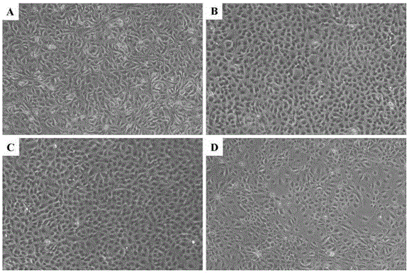
[0023] Example 1: Isolation, preparation, morphological and phenotypic characteristics of human amniotic epithelial cells

[0024] The amniotic membrane was aseptically stripped from the fresh placental tissue, washed repeatedly with the newly prepared D-Hank's solution containing double antibodies (final concentration of penicillin 100 U / mL, final concentration of streptomycin 0.1 mg / mL) to remove residual blood stains, and the amniotic membrane was cut into pieces. Add 0.05% trypsin digestion solution containing 0.02% EDTA, 37°C, 200 rpm rotary digestion for 10 min, discard the digestive solution, then add fresh digestion solution to the amnion tissue, 37°C, 200 rpm rotary digestion for 30 min, 300 mesh stainless steel The single cell suspension was collected by mesh filtration, and the digestion was terminated by adding medium containing 10% fetal bovine serum. Treated twice in the same way, combined the collected single cell suspension, centrifuged at 1500 rpm for 10 min, ...

Embodiment 2

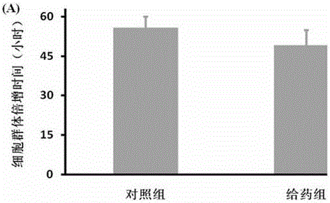
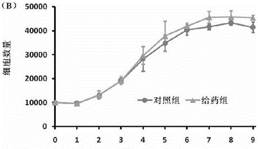
[0026] Example 2: Effect of Nutritional Composition on Proliferation of Human Amniotic Epithelial Cells

[0027] Take P1 human amniotic epithelial cells in the logarithmic growth phase, digest and resuspend, and count the cells at 1.0×10 4 The cells / well were planted in a 96-well plate, and the medium was replaced after 48 h, and a control group and a nutritional composition group were set up (the dose of 300 kDa hyaluronic acid was 1 mg / mL concentration gradient, and the concentration of epidermal growth factor was 10 ng / mL, the concentration of vitamin C is 50 µg / mL, the concentration of GlutaMAX™-I supplement is 1% volume ratio factor, β -The concentration of mercaptoethanol is 1% volume ratio coefficient, the concentration of glycine is 7.5 mg / mL, the concentration of L-alanine is 8.9 mg / mL, the concentration of L-aspartic acid is 13.2 mg / mL, the concentration of L- The concentration of asparagine was 13.3 mg / mL, the concentration of L-glutamic acid was 14.7 mg / mL, the c...

Embodiment 3

[0034] Example 3: Effects of Nutritional Compositions on Human Amniotic Epithelial Cell Proliferation-Related Genes and Stemness Gene Transcription Levels

[0035] Take the P1 human amniotic epithelial cells in the logarithmic growth phase, and use 3×10 5 The cell density of cells / well was inoculated in 6-well plates, and the fresh medium was replaced after 3 days, and the control group and nutrition combination group (300 kDa hyaluronic acid 1 mg / mL) were set up, and the culture was continued for 48 h. Total RNA was extracted according to the kit instructions, and cDNA was synthesized by reverse transcription, that is, the concentration of total RNA in each group was uniformly adjusted to 50 ng / µL, and the reaction conditions were: 37°C, 15min; 85°C, 5s. The reverse transcription product (cDNA) was frozen and stored at -80°C for future use. Real-time fluorescence quantitative PCR, the reaction conditions are: 95°C, pre-denaturation for 30 s, then 40 cycles of PCR (denaturati...


PUM


Abstract

The invention relates to a method for promoting in-vitro expansion of human amniotic epithelial cells. The method comprises adding a nutritional composition during human amniotic epithelial cell culture, wherein the nutritional composition mainly comprises hyaluronic acid, an epidermal cell growth factor, vitamin C, a GlutaMAX-I additive, beta-mercaptoethanol, glycine, L-alanine, L-aspartic acid, L-asparagine, L-glutamic acid, L-proline and L-serine. The method can significantly promote human amniotic epithelial cell proliferation, reduce the doubling time, maintain biological characteristics such as human amniotic epithelial cell surface marker expression, multi-directional differentiation potential and immune tolerance, and significantly enhance the expression level of a human amniotic epithelial cell related gene and an immunosuppressive factor. Therefore, the nutritional composition as a human amniotic epithelial cell proliferation regulator has obvious advantages and has a great clinical significance to alleviate the shortage of seed cells in the field of regenerative medicine.

Description

technical field [0001] The invention belongs to the field of stem cell biology technology, and in particular relates to a method and application for promoting human amniotic epithelial cell expansion in vitro. Background technique [0002] Human amniotic epithelial cells are derived from the epithelial layer of the amniotic membrane tissue of the placenta. Since the amniotic membrane is a waste product after childbirth, it does not involve medical ethics and legal issues. Moreover, human amniotic epithelial cells have the characteristics of undifferentiated embryonic stem cells, can express all molecular markers of embryonic stem cells, and have the potential to differentiate into three germ layer tissues. And because of the lack of telomerase, the risk of tumorigenesis after transplantation is avoided. Human amniotic epithelial cells do not express HLA antigens, have no immune rejection after transplantation, have excellent transplant immune tolerance characteristics, and ...

Claims


Application Information

Patent Timeline
IPC IPC(8): C12N5/073C12N5/071
CPCC12N5/0605C12N5/0625C12N2500/32C12N2500/38C12N2500/44C12N2501/11C12N2501/905
Inventor 肖建辉田亚冰
Owner AFFILIATED HOSPITAL OF ZUNYI MEDICAL COLLEGE
Why Eureka
Try Eureka