Lysosome targeted pH fluorescent probe for monitoring cell autophagy as well as preparation and application thereof

A fluorescent probe and lysosome technology, applied in the field of pH fluorescent probes, can solve the problems of unsuitability for long-term detection, large cytotoxic side effects, strong background fluorescence, etc., and achieves good cell membrane permeability and easy mass production. , the effect of highly sensitive detection

Inactive Publication Date: 2020-04-03
SHANXI UNIV
View PDF1 Cites 7 Cited by
  • Summary
  • Abstract
  • Description
  • Claims
  • Application Information

AI Technical Summary

Problems solved by technology

[0004] At present, many fluorescent probes have been reported in the literature to detect the pH changes of lysosomes. However, since the excitation and emission wavelengths of these probes are mostly in the ultraviolet region, they overlap with cell autofluorescence, resulting in a strong background fluorescence, and ultraviolet light is harmful to cells. The toxic and side effects are relatively large, so it is not suitable for long-term detection
In addition, the existing lysosome targeting groups are concentrated on the traditional morpholine ring and dimethylamino structure, and such weakly basic groups are easy to gather in weakly acidic lysosomes to cause the "alkalinization effect" of lysosomes , long-term action can lead to cytotoxicity

Method used


Image

  • Lysosome targeted pH fluorescent probe for monitoring cell autophagy as well as preparation and application thereof
  • Lysosome targeted pH fluorescent probe for monitoring cell autophagy as well as preparation and application thereof
  • Lysosome targeted pH fluorescent probe for monitoring cell autophagy as well as preparation and application thereof

Examples


Embodiment 1

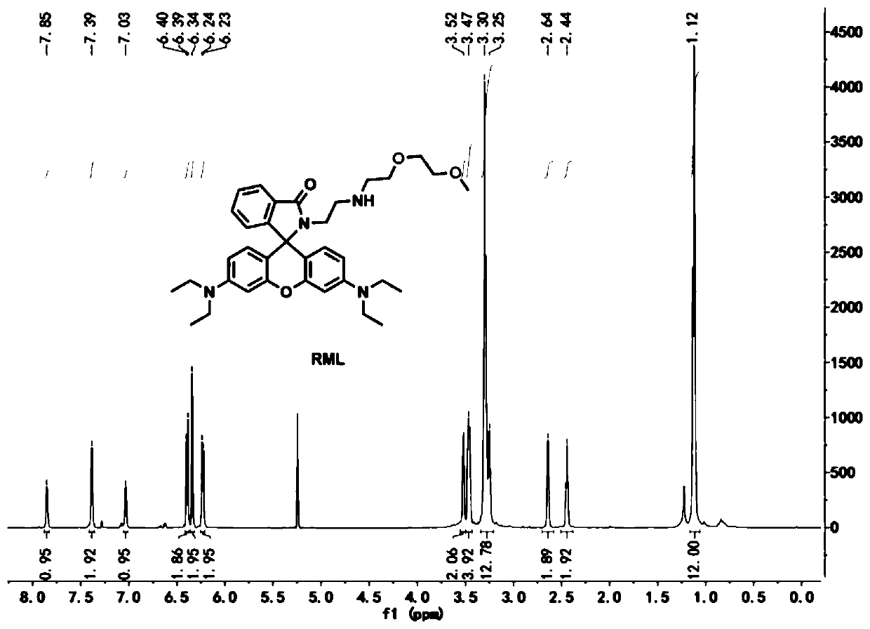
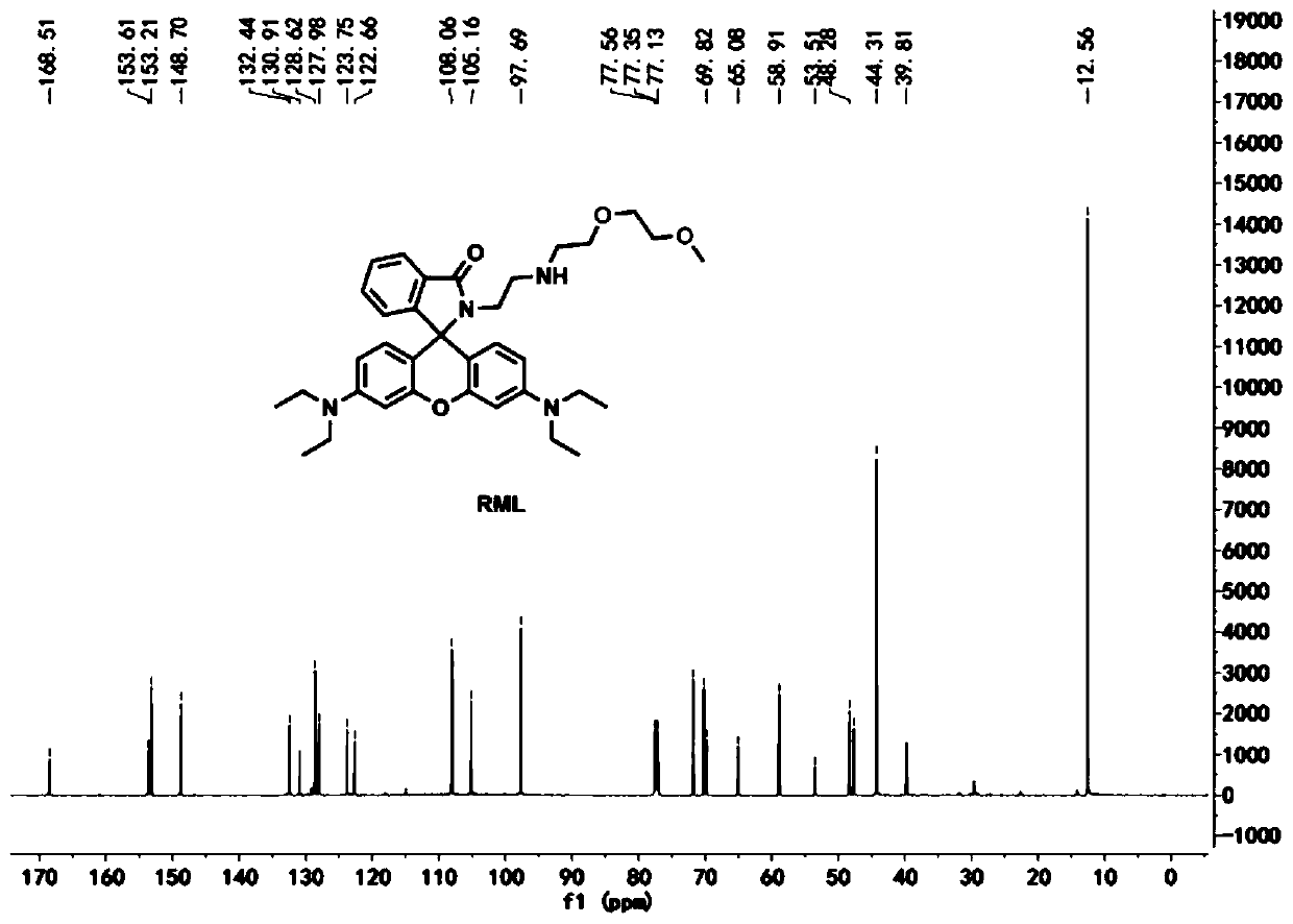
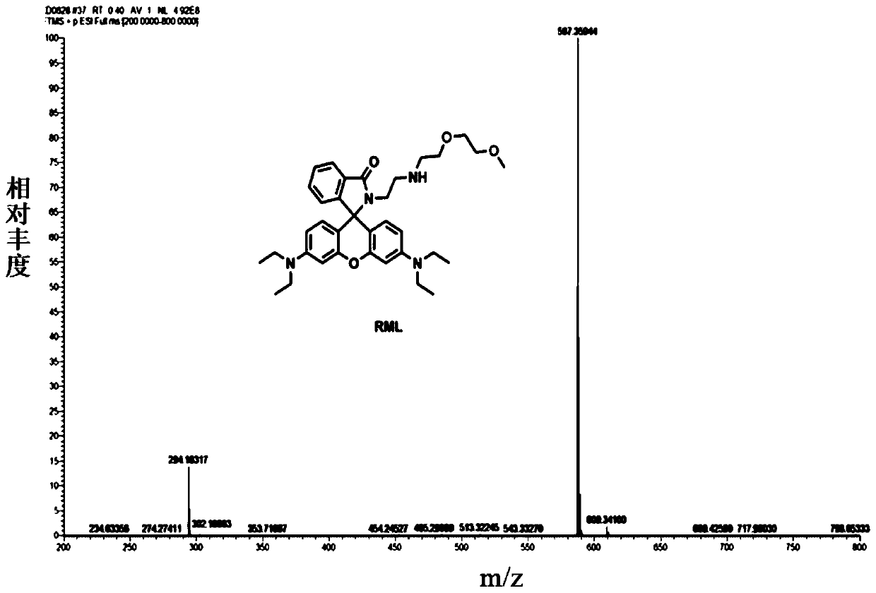
[0031] 3',6'-bis(diethylamino)-2-(2-((2-(2-methoxyethoxy)ethyl)amino)ethyl)spiro[isoindoline-1, Preparation and characterization of 9'-xanthene]-3-one (RML):

[0032]

[0033] In a round bottom flask, 2-(2-aminoethyl)-3',6'-bis(diethylamino)spiro[isoindole-1,9'-xanthogen]-3-one (170mg , 0.62mmol) and 2-(2-methoxyethoxy) ethyl 4-methylbenzenesulfonate (300mg, 0.93mmol) were dissolved in N,N-dimethylformamide, heated to reflux for 12h to react Complete; the system was cooled to room temperature, and the solvent was removed by rotary evaporation under reduced pressure to obtain a crude product; the crude product was purified by silica gel column (eluent chloroform:methanol=20:1 by volume) to obtain a light yellow solid as the target compound RML.

[0034] 1 H NMR (600MHz, DMSO-d6 , Figure 1a )δ(ppm):1.11-1.13(t,12H,-CH3-),2.44(s,2H,-CH2-),2.64(s,2H,-CH2-),3.32-3.30(m,13H,- CH 2 -),3.47(m,4H,-CH 2 -),3.52(m,2H,-CH 2 -),6.23-6.24(d,2H,Ar-H),6.34(s,2H,Ar-H),6.39-6.40(d,2H,...

Embodiment 2

[0038] The fluorescent probe RML in Example 1 was prepared as a stock solution with a concentration of 1 mM in dimethyl sulfoxide and preserved. In the experiment, the probe was diluted to a final concentration of 25 μM with Britton-Robinson buffer (BR) at different pH values, and the UV absorption spectrum ( image 3 ). As the pH value decreased from 7.4 to 4.5, the absorption peak at 561nm gradually increased. At the same time, the color of the solution changed from colorless to pink ( Figure 4 ).

Embodiment 3

[0040] Also dilute the probe to a final concentration of 10 μM with Britton-Robinson buffer with different pH values, fix the excitation wavelength at 560 nm, and record the change of the fluorescent probe RML with pH in the DMSO / BR (1 / 99, v / v) system Fluorescence emission spectrum changes. As the pH value decreased from 7.4 to 4.4, the fluorescence intensity at 583nm gradually increased ( Figure 5 ). At the same time, the fluorescent color of the solution changed from colorless to red ( Figure 6 ). According to the Singmoidal fitting curve calculation of the fluorescence intensity value of the fluorescent probe RML at 583nm as a function of pH, the pKa value is 4.96 ( Figure 7 ), the pH response linear range is 4.5-5.7, which is very suitable for the detection of pH changes in the weakly acidic environment of lysosomes.


PUM


Abstract

The invention relates to the technical field of pH fluorescent probes and particularly relates to a lysosome targeted pH fluorescent probe for monitoring cell autophagy as well as preparation and application of the lysosome targeted pH fluorescent probe. The preparation method comprises the following steps of dissolving 2-(2-aminoethyl)-3',6'-bis(diethylamino) spiro [isoindole-1,9'-xanthan]-3-ketone and 2-(2-methoxyethoxy)4-methyl benzene sulfonic acid ethyl ester in N, N-dimethylformamide, and carrying out heating reflux to obtain a crude product; and removing a solvent from the crude product, and separating through a silica gel column to obtain a pure product. Cytotoxicity tests show that the probe has almost no toxic or side effect on cells, a cell co-localization experiment determinesthat the probe can specifically target a cell lysosome, and a laser confocal microimaging experiment shows that the probe has good cell membrane permeability and can perform high-sensitivity detectionon pH change in the lysosome. The probe provided by the invention can monitor the autophagy process of the cells by detecting the change of pH in the lysosome.

Description

technical field [0001] The invention relates to the technical field of pH fluorescent probes, in particular to a lysosome-targeted pH fluorescent probe for monitoring cell autophagy and its preparation and application. Background technique [0002] Autophagy (autophagy) was proposed by Ashford and Porter in 1962 after they discovered the phenomenon of "self-eating" in cells. The components such as cell organelles and proteins that need to be degraded in the cell form autophagosomes (autophagosomes), and fuse with lysosomes to form autophagolysosomes, which degrade the contents wrapped in them, so as to meet the metabolic needs of the cells themselves and some Cellular renewal. As a very important metabolic pathway in organisms, autophagy is the basis for the degradation and recycling of cellular components, and plays the role of "scavenger" in cells. Traditional strategies for autophagy monitoring are based on scanning electron microscopy (TEM) to monitor autophagosome-ass...

Claims


Application Information

Patent Timeline
Patent Type & Authority Applications(China)
IPC IPC(8): C09K11/06C07D491/107G01N21/64
CPCC07D491/107C09K11/06C09K2211/1033G01N21/6428
Inventor 王晓东樊丽董川双少敏
Owner SHANXI UNIV
Why Eureka
Try Eureka