[0056] Example 2: Preparation method of recombinant fibronectin with anti-wrinkle repair function
[0057] 1. Preparation method of recombinant fibronectin with anti-wrinkle repair function
[0058] Include the following steps:
[0059] (1) Vector construction
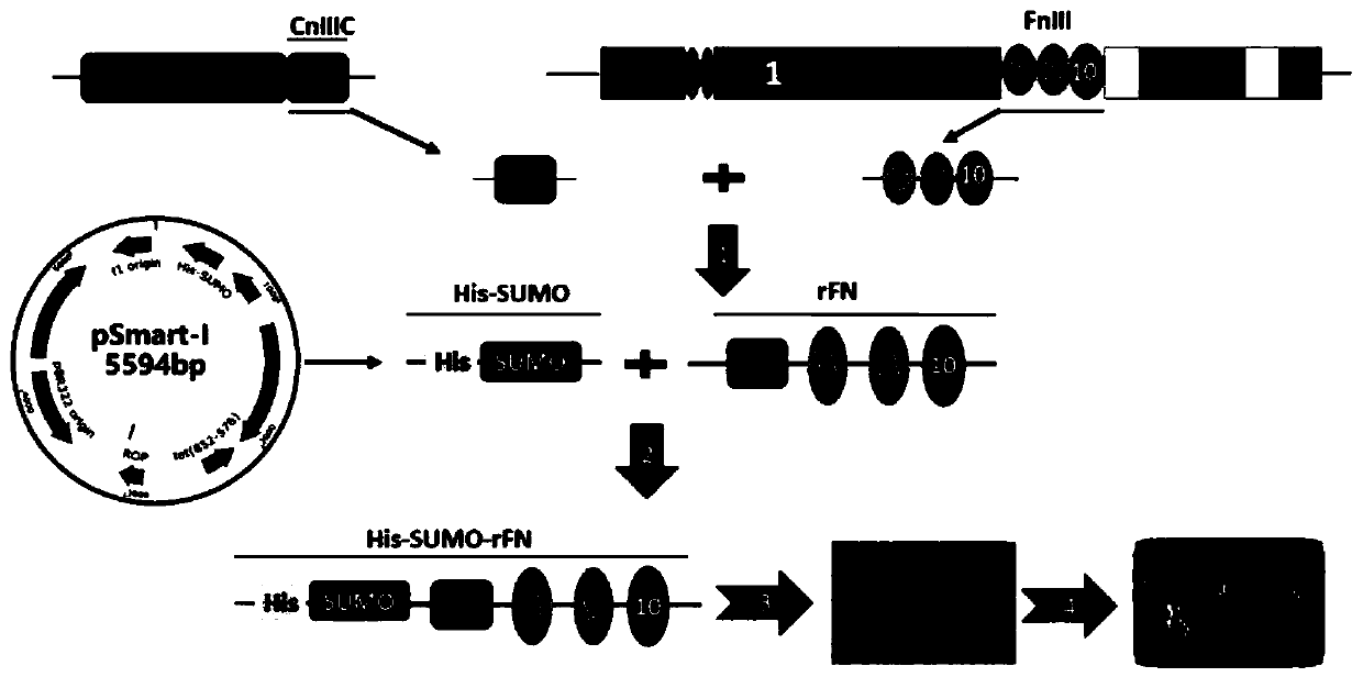
[0060] like Figure 2A Indicated by the arrow 1, the FnIII-8, FnIII-9, FnIII-10 domains of natural fibronectin are intercepted, and the Mu-CnIIIC fragment of the natural conus peptide is intercepted, and the FnIII The -10 domain is connected to the Mu-CnIIIC fragment, and the FnIII-8, FnIII-9, FnIII-10 domain and Mu-CnIIIC fragment are seamlessly spliced by whole gene optimization and synthesis to obtain the coding DNA of recombinant fibronectin rFN.
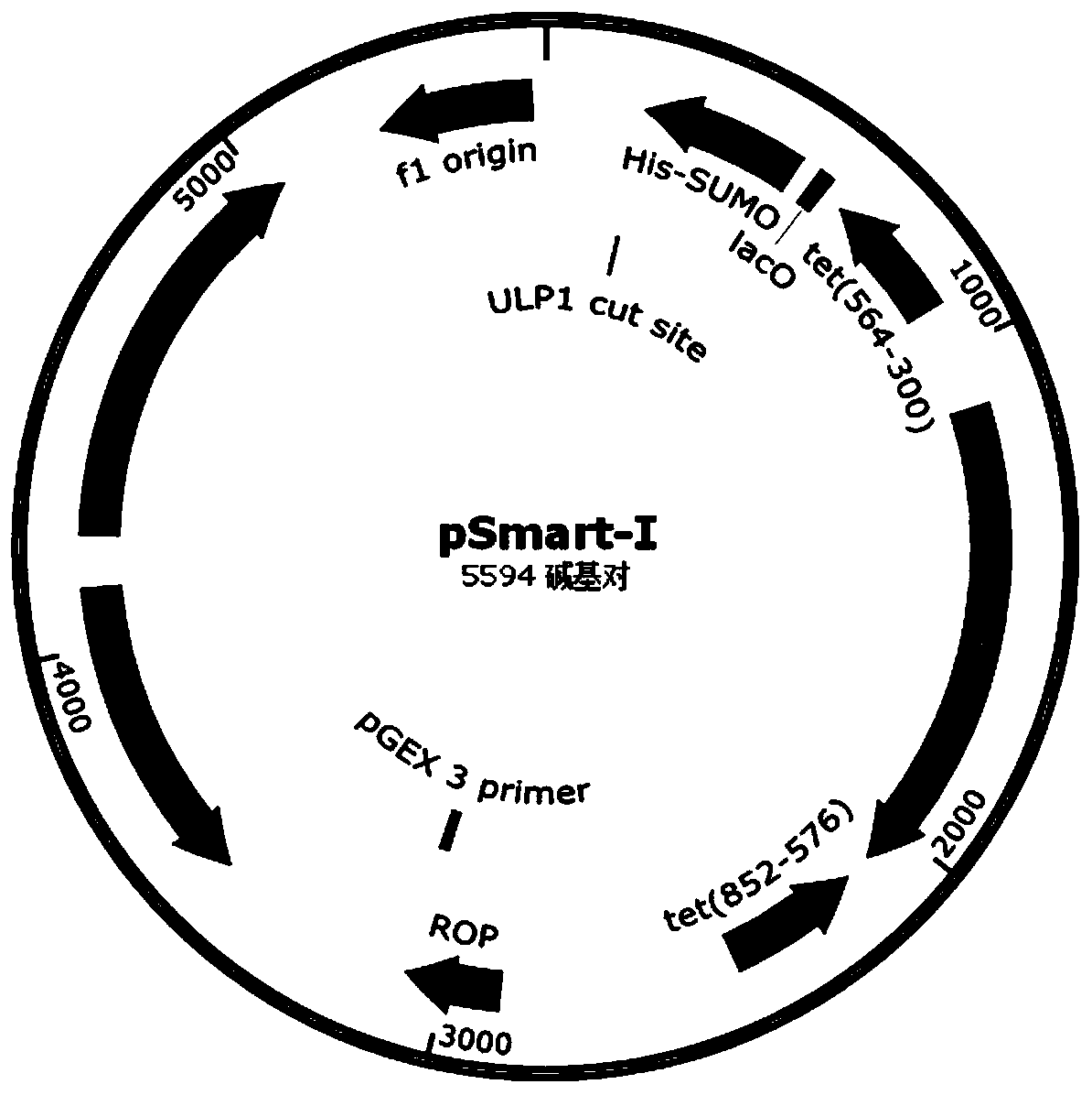
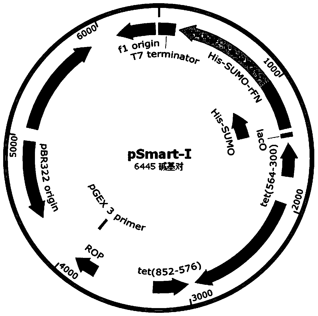
[0061] like Figure 2A Indicated by the arrow 2, the DNA encoding the recombinant fibronectin rFN was inserted into the appropriate reading frame position of the pSmart-I vector. The pSmart-I vector was purchased from Changzhou Tiandi Renhe Biotechnology Co., Ltd. to obtain the fusion protein His - The recombinant expression vector pSmart-I-rFN plasmid of SUMO-rFN.
[0062] The framework map of the pSmart-I vector and the frame map of the recombinant expression vector pSmart-I-rFN plasmid are shown in Figure 1A and Figure 1B , wherein the original pSmart-I vector contains the coding frame of His-SUMO, and the coding DNA sequence of recombinant fibronectin rFN is inserted into the coding frame of His-SUMO, and a protein encoding a fusion of His-SUMO and recombinant fibronectin is obtained. rFN expression plasmid pSmart-I-rFN.
[0063] The amino acid sequence of the fusion protein His-SUMO-rFN is shown in SEQ ID NO.11, the nucleic acid sequence of the pSmart-I vector is shown in SEQ ID NO.12, and the nucleic acid sequence of the recombinant expression vector pSmart-I-rFN plasmid is shown in SEQ ID Shown in NO.13.
[0064] (2) Shake flask expression
[0065] like Figure 2A Indicated by the arrow 3, the recombinant expression vector pSmart-I-rFN plasmid was transferred into the Escherichia coli expression strain, and the shake flask was identified to obtain a positive genetically engineered bacterium BL21(DE3)/pSmart-I-rFN.
[0066] Specifically: transfer the recombinant expression vector pSmart-I-rFN plasmid into Escherichia coli BL21(DE3), obtain positive genetically engineered bacteria BL21(DE3)/pSmart-I-rFN, pick positive transformants and inoculate them into 600ml LB medium Cultivate until the OD600 reaches 0.6, add isopropylthiogalactopyranoside (IPTG) at a final concentration of 1 mmol/L to induce fermentation, IPTG is the inducer, induce at 37°C for 4 hours, or at 22°C for 6 hours, 4 hours Centrifuge at 7000r/min for 10min to collect the cells, add 30mL of TBS (Tris-HCl 25mM, NaCl 150mM, pH7.4) to resuspend the cells, ultrasonically break the cell suspension until the solution is clear, centrifuge at 12000r/min for 10min, and collect the whole cells The lysate and supernatant were analyzed by SDS-PAGE to detect the expression of fusion protein His-SUMO-rFN.
[0067] Among them, the recombinant expression vector pSmart-I-rFN itself has kanamycin resistance and a T7 promoter. After being transferred into the E. coli BL21 (DE3) expression strain, the T7 RNA polymerase contained in the strain can initiate fusion Protein gene transcription, under the induction condition of isopropylthiogalactopyranoside (IPTG), can utilize the protein translation system of Escherichia coli itself, can express fusion protein His-SUMO-rFN, this fusion protein His-SUMO-rFN carries His-tag tags can be used for metal affinity purification; with a SUMO tag protein for specific cleavage by proteases, and with a functional fragment rFN from natural fibronectin, after screening, a small amount of expression in shake flasks can The soluble portion of the fusion protein His-SUMO-rFN reaches 40mg/ml, and the soluble portion of the fusion protein His-SUMO-rFN reaches more than 90%. After affinity purification by nickel column, the His-SUMO tag and rFN fragment can be efficiently cut with ULP1 protease.
[0068] (3) Scale fermentation
[0069] The positive genetically engineered bacterium BL21(DE3)/pSmart-I-rFN obtained in the step (2) is inoculated into 37 ℃ of cultivating in 600mlLB substratum overnight as seed liquid, 600ml seed liquid is inoculated in 50L fermentation medium, at 37 Fermentation was carried out by constant temperature culture at ℃ temperature. After 4 hours of fermentation culture, the fermentation feed was added at a rate of 800ml/h. In the 8th hour, the OD600 of the fermentation broth reached more than 30. At this time, 15g of inducer isopropylthio was added to the fermentation broth. Induced expression of galactoside (IPTG), after adding the inducer, continue to add fermentation feed to the fermentation broth at a rate of 1000ml/h, the 14th hour (6 hours after induction) the OD600 of the fermentation broth reaches more than 50, and the fermentation is terminated , centrifuged to obtain fermentation sludge.
[0070] And after adding the inducer IPTG for induction, it is necessary to take samples every hour and use SDS-PAGE analysis to detect the expression of the fusion protein His-SUMO-rFN, the results are shown in image 3 shown.
[0071]The formula of the fermentation medium is: 2.5g/L ammonium sulfate, 6.25g/L potassium dihydrogen phosphate, 5g/L yeast extract powder and 10g/L peptone, the formula of the fermentation feed is: 400g/L glycerin, 200g/L L peptone.
[0072] (4) Super nickel-resistant affinity purification
[0073] like Figure 2A The purification process flow diagram of the recombinant fibronectin rFN indicated by the arrow 4, such as Figure 2B As indicated by the arrow in 4.1, the fermentation sludge obtained in step (3) was homogeneously crushed, treated by centrifugal filtration, and subjected to third-generation ultra-nickel-resistant affinity purification on a chromatographic column to obtain the eluted fusion protein His- SUMO-rFN.
[0074] Specifically: 6000g of fermented bacteria sludge is collected by centrifugal force, the supernatant is discarded, and the remaining bacteria sludge is suspended and dispersed in TBS buffer solution pre-cooled by ice water, and the bacteria are homogeneously broken under 60MPa pressure and high pressure, and then at a low temperature of 4, 10000g Centrifugal high-speed centrifugation to remove bacterial fragments, 0.22um membrane filtration to further process the supernatant of the lysate sample before loading into the column;
[0075] The supernatant was applied to a chromatographic column for super-nickel-resistant affinity purification. The packing type of the nickel column was Smart-Ni 6FF, which was purchased from Changzhou Tiandi Renhe, and the radial flow rate of the sample was 200cm/hour. Finally, the following three The eluent was eluted in the order of imidazole concentration from low to high, and the eluted fusion protein His-SUMO-rFN was obtained, and the SDS-PAGE electrophoresis test of the eluted sample His-SUMO-rFN was carried out. The test results are shown in Figure 5 shown. The eluted fractions with purity reaching the standard were combined for the next step of purification.
[0076] Among them: the balance solution is TBS buffer solution (Tris-HCl 25mM, NaCl 150mM, pH7.4);
[0077] Washing solution: TBS buffer containing 5mM imidazole (Tris-HCl 25mM, NaCl 150mM, imidazole 5mMpH7.4);
[0078] Eluent: TBS buffer containing different concentrations of imidazole
[0079] The first eluent: TBS buffer containing imidazole concentration of 20mM (Tris-HCl 25mM, NaCl 150mM, imidazole 20mM, pH 7.4);
[0080] The second eluent: TBS buffer (Tris-HCl 25mM, NaCl 150mM, imidazole 50mM, pH 7.4) containing imidazole concentration of 50mM;
[0081] The third eluent: TBS buffer containing 250 mM imidazole (Tris-HCl 25 mM, NaCl 150 mM, imidazole 250 mM, pH 7.4).
[0082] (5) Enzyme cutting and hanging back to remove tags, ion column purification
[0083] Enzyme cut back to remove tags: eg Figure 2B As indicated by the arrow 4.2 in the figure, take the fusion protein His-SUMO-rFN obtained in step (4) after being eluted from a nickel column, and add ULP1 protease to cut off the His-SUMO tag. Figure 4 , the molar ratio of fusion protein His-SUMO-rFN to ULP1 protease is 1000:1, ULP1 protease was purchased from Zhenjiang Ruihua Biotechnology Co., Ltd., its amino acid sequence is shown in SEQ ID NO.7, and its coding nucleic acid sequence is shown in SEQ ID NO .8, digested at 4°C for 2 hours to obtain the digested recombinant fibronectin rFN buffer.
[0084] Dilute the recombinant fibronectin rFN buffer with TBS buffer that does not contain imidazole, hang it back on the ultra-resistant nickel column, wash and collect the recombinant fibronectin rFN that is partially retained in the chromatography column with 10 times the column volume of TBS buffer, Combine the washout and washout.
[0085] Ion column refining and purification: such as Figure 2B As indicated by the arrow 4.3, the above-mentioned combined permeate and flushing solution are switched to low-salt phosphate buffer (PB) buffer in the hollow fiber system for ion exchange packing purification. The packing type is cation exchange packing CM 6FF Beads , that is, carboxymethyl high-flow rate agarose was purchased from Changzhou Tiandi Renhe, and finally eluted with a salt gradient elution solution containing 100mM/200mM/500mM NaCl. All eluted samples were tested by SDS-PAGE electrophoresis. The test results are shown in Image 6 shown.
[0086] PB buffer: sodium dihydrogen phosphate (NaH 2 PQ 4 ) 25mM, pH 6.5;
[0087] The eluent is PBS buffer containing different concentrations of sodium chloride;
[0088] The first eluent: PBS buffer containing 100mM sodium chloride (25mM NaH 2 PQ 4 , 100mM NaCl, pH 6.5);
[0089] The second eluent: PBS buffer containing 200mM sodium chloride (25mM NaH 2 PQ 4 , 200mM NaCl, pH 6.5);
[0090] The third eluent: PBS buffer containing 500mM sodium chloride (25mM NaH 2 PQ 4 , 500mM NaCl, pH 6.5).
[0091] In this application, the N-terminus of the SUMO part of the purification tag His-SUMO contains a histidine tag for purification. The histidine tag contains greater than or equal to 6 His amino acid residues, and the amino acid sequence of the purification tag His-SUMO As shown in SEQ ID NO.5, the coding nucleic acid sequence is shown in SEQ ID NO.6.
[0092] 2. Results
[0093] 1. Time-series electrophoresis test of the expression level of His-SUMO-rFN large-scale fermentation
[0094] like image 3 It is the time-series electrophoresis diagram of the fusion protein His-SUMO-rFN large-scale fermentation expression, in which the first well is the sample before induction, and the second, third, fourth, fifth, and sixth wells are the induction time of 2h, 3h, 4h, and 5h, respectively. , 6h sample, M is the pre-stained molecular weight protein marker.
[0095] according to image 3 As shown, in the large-scale fermentation process, the background expression before induction was controlled at a very low level, and there was no obvious fusion protein His-SUMO-rFN band in the sample before induction, and the molecular weight was about 54kDa. After adding the inducer IPTG After 2 hours, the proportion of the fusion protein His-SUMO-rFN to the total bacterial protein content increased, and reached a plateau after 5 hours, with a molecular weight of about 54kDa. After 6 hours of induction, it can be placed in the tank and the fermentation broth harvested.
[0096] According to the amount of sample loaded (1ul fermentation broth sample amount per well) and the gray scale of the stained bands, the expression level per unit volume of fermentation broth can reach more than 8g/L 6 hours after induction. The concentration can continue to rise, increasing the cell density by 2-3 times, and the OD600 of the final fermentation product exceeds 100. Correspondingly, the expression amount per unit volume of fermentation broth exceeds 10g/L.
[0097] 2. Electrophoresis test of His-SUMO-rFN digestion efficiency
[0098] like Figure 4 As shown in the electrophoresis diagram of the digestion efficiency of His-SUMO-rFN, the first well is the fusion protein His-SUMO-rFN before digestion, the second well is the sample digested for 1 hour, and the third hole is the sample digested for 2 hours. Sample, M is the pre-stained molecular weight protein marker.
[0099] The results show that ULP1 protease can efficiently cut the His-SUMO-rFN fusion protein (molecular weight is about 54kDa), and the digestion efficiency is greater than 95% in 1 hour and greater than 99% in 2 hours. The two fragments His-SUMO fragment (approximate molecular weight about 20kDa) and rFN fragment (apparent molecular weight about 34kDa) after enzyme digestion were of correct size.
[0100] 3. Purification test of His-SUMO-rFN metal affinity filler (Smart Ni)
[0101] Figure 5 The electrophoresis test chart of the samples in each stage of the metal affinity filler purification process for the fusion protein His-SUMO-rFN, the first hole is the lysate supernatant before loading the fusion protein, the second hole is the flow-through, The 3rd, 4th, and 5th wells were used respectively with the first eluent (TBS buffer containing imidazole concentration of 20mM), the second eluate containing imidazole concentration (TBS buffer containing imidazole concentration of 50mM), the second eluate The eluted fractions obtained by elution with three eluents (TBS buffer containing imidazole concentration of 20mM), the sixth well is the residual fusion protein His-SUMO-rFN on the filler, and M is the pre-stained molecular weight protein Marker.
[0102] The results of electrophoresis show that the fusion protein His-SUMO-rFN can be efficiently captured by the filler Smart-Ni 6FF, and the recovery rate of one-step affinity chromatography can reach more than 90%. In dehydration. The SDS-PAGE electrophoresis was analyzed using grayscale scanning analysis software, and the purity of the harvested fusion protein His-SUMO-rFN was greater than 95%.
[0103] 4. Purification test of recombinant fibronectin rFN cation exchange medium (CM)
[0104] Image 6 Cation exchange medium (CM) for recombinant fibronectin rFN, that is, the electrophoresis test chart of samples at each stage of the purification process. The first hole in the figure is the recombinant fibronectin rFN after Smart Ni back-hanging to remove the label, and the second hole The first eluent (PBS buffer containing 100mM sodium chloride) and the second eluent (PBS buffer containing 200mM sodium chloride) were used for the 3rd, 4th, and 5th wells respectively. 1. The elution fraction obtained by eluting with the third eluent (PBS buffer solution containing 500 mM sodium chloride), the sixth well is the recombinant fibronectin rFN remaining on the filler, and M is the pre-stained molecular weight protein Marker.
[0105] The results showed that recombinant fibronectin rFN could be efficiently adsorbed by cation exchange packing CM, and there was a small amount of recombinant fibronectin rFN in the permeate, accounting for less than 5%.
[0106] After high-salt elution, recombinant fibronectin rFN is mainly located in the eluent containing 200mM and 500mM NaCl concentration, and the residual ratio on the column is less than 5%, and the recombinant fibronectin rFN recovered by high-salt elution has no obvious Miscellaneous bands can be seen, and the electrophoretic purity is greater than 99%.
[0107] Purified by His-SUMO-rFN metal affinity filler and recombinant fibronectin rFN cation exchange filler, the total recovery rate of the obtained recombinant fibronectin rFN is greater than 90%, the His-SUMO tag is completely removed, and the recombinant fibronectin rFN Toxin levels are less than 0.001 EU/ug.