

A kind of exosome drug delivery system and its preparation method and application
A delivery system and exosome technology, applied in the field of medicine, can solve the problems of low drug loading and achieve the effects of good biocompatibility, good safety and stable properties
- Summary
- Abstract
- Description
- Claims
- Application Information
AI Technical Summary
Problems solved by technology
Method used
Image

Examples
preparation example Construction
[0043] The preparation method of cisplatin prodrug, concrete steps are as follows:
[0044] Step 1 uses H at a concentration of 30% (W / V) 2 o 2 Activate cisplatin (CDDP), the reaction temperature is 55-80°C, stir in the dark for 4.5-7.5h, cool at 4°C for 2h, filter under reduced pressure to obtain a light yellow solid, and wash with water, ethanol, and ether in sequence to obtain di Hydroxydiamine dichloroplatinum;
[0045]
[0046] Step 2 uses 4-dimethylaminopyridine (DMAP) as a catalyst to react dihydroxydiamine dichloroplatinum with succinic anhydride, and the molar ratio of dihydroxydiamine dichloroplatinum to succinic anhydride is 1:1-3; The temperature is 60-80°C, stirring for 3 hours to obtain succinic acid hydroxydiamine dichloroplatinum;
[0047] Step 3 uses N-hydroxysuccinimide (NHS) and 1-(3-dimethylaminopropyl)-3-ethylcarbodiimide hydrochloride (EDC) as a catalyst to convert succinic acid hydroxydiamine di Chloroplatinum and phosphatidylethanolamine undergo ...
Embodiment 1
[0058] Embodiment 1: the preparation of cisplatin prodrug
[0059] Synthesis of dihydroxy diamine dichloroplatinum: CDDP (2g) and 30ml 30% hydrogen peroxide (w / v) were added to a round bottom flask, stirred at 70°C in the dark for 6 hours, cooled at 4°C for 2 hours, and decompressed Light yellow crystals were obtained by filtration, washed with water, ethanol and ether respectively, and dried to obtain the product dihydroxydiammine dichloroplatinum.
[0060] Synthesis of cisplatin prodrug: In a round bottom flask, dihydroxydiamine dichloroplatinum (100 mg, 0.3 mmol) and succinic anhydride (300 mg, 0.3 mmol) were added, dissolved in (DMF), and 4-dimethyl Aminopyridine (DMAP, 0.3 mmol). Stir at 75°C for 3 hours. After the reaction, add water to precipitate a light yellow solid; then wash with water and petroleum ether to obtain the product.
[0061] The above yellow solid of Pt(IV) (129 mg, 0.3 mmol) was dissolved in ultrapure water, and EDC (34 mg, 0.3 mmol) and NHS (56.7 mg,...
Embodiment 2
[0062] Example 2: Detection of fusion efficiency of DSPE liposomes and milk exosomes
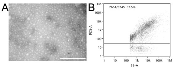
[0063] Dissolve DSPE in 1ml CH 3 OH, N 2 Blow dry the solvent and vacuum dry for 24h. Then add 2ml of PBS solution to ultrasonically dissolve, then repeatedly extrude 20 times through a 30nm extruder to collect DSPE liposomes. Use DID dye to stain the liposome membrane, and use a 10KD ultrafiltration tube to remove unbound DID dye to obtain red-stained DSPE liposomes. Liposomes were characterized morphologically by TEM (Transmission electron microscopy), and the proportion of stained liposomes in the total was detected by nano flow cytometry. Such as figure 1 As shown, the particle size distribution of the prepared liposomes was all about 30nm, and the distribution was uniform, and the results of nano flow cytometry showed that 87.5% of the liposomes were successfully stained.
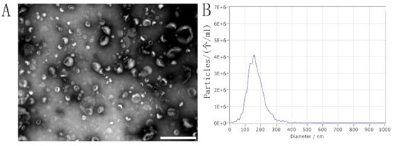
[0064] Milk exosomes were separated from fresh milk by acid precipitation, centrifugation, and ultracentrifug...
PUM
| Property | Measurement | Unit |
|---|---|---|
| diameter | aaaaa | aaaaa |
| purity | aaaaa | aaaaa |
Abstract
Description
Claims
Application Information