
Orthopedic implant having a crystalline calcium phosphate coating and methods for making the same

a technology of calcium phosphate and orthopedic implants, which is applied in the field of orthopedic implants having a solution-deposited calcium phosphate coating, can solve the problems of increased wear, inability to uniformly dissolve or degrade psha films in vivo, and inflammatory cascades leading to osteolysis, and achieve the effect of mitigating surface cracks

Active Publication Date: 2020-01-21
DEPUY SYNTHES PROD INC
View PDF189 Cites 6 Cited by
  • Summary
  • Abstract
  • Description
  • Claims
  • Application Information

AI Technical Summary

Benefits of technology

The solution achieves a calcium phosphate coating that degrades uniformly over several weeks without particulate formation, enhancing implant stability and biocompatibility, while maintaining strong adhesion and cohesion properties.

Problems solved by technology

As a result, plasma sprayed hydroxyapatite (PSHA) films do not uniformly dissolve or degrade in vivo.
This non-homogenous degradation can generate particulates in the vicinity of the implant which can result in an inflammatory cascade leading to osteolysis.
The particles may also find their way into joint articular surfaces, resulting in increased wear.
Finally, the process is not well suited for coating porous structures of cementless implants because it is a “line of sight” process.
This attribute gives rise to concerns over long term delamination of these relatively thick stable coatings.
These physical methods do not reproduce the nano-crystallinity and high surface area of biological apatites, and the resulting coatings may not uniformly dissolve and may release particulates.
High surface area, microcrystalline coatings are produced, but their adhesion to the substrate is poor.
Poor adhesion is also a concern with these films.
Finally, control of electrochemical currents on porous implants with irregular particles is challenging, making this process difficult to scale.
These processes can yield bone-like apatite but require days or weeks to produce films a few microns thick.
Attempts to increase rates associated with such methods have led to complications in reproducibly controlling pH, deposition rate, and accretion rate compared to crystalline growth on the surface of the implant.
Uncontrolled deposition rate also makes it difficult to achieve target coating weights or thicknesses.
As described above, hydroxyapatite coatings may be applied to orthopedic implants to enhance osteoconductivity using methods that are either rapid but lead to coatings having certain undesirable or unpredictable properties or lead to more desirable products but can take days to form.

Method used


Image

  • Orthopedic implant having a crystalline calcium phosphate coating and methods for making the same
  • Orthopedic implant having a crystalline calcium phosphate coating and methods for making the same
  • Orthopedic implant having a crystalline calcium phosphate coating and methods for making the same

Examples


example 1

Activation of CoCr by Atomic Layer Deposition of TiO2 Films

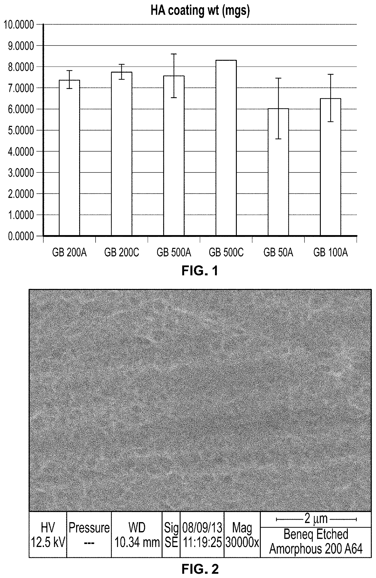
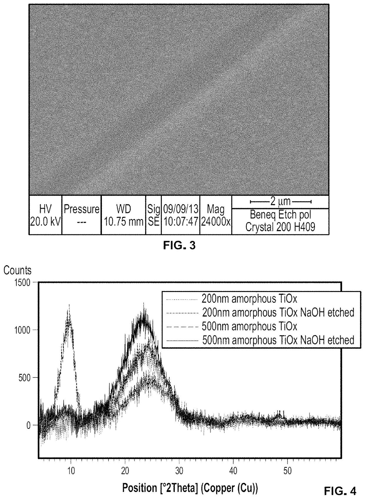
[0284]TiO2 films were prepared on CoCr grit blast surfaces by Beneq (Helsinki, Finland) by atomic layer deposition. The films were produced from TiCl4 and H2O precursors. Amorphous films were produced at 90° C. and crystalline (Anatase plus Rutile) films were produced at 200° C. Films were activated with hydroxide, according to the methods described in Example 3 (4 hrs, 5M NaOH, 60° C.). 500, 200, 100, and 50 nm amorphous films (500 A, 200 A, 100 A, and 50 A) and 500 and 200 nm crystalline films (500° C., 200° C.) were evaluated for their ability to form hydroxyapatite coatings according to the SoDHA method using nominal concentrations, as further described in Examples 5 and 6.

[0285]HA coating weight for the samples ranged from about 6 mg to about 8 mg, as shown in FIG. 1. Both amorphous and crystalline films were able to nucleate HA after activation by strong base. Coating experiments showed that deposition occurred for as ...

example 2

Electrolytic Formation of TiO2 Films on CoCr Alloy

[0288]An electrolyte solution of 0.05 M TiCl4 and 0.25 M H2O2 in a mixed methanol / water (3 / 1 vol %) solvent was prepared. The pH of the solution was fixed between 0.9 and 1.0. All chemicals were ACS grade. To prepare the electrolyte solution, TiCl4 was slowly added to the solvent, followed by H2O2. During the addition of H2O2, an immediate color change from transparent to dark orange was observed, indicating the formation of a peroxo-complex. Electrolytes were stored at about 4° C. after preparation and pH measurement and, if necessary, pH adjustment. Electrodeposition was performed galvanostatically up to the desired charge density. The temperature was fixed by a cryostat to 0° C. A homemade Labview program (controlling a Xantrex XDC 300-20 power source) was built to control current charge density while recording voltage-time curves. The applied charge density was fixed in the range form 2.5 C / cm2 to 40 C C / cm2. The current density ...

example 3

Titanium Surface Activation

[0290]1 inch diameter Ti6-4 disks coated with DePuy Gription porous metal coating were cleaned and activated by the following steps. Disks were set in a container with reverse osmosis (RO) water and alkaline detergent for 15 min with a sonicator 2 times. Next, disks were set in a container with only RO water for 15 minutes with a sonicator 2 times. 4 disks were placed in a 500 mL beaker and 200 mL of 5M NaOH was added. The disks were placed in secondary containment and a loose cap was set over the top of the beaker. The temperature of the beaker was set to 60° C. and held for 4 hrs. The desks were removed from the beaker and rinsed in RO water in a sonicator for 15 minutes 4 times. Disks were left in a 60° C. oven overnight to dry. No effect of heat treatment of this activation layer on the tendency to promote nucleation and growth of CaP coatings was found in the methods disclosed herein, and examples reported below were performed without heat treatment o...


PUM

PropertyMeasurementUnit
crystallite sizeaaaaaaaaaa
shear strengthaaaaaaaaaa
tensile strengthaaaaaaaaaa

Abstract

An orthopedic implant having a metal surface and a calcium phosphate layer disposed on at least part of the metal surface is described. The calcium phosphate layer has an average crystallite size of less than about 100 nm in at least one direction and dissolves for more than 2 hours in vitro. The calcium phosphate layer is substantially free of carbonate. The coating, which is formed on a sodium titanate surface, has increased shear strength and tensile strength. The coating is formed by a solution deposited hydroxyapatite process under inert conditions. The pH of the solution varies by less than 0.1 pH unit / hour during coating formation.

Description

CROSS-REFERENCE TO RELATED APPLICATIONS[0001]Cross reference is made to copending U.S. patent application Ser. No. 15 / 472,186 entitled “ORTHOPEDIC IMPLANT HAVING A CRYSTALLINE GALLIUM-CONTAINING HYDROXYAPATITE COATING AND METHODS FOR MAKING THE SAME”, which is hereby incorporated by reference.FIELD OF THE INVENTION[0002]The present invention relates generally to a calcium phosphate coating, and more particularly to an orthopedic implant having a solution deposited calcium phosphate coating and methods for making the same.BACKGROUND OF THE INVENTION[0003]Bone repair often involves the use of orthopedic implants to replace missing bone or support bone during the healing process. It is typically desirable to coat such orthopedic implants with osteoconductive materials to encourage bone growth or biological fixation.[0004]Hydroxyapatite (HA) is a naturally occurring mineral found in bones and teeth. Studies have shown that HA is osteoconductive, and orthopedic implants have been coated ...

Claims


Application Information

Patent Timeline
Patent Type & Authority Patents(United States)
IPC IPC(8): A61L27/32A61L27/54B05D1/18B05D3/00C23C16/40C23C16/455C25D9/04A61L27/56A61L27/58A61L27/06
CPCA61L27/06A61L27/56A61L27/54A61L27/58A61L27/32A61L2420/02A61L2300/606A61L2430/02C23C16/45525A61L2300/112B05D3/002C25D9/04A61L2400/12A61L2300/412B05D1/18C23C16/405A61L27/50A61L2400/18A61L27/045A61L27/00A61L27/04A61L31/08C01B25/32
Inventor KASINATH, RAJENDRAERNSBERGER, CRAIGVASS, STEPHANIEGINN, STEVEN N.QU, HAIBOTONG, WEIDONG
Owner DEPUY SYNTHES PROD INC
Learn More