Use of poly-nitroxyl-functionalized dendrimers as contrast enhancing agent in mri imaging of joints

Inactive Publication Date: 2003-11-20
NITROSCI PHARMA
View PDF1 Cites 9 Cited by
  • Summary
  • Abstract
  • Description
  • Claims
  • Application Information

AI Technical Summary

Benefits of technology

[0017] In another method aspect, this invention relates to an improvement in a method of enhancing the image obtained by MRI of a joint of a human exhibiting symptoms of osteoarthritis or rheumatoid arthritis by injecting into the joint, prior to the MRI scan thereof, in admixture with a non-toxic injectable pharmacologically acceptable vehicle, an image enhancing amount which is both non-toxic and effective to enhance the MR image produced by the scan, of a water soluble organic nitroxide, which improvement comprises employing as the nitroxide, a poly-nitroxyl-functionalized dendrimer of Formula (I).
[0055] The amount of dendrimer injected into the joint is influenced by the volume of the joint void, the concentration of the dendrimer in the solution injected into the joint and the reflectivity of the dendrimer. Generally speaking, at a concentration of 100 micromolar, a dose from a low of about 0.17 mg / mL for a dendrimer containing 16 nitroxyl groups injected into one of the joints of the hand to a high of about 0.89 mg / mL for a dendrimer containing 32 nitroxyl groups injected into the shoulder joint is employed. Knowing the in vitro image enhancing activity of the selected dendrimer and the dose required to obtain satisfactory MRI contrast enhancement in the joint with, for example, Gd-DTPA, a person knowledgeable MRI can readily predict the optimum dose for obtaining the desired degree of MRI contrast enhancement in a scan of that joint.

Problems solved by technology

To date, radiography has been limited in its ability to accurately evaluate cartilage disorders, because cartilage can only be imaged indirectly.
The osseous abnormalities (such as osteophyte formation) that eventually develop in patients with osteoarthritis are not sensitive indicators of early disease, nor are they reliable measures of disease progression.
Unfortunately, these studies have yielded variable, contradictory and often disappointing results.
As a consequence, there is no universally accepted "best sequence or technique" for accurate MR imaging of articular cartilage.
However, inclusion of an intraarticular injection of gadolinium (Gd)-DTPA in the protocol of an MRI evaluation of the progression of osteoarthritis in a joint of a patient exhibiting the symptoms of the disease only modestly improves the evaluation of proteoglycan status and cartilage morphology.
This is due to the inability of Gd-DTPA to effectively bind to proteoglycans in the joint.
However, this technique is limited in that the diffusion of the contrast into the cartilage is quite slow.
Additionally, with this technique, the abnormal cartilage appears bright while the normal cartilage appears dark, potentially leading to confusion between incomplete diffusion of the contrast into normal cartilage and abnormal superficial portion of the cartilage.
Thus, Gd-DTPA, although very potent as a MRI contrast enhancing agent generally, is very poor as a tool for evaluating degenerative joint diseases, such as osteoarthritis, due to its lack of specificity for proteoglycans, which results in a high concentration of the Gd-DTPA in equilibrium within the joint space and the surface of the joint where the proteoglycans are located, which has prompted the commercially unattractive manipulations as noted above.
However, nitroxides as a class are not a likely candidate because they are rapidly reduced enzymatically in tissues into products which do not enhance an MR image (Griffeth, et al., Invest. Radiol. 19: 553-562, 1984) and, like Gd-DTPA, have no specificity with respect to cartilage proteoglycans in joints.
Therefore, as a class nitroxides are not suitable for use in evaluating the progress of a joint degenerative disease by MRI of a joint of a patient with such a disease.
However, the potency of these positively charged nitroxides as an MRI contrast enhancing agent is very low compared to a gadolinium-based contrast agent because of their low relaxivity (.sup.-0.2 mM.sup.-1 sec.sup.-1 for nitroxides vs.
Such high doses are not feasible clinically.
Moreover, even if this fact alone did not preclude their use in joints to evaluate the progress of a joint degenerative disease, the high dose required to achieve a satisfactory level of image contrast enhancement would result in such a high concentration of the nitroxide in equilibrium between the joint space and the surface of the joint where the proteoglycans are located that the requisite selectivity of binding to the proteoglycans, which is necessary to permit evaluation of the progress of the degenerative disease in a joint cannot be achieved with these charged nitroxides, would not be present.
Therefore, even the charged nitroxides, which are useful as MRI contrast enhancing agents generally, including in joints, are of little practical value in assisting in the evaluation of the degenerative progress in the joint of a patient afflicted with a joint degenerative disease, because this end use requires a dose which is low enough to achieve selective binding to the proteoglycans while still enhancing to a clinically acceptable level the MR image achieved.
However, because of the loss of selectivity which occurs at the high concentration in the joint required to achieve a clinically acceptable level of MRI contrast enhancement in the case of the corresponding mono-nitroxides (due to their low relaxivity), one would expect that the increased concentration of such nitroxyl groups in a joint provided by the presence of a plurality of these nitroxyl groups in a calixarene or dendrimer molecule as functionalizing groups would have the same adverse effect on the selectivity of the MRI imaging achieved in a degenerative joint as reported for the mono-nitroxides of my prior patents.

Method used


Image

  • Use of poly-nitroxyl-functionalized dendrimers as contrast enhancing agent in mri imaging of joints
  • Use of poly-nitroxyl-functionalized dendrimers as contrast enhancing agent in mri imaging of joints
  • Use of poly-nitroxyl-functionalized dendrimers as contrast enhancing agent in mri imaging of joints

Examples


example 1

[0060] Synthesis of Dendrimer-Containing Nitroxides

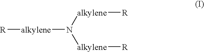
[0061] 3-[(2,5-Dioxo-1-pyrrolidinyl)oxy]carbonyl]-2,2,5,5-tetramethyl-1-py-rrolidinyloxyl [3]. To a solution of 3-carboxy-2,2,5,5-tetramethyl-1-pyrro-lidinyloxyl [1], prepared as described in the literature, E. G. Rozantsev. Free Nitroxyl Radicals, Plenum Press, N.Y., pp. 203-206, (500 mg, 2.69 mmoles) dissolved in THF (50 mL) was added N-hydroxysuccinimide [2] (Aldrich Chemical Company, Milwaukee, Wis., 310 mg, 2.69 mmoles). After dissolution, 1,3-dicyclohexylcarbodiimide (Aldrich Chemical Company, 610 mg, 2.96 mmoles) was added. The reaction mixture was stirred for 2 days at room temperature, filtered and evaporated, in vacuo. The remaining oil was taken up in methylene chloride, filtered and evaporated in vacuo to dryness. The residue material was then washed with hot cyclohexane (3 times, 10 mL). The residual oil, dissolved in chloroform:hexane (80:20), was passed through a chromatographic column containing silica gel (Aldrich C...

example 2

[0066] In Vivo Pharmacokinetics of Joint Injected With Nitroxyl-Containing Dendrimer Using Magnetic Resonance Imaging (MRI)

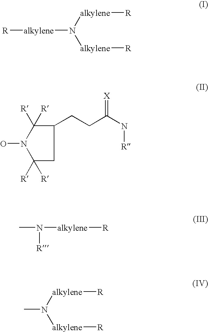
[0067] Inject into both knee joints of a geriatric male (ca. 65-75 years; 70-80 kg), who has been diagnosed as osteoarthritic and is about to be treated medically for chronic severe pain and inflammation in both knees, 0.5 mL of a stock 100 .mu.M sterile solution sterile solution in 0.9% normal saline of a dendrimer of Formula (I) containing 32 terminal PROXYL-3-carbamido nitroxide groups [9] using a (gauge 25) needle. Then position the patent in a MR imager (1.5 Tesla MR system, SIGNA; General Electric Medical Systems, Milwaukee, Wis.) and obtain a scan of both knee joints in the conventional manner. At periodic intervals of about a month, repeat the procedure. Compare the succession of MR images thus obtained to evaluate the response of the knee joints to the treatment protocol.

[0068] Follow the above procedure in a peri-menopausal female whose blood chemistry...

example 3

[0072] In Vitro Stability of Dendrimer-Containing Nitroxides (Rate of Nitroxide Reduction).

[0073] In vitro rate of nitroxide reduction (Table 1) was undertaken using superoxide in the presence of a thiol, as an in vitro model for activated phagocytic cells. G. M. Rosen, B. E. Britigan, M. S. Cohen, S. P. Ellington and M. J. Barber, Biochim. Biophys. Acta, 969:236-241, 1988; G. M. Rosen and E. J. Rauckman. Biochem. Pharmacol. 26: 675-678, 1977; S. Pou, P. L. Davis, G. L. Wolf and G. M. Rosen, Free Rad. Res. 23: 353-364, 1995; and E. Finkelstein, G. M. Rosen and E. J. Rauckman, Biochim. Biophys. Acta, 802:90-98, 1984. For these determinations, nitroxides [S. Pou, et al, E. Finkelstein, et al., supra, and H. Kuthan, V. Ullrich and R. Estabrook, Biochem. J. 203: 551-558, 1982, (50 micromolar) were added to cysteine (200 micromolar) in the presence of a continued flux of superoxide (10 micromolar / min), generated by the action of xanthine oxidase on xanthine (400 micromolar) in Chelexed s...


PUM


Abstract

The image obtained by MR imaging of the joint of an animal, including humans, is enhanced by injecting into the joint prior to the scan of an imaging enhancing amount of a nitroxyl functionalized dendrimer containing at least 6 nitroxyl terminal functional groups, e.g, of Formula (I) wherein each R independently is either a nitroxyl group of Formula (II) wherein X is an oxygen atom or two hydrogen atoms, R' is lower alkyl and R'' is H or lower alkyl, or a group of Formula (III) wherein R''' is lower alkyl, or a group of Formula (IV): wherein each alkylene in Formulae (I), (III) and (IV) is a bridging divalent alkylene group of 2-6 carbon atoms with 2-3 carbon atoms in the chain and R is as defined herein, with the proviso that the total number of R groups in the molecule which are nitroxyl groups of Formula (II) is at least 6.

Description

[0001] This invention relates to the use of poly-nitroxyl-functionalized dendrimers as contrast enhancing agents for Magnetic Resonance Imaging (MRI) for the evaluation of degenerative joint diseases, such as osteoarthritis or rheumatoid arthritis.[0002] Normal articular cartilage is a critical component of efficient, painless joint function. Pathologically, osteoarthritis is characterized by progressive loss of articular cartilage. Recent advances in understanding the pathogenesis of osteoarthritis have suggested that, in the near future, the development of drugs that will be able to reverse pre-existing abnormalities in articular cartilage or prevent their progression may be possible, if therapy is started early during the course of the disease. As a result, there is a tremendous need for a non-invasive technique that can accurately detect the presence of early cartilage abnormalities and the progression of those abnormalities in patients with osteoarthritis or rheumatoid arthriti...

Claims


Application Information

Patent Timeline
IPC IPC(8): C07D207/16A61K49/00A61K49/20
CPCA61K49/20
Inventor ROSEN, GERALD M.
Owner NITROSCI PHARMA
Why Eureka
Try Eureka