Uses of vascular endothelial growth factor and type I collagen inducible protein (VCIP)

a growth factor and vascular endothelial technology, applied in the field of cell cell interaction, can solve the problems of preventing metastasis and impeded wound healing, and achieve the effects of promoting adhesion, spreading and tyrosine phosphorylation, and increasing p120 catenin expression

Inactive Publication Date: 2005-01-06
TEXAS A&M UNIVERSITY
View PDF2 Cites 8 Cited by
  • Summary
  • Abstract
  • Description
  • Claims
  • Application Information

AI Technical Summary

Benefits of technology

[0010] Vascular endothelial growth factor and type I collagen inducible protein (VCIP), also known as phosphatidic acid phosphatase 2b (PAP2b), was identified in a functional assay of angiogenesis. VCIP / PAP2b exhibits an Arg-Gly-Asp (RGD) cell adhesion sequence. Immunoprecipitation and fluorescence-activated cell sorting analyses demonstrated that VCIP-RGD is exposed to the outside of the cell surface. Retroviral transduction of VCIP induced cell aggregation / cell-cell interactions, modestly increased p120 catenin expression and promoted activation of the Fak, Akt and GSK3β protein kinases. Furthermore, expression of recombinant VCIP promoted adhesion, spreading and tyrosine phosphorylation of Fak, Shc, Cas and paxillin in endothelial cells. GST-VCIP-RGD, but not GST-VCIP-RGE, specifically interacted with a subset of integrins, and these interactions were effectively blocked by anti-αvβ3 and anti-α5β1 integrin antibodies, and by PAP2b / VCIP-derived peptides. Interestingly, PAP2b / VCIP is expressed in close proximity to vascular endothelial growth factor, von Willebrand factor and αvβ3 integrin in tumor vasculatures. These findings demonstrate an unexpected function of PAP2b / VCIP, and represent an important step towards understanding the molecular mechanisms by which PAP2b / VCIP-induced cell-cell interactions regulate specific intracellular signaling pathways.
[0011] The present invention also provides evidence that the cytoplasmic domain of VCIP interacts with p120catenin that alters β-catenin localization and LEF-1 transcriptional activation. In particular, retroviral-mediated over-expression of wild-type VCIP in primary endothelial cells impeded wound healing without affecting proliferative potential of these cells. Reciprocal co-immunoprecipitation and western immunoblot analyses showed that VCIP binds to p120catenin on endothelial cells, but not VE-cadherin or other Armadillo domain-containing proteins such as β-catenin or γ-catenin (plakoglobin). VCIP immunocomplex prepared from E-cadherin-deficient SW480 cell line contained p120catenin immunoreactivity, suggesting VCIP and p120catenin interaction may be E cadherin-independent. Moreover, a truncated VCIP without C-terminal cytoplasmic domain failed to coprecipitate p120catenin. Far-western analyses suggested that the cytoplasmic domain of VCIP interacts with p120catenin. Furthermore, it was demonstrated that elevated expression of VCIP in SW480 cells induced recruitment of p120catenin directly and promoted redistribution of β-catenin indirectly. These results are consistent with the observations that elevated expression of wild-type VCIP in SW480 cells caused increased cell-cell contact formation, decreased phosphorylation of β-catenin, and moderate inhibition of LEF-1-mediated transcription. Taken together, these results show that in endothelial and SW480 cells VCIP mediates cell-cell adhesion and modulates Wnt signaling pathway in an unprecedented manner.
[0013] In one embodiment of the present invention, there is provided a method of enhancing cell-cell interactions by over-expressing vascular endothelial growth factor and type I collagen inducible protein (VCIP) in a cell.

Problems solved by technology

In particular, retroviral-mediated over-expression of wild-type VCIP in primary endothelial cells impeded wound healing without affecting proliferative potential of these cells.
Phosphatase inactive VCIP / PAP2b and delta-C-cyto mutants also promoted tumor growth and neovascularization, but did not support metastasis.

Method used


Image

  • Uses of vascular endothelial growth factor and type I collagen inducible protein (VCIP)
  • Uses of vascular endothelial growth factor and type I collagen inducible protein (VCIP)
  • Uses of vascular endothelial growth factor and type I collagen inducible protein (VCIP)

Examples


example 1

[0051] Cells and Reagents

[0052] Human umbilical vein endothelial cells (HUVECs), human dermal microvascular endothelial cells (HdMVECs), carotid artery smooth muscle cells (CASMCs) and aortic smooth muscle cells (AoSMC) were obtained from Clonetics. ECM molecules, endotoxin-free fetal bovine serum, antibiotics, heparin, 100× ITS (insulin, transferrin and selenium), M199 media, anti-α5β1 (P1D6) and anti-α3β1 (P1B5) antibodies and Superscript II reverse transcriptase enzyme were obtained from Invitrogen. Basic Fibroblast growth factor (bFGF) and human recombinant vascular endothelial growth factor (hrVEGF165) were purchased from R&D systems. Bovine skin-derived type I collagen (3.0 mg / ml) solution was purchased from Cohesion Inc. Multiple tissue northern blot, cDNA amplification kit and human placental cDNA library in _Triple-Ex vector were purchased from Clontech Laboratories, Inc. Anti-phosphospecific antibodies were purchased from New England Biolabs. Hybridomas producing the anti...

example 2

[0053] Monolayer and Three-Dimensional Cell Culture

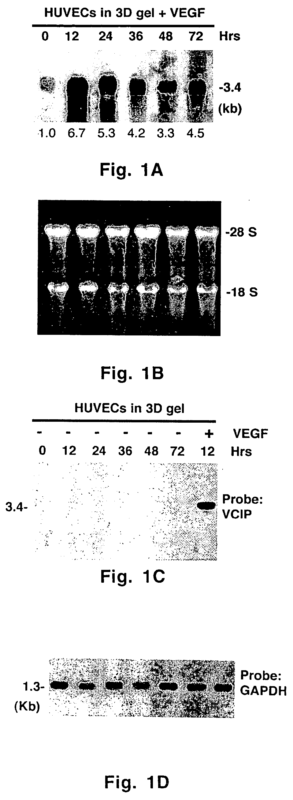
[0054] Monolayer cell cultures were carried out as described previously. Three-dimensional matrix gel was prepared by gently mixing a cold solution of bovine skin-derived type I collagen solution (2.1 mg / ml) with media M199, 1× ITS, hrVEGF165 (100 μg / ml) and glutamine (2.4 mM). The pH was adjusted to 7.5 with 0.1 N sodium hydroxide and sterile water was used to adjust the final volume. Proliferating endothelial cells in the third or fourth passage were cultured in complete media and gently resuspended in complete M199 media at a concentration of 4×10⁵ cells / ml. Twenty four-well tissue culture dishes were filled with 300 μl of cold 3D gel solution, and placed at 37° C. in a CO2 incubator for 30-45 min to polymerize and solidify. Resuspended cells (2×10⁵ cells in 500 μl) were seeded onto 3D gel and the dishes were returned to the CO2 incubator at 37° C. to allow the cells to attach for 2-3 h. At the end of this period, unattached cel...

example 3

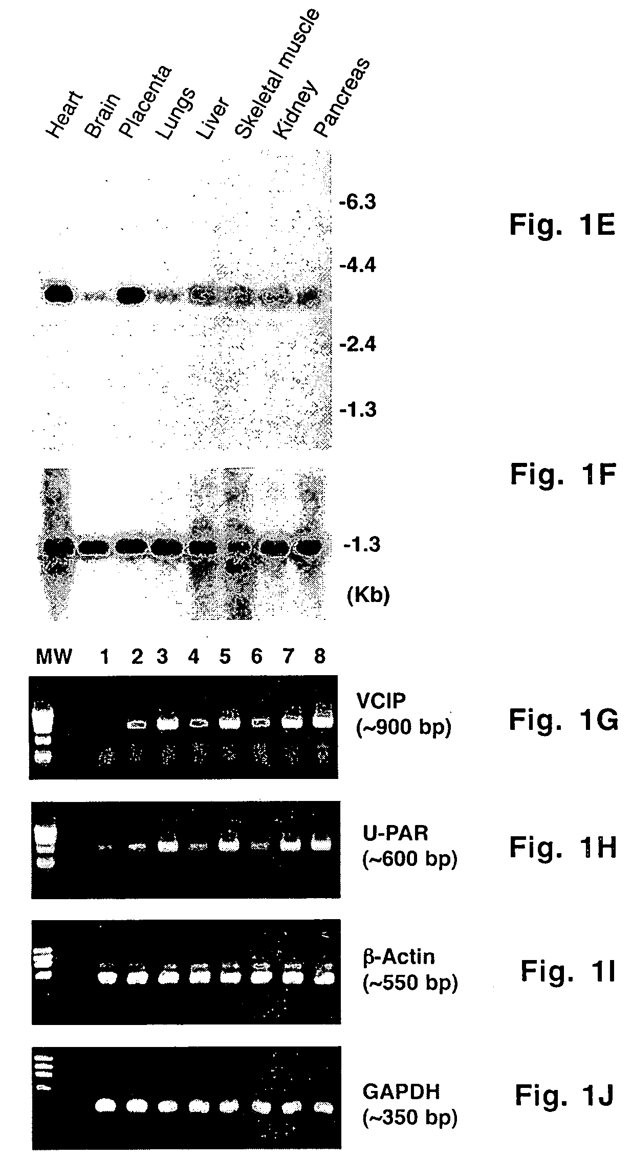
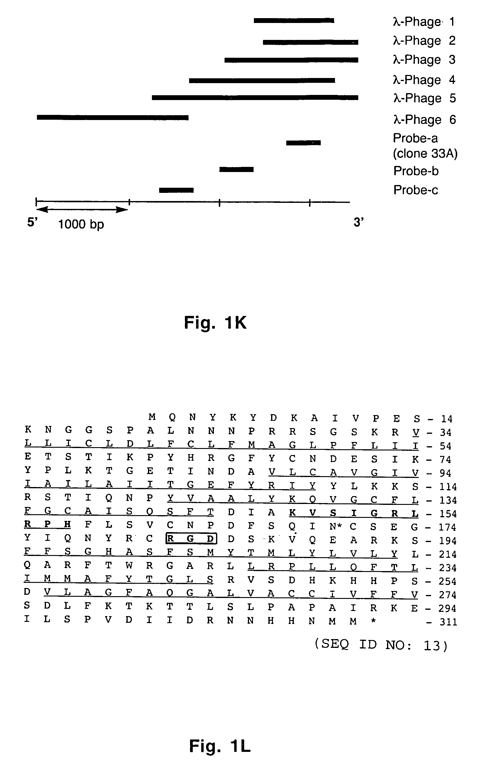
[0056] cDNA Library Screening, Northern Blot Analysis, PCR and RT-PCR

[0057] A _TripleEx phage cDNA library prepared from human placenta (Clontech) was screened as described previously (Wary et al., 1993). Plasmids were extracted, purified by Qiagen affinity column and then digested with EcoRI and XbaI to confirm the presence of the insert. Six overlapping clones were subjected to DNA sequencing. All northern blot analyses were performed as described previously (Wary et al., 1993). In brief, 20 μg of total RNA or 2 μg poly(A)+ mRNA from control cells (i.e. endothelial cells embedded in three-dimensional type I collagen in the presence of 20% human adult serum-AB±100 ng / ml hrVEGF165 supplied every 6 h) were fractionated on an agarose gel containing formaldehyde. To analyze various mRNA levels by RT-PCR, the following primers were used: VCIP-forward 5′-GGAGGATCCCTCGCGCCGCAGCCAGCGCCATGC-3′ (SEQ ID NO:3) and -reverse 5′-GTGGCACCTACATCATGTTGTGGTG-3′ (SEQ ID NO:4); human uPAR-forward 5′-C...


PUM


Abstract

Vascular endothelial growth factor and type I collagen inducible protein (VCIP), also known as phosphatidic acid phosphatase 2b (PAP2b), was identified in a functional assay of angiogenesis. Previously, VCIP was not known to function as an integrin ligand. The present invention discloses VCIP-derived peptides and proteins act as integrin ligands. Since VCIP-derived peptides or proteins are capable of inhibiting specific cell-cell interactions, such inhibitors of cell-cell interactions would be useful for developing novel therapeutic approaches to treat diseases where these interactions have clear pathological consequences. For example, VCIP / PAP2b can be a novel target for anti-angiogenic, anti-cancer and anti-metastatic therapy.

Description

CROSS-REFERENCE TO RELATED APPLICATION [0001] This non-provisional patent application claims benefit of provisional patent application U.S. Ser. No. 60 / 458,164, filed Mar. 27, 2003, now abandoned.BACKGROUND OF THE INVENTION [0002] 1. Field of the Invention [0003] The present invention relates generally to the field of cell-cell interaction. More specifically, the present invention discloses novel functions for vascular endothelial growth factor and type I collagen inducible protein (VCIP) in cell-cell interaction and intracellular signaling. [0004] 2. Description of the Related Art [0005] Cell-cell and cell-matrix interactions play fundamental roles in embryonic development and in wound healing, and these interactions are known to be altered in many pathological processes. Endothelial cells, which line the walls of blood vessels, are able to promote both ‘homotypic’ and ‘heterotypic’ cell-cell interactions. Such interactions are critical for angiogenesis, which proceeds through seve...

Claims


Application Information

Patent Timeline
Patent Type & Authority Applications(United States)
IPC IPC(8): A01K67/027A61K35/12A61K35/28A61K35/54A61K49/18
CPCA01K67/0271G01N2333/916A01K2217/05A01K2267/03A61K35/12A61K35/28A61K35/54A61K49/1896C12N2510/02A61K38/1709A61K38/1866A01K67/0275G01N2333/515G01N33/68C12N9/16C07K14/52A61K2300/00A61P35/00
Inventor WARY, KISHOREHUMTSOE, JOSEPH
Owner TEXAS A&M UNIVERSITY
Why Eureka
Try Eureka