Biodegradable scaffolds and uses thereof

Inactive Publication Date: 2006-01-05
NORTHWESTERN UNIV
View PDF5 Cites 92 Cited by
  • Summary
  • Abstract
  • Description
  • Claims
  • Application Information

AI Technical Summary

Benefits of technology

[0021] In its broadest aspect, the present invention provides a biodegradable polymeric scaffold to provide a mechanical support for tissue regeneration. The scaffold comprises a polymeric matrix prepared by a gas foaming/particulate leaching procedure, and includes a wet granulation step prior to gas foaming that allows for a homogeneous mixture of porogen and polymer and for sculpting the scaffold into the desired shape. The polymeric matrix in the scaffold acts as a substrate permissible for cell growth or for incorporation of DNA or protein and for controlled or sustained release of such DNA, protein or cells at the site of injury. In addition, such protein or DNA may be taken up by cells at the site of injury to provide genes encoding proteins such as enzymes or growth factors to aid in growth of cells at the site of injury. The present invention also provides a nerve guidance tube that is fabricated in a manner that allows for spatially controlled attachment of the cells, proteins or DNA for release over a period of time sufficient to allow enhanced cell or tissue growth or regrowth. The guidance tube may contain one or more lumens. The biodegradable polymer used

Problems solved by technology

Disorder is seen in degenerative processes and is also seen in the failure of regeneration to create fully normal tissue.
When it occurs in deeper structures, function can be severely compromised.
While in general the field of tissue repair and tissue engineering has grown over the past decade to include various means for providing for new cell growth and new tissue formation, the area of nervous tissue regeneration has still proven to be perhaps the most complex.
Nerve injuries complicate successful rehabilitation more than any other form of trauma.
Painful neuroma formation, often more disabling than its associated sensory deficits, commonly causes major disability.
However, the growth cones and the dendrites on the proximal stump typically grow in many directions and unless the injury is small, the growth cones may never reach the distal segment.
Nerve autograft procedures are difficult, expensive, and offer limited success.
Often, a second surgical procedure is required and may lead to permanent denervation at the nerve donor site.
However, use of artificial nerve grafts has m

Method used


Image

  • Biodegradable scaffolds and uses thereof
  • Biodegradable scaffolds and uses thereof
  • Biodegradable scaffolds and uses thereof

Examples


example 1

Fabrication of Cylindrical Scaffolds by the Gas Foaming Procedure

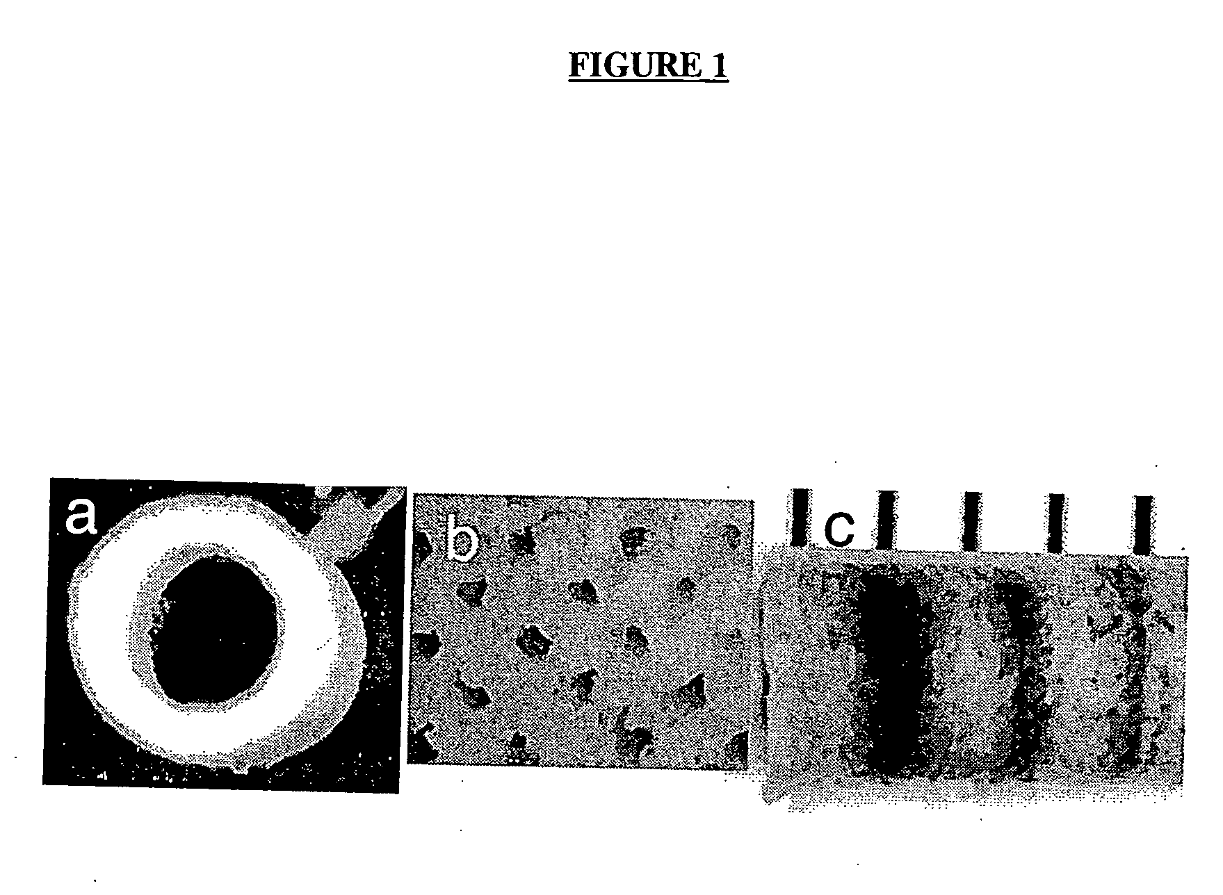
[0172] A gas foaming process has been employed to fabricate cylindrical scaffolds, with either a single (FIG. 1a) or multiple (FIG. 1b) lumens, by assembly and fusion of assembled microspheres. The copolymer of lactide and glycolide (PLG) are used for fabrication, which as been widely used in tissue engineering and neural regeneration, and is well known to the skilled artisan. Microspheres are loaded into a custom built mold (O.D.=3 mm) and fused using a gas foaming process as described in U.S. Pat. No. 6,281,256. Molds are equilibrated in high pressure CO2 (800 psi), and pressure quenching leads to the nucleation and growth of gas pores in the polymer, resulting in fusion of adjacent microspheres. PLG copolymers (50:50, 75:25, and 85:15) with a range of molecular weights (i.v.=0.2 to 1.4 dL / g) can be processed, allowing scaffolds to be fabricated that degrade over times ranging from weeks to months.

[0173] For applic...

example 2

Drug Incorporation and Release from Cylindrical Scaffolds

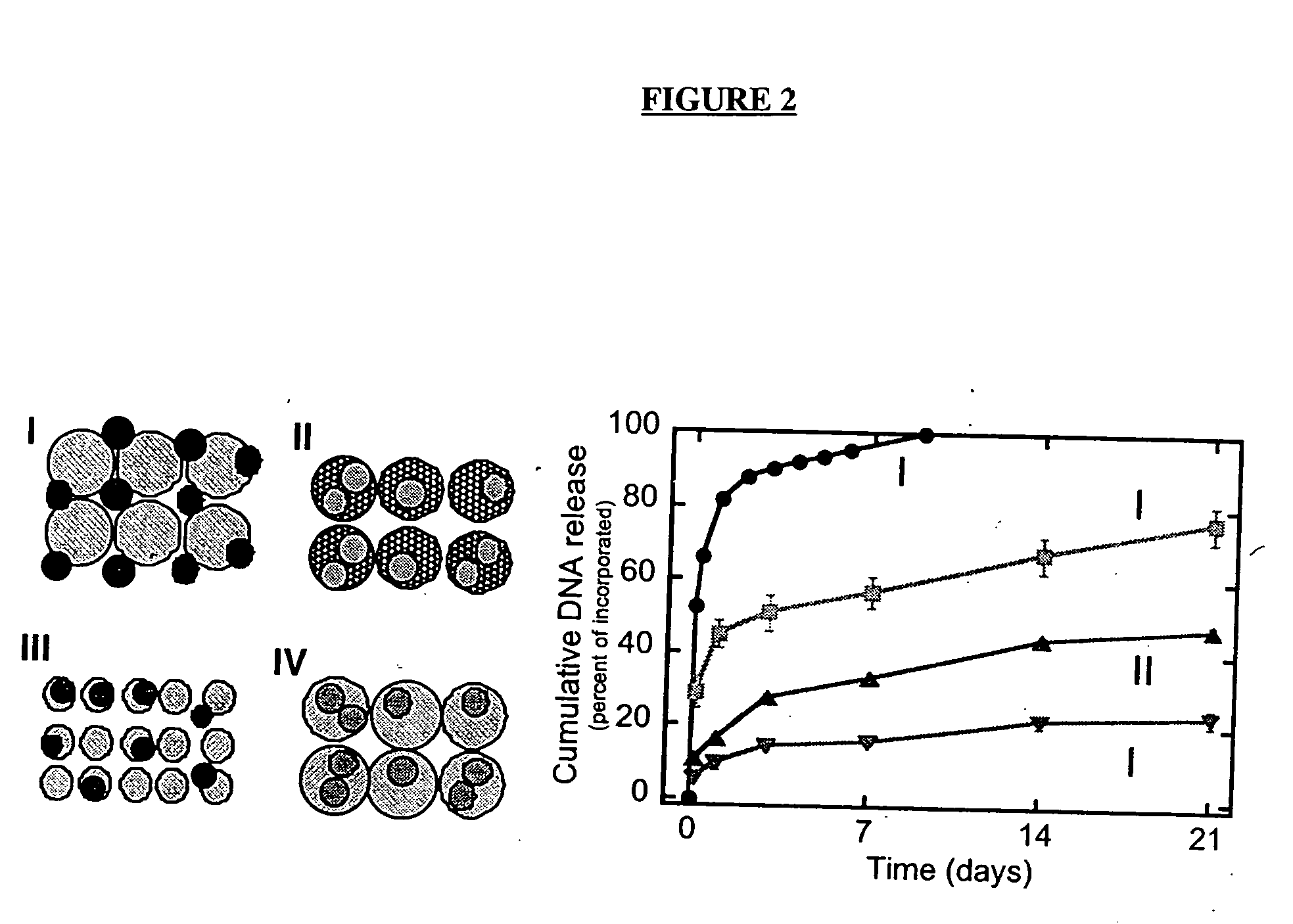
[0175] The gas foaming procedure allows for the incorporation of bioactive factors into polymers by two methods shown schematically in FIG. 2. In the first method (FIG. 2-I), lyophilized drug is mixed with polymer microspheres, compression molded, and foamed into a structure. Proteins and DNA can be lyophilized in the presence of stabilizers (e.g., sucrose, lactose 0.1 M), which retain their integrity and activity during the freezing and dehydration processes. Alternatively, the factors can be incorporated directly into the polymer microspheres using a double emulsion process, and then formed into the cylindrical structure (FIG. 2, II-IV).

[0176] The two techniques for incorporation can be used to differentially regulate the release kinetics of drug from the polymer scaffold (FIG. 2). Scaffolds with incorporated DNA were fabricated as shown in FIG. 2, immersed in PBS, and the concentration of DNA released into the surrounding...

example 3

Transfection with Release DNA Complexes Encoding for Reporter Genes

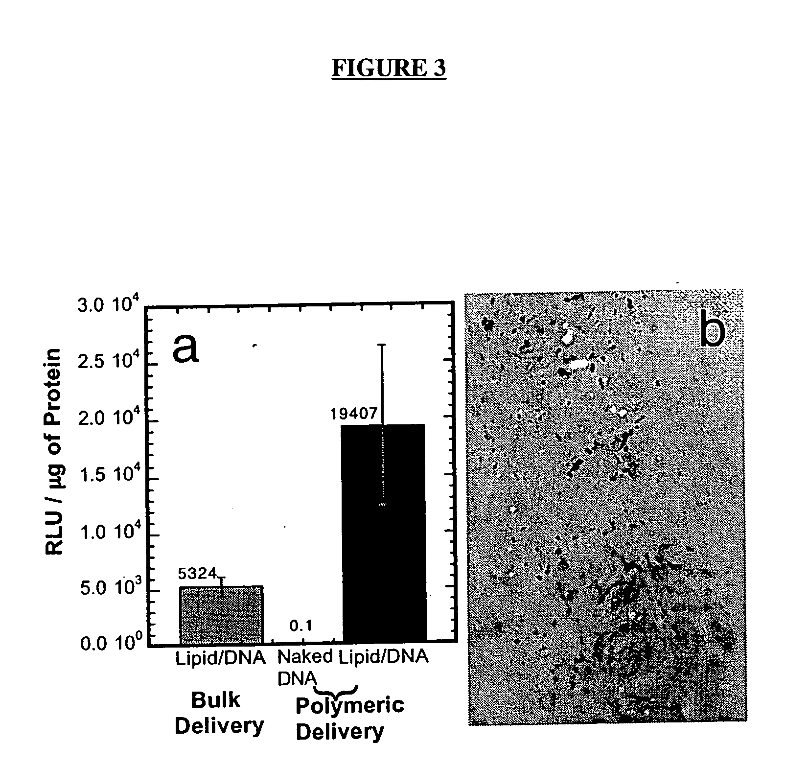
[0177] The ability of DNA complexes released from the polymer scaffolds to transfect cells in vitro was subsequently examined. The reporter genes luciferase and b-galactosidase were used to characterize transfection through measurements of the quantity of protein production (luciferase assay) and the number of cells transfected (light microscopy). The release of naked DNA from the polymer scaffolds resulted in low quantities of protein production (FIG. 3a). The release of lipid / DNA complexes, termed lipoplexes, resulted in expression by NIH / 3T3 cells at levels similar to bolus delivery of freshly prepared lipoplexes. Polymeric tubes releasing DNA lipoplexes (10 μg) transfected cells cultured within the lumen. NIH / 3T3 cells were seeded onto collagen matrices using a dynamic seeding procedure and cultured within the lumen. X-gal staining of the collagen showed transfected cells throughout the collagen (FIG. 3b).


PUM

PropertyMeasurementUnit
Diameteraaaaaaaaaa
Diameteraaaaaaaaaa
Diameteraaaaaaaaaa

Abstract

The invention is directed to scaffolds containing porous polymer material prepared by a process of gas foaming/particulate leaching and a wet granulation step prior to gas foaming and particulate leaching, particularly having a characteristic interconnected pore structure, as well as sustained release of protein, DNA or cells, and to methods for using such porous polymer material for preparation of scaffolds, particularly for tissue engineering.

Description

CROSS-REFERENCE TO RELATED APPLICATIONS [0001] The present application is a non-provisional application claiming the priority of copending provisional application Ser. No. 60 / 578,785, filed Jun. 10, 2004, the disclosure of which is incorporated by reference herein in its entirety. Applicants claim the benefits of this application under 35 U.S.C. §119 (e).FIELD OF THE INVENTION [0002] This invention relates generally to devices for the preparation and delivery of proteins, DNA or cells to effect growth, regrowth, or repair of tissues damaged or destroyed by disease, accident or surgery. More particularly, the invention is directed to a device fabricated as a biodegradable polymeric scaffold that is particularly useful for the controlled or sustained release of DNA and / or protein or cells to promote regrowth of tissue in vivo. The invention is also directed to the use of the devices for the growth of multifilamentous tissue such as nerve tissue, and can also be used for the delivery o...

Claims


Application Information

Patent Timeline
IPC IPC(8): A61K38/17A61F2/00
CPCA61K38/18A61L27/18A61L27/54A61L27/56A61L2300/252A61L2300/258A61L2430/32A61L2300/414A61L2300/602A61L2300/64C08L67/06
Inventor SHEA, LONNIESHEA, LONNIEWHITTLESEY, KEVINYANG, YANGRIVES, CHRISTOPHERDE LAPORTE, LAURAJANG, JAE-HYUNG
Owner NORTHWESTERN UNIV
Why Eureka
Try Eureka