Method for the validatable inactivation of pathogens in a biological fluid by irradiation

a biological fluid and pathogen technology, applied in the field of batch irradiation, can solve the problems of ineffective uv-b in pathogen inactivation, too little uv-c energy may actually reach the pathogen, and require excessively high doses of inactivation

Active Publication Date: 2006-11-16
TAKEDA PHARMA CO LTD
View PDF19 Cites 32 Cited by
  • Summary
  • Abstract
  • Description
  • Claims
  • Application Information

AI Technical Summary

Benefits of technology

[0052] The biological fluid can be contained, for example in stirred-batch photoinactivation reactors. The biological fluid may comprise at least one additive to reduce damage and loss of biological activity of said fluid.

Problems solved by technology

(1989) confirmed that UV-C alone would require excessively high doses to inactivate HIV in blood plasma, and UV-B was ineffective for pathogen inactivation (Prodouz et al.
However, if these additives supplement an additional absorbance at the wavelength used, too little UV-C energy may actually reach the pathogens.
However, an excess of applied energy readily damages the proteins, and it is therefore necessary to not exceed the UV or visible light dose required for sufficient pathogen inactivation, and to calculate or determine this dose as accurately as possible.
1941) are only of limited use in the irradiation of absorbing biological fluids, and the use of radiometer sensor measurements or signals to determine a or calculate a fluence (dose) or fluence rate (dose rate) distribution in a photochemical or pathogen photoinactivation reactor is limited by the laws of optics.
However, models have only been established for very simple systems, such as tubular cells or the elliptical photoreactor (Alfano et al.
These sensors, however, measure only the fraction of radiation incident on them, but not the average fluence (light dose) effective on the fluid to be irradiated while contained within the irradiation reactor.
Apparently, no definitive results for the effective dose were obtained from this validation approach, as remarked upon the experimental treatment of liquid egg white in the Centrifilmer irradiator for the UV-C inactivation of Salmonella spp.
The proposed addition of an actinometer substance to an already absorbing medium is therefore not capable to accomplish an exact effective dose measurement.
However, the reduction equivalent dose (RED), which is the standard parameter determined in water UV-C disinfection, is not the same as the average dose effective in the fluid.
Nevertheless, an electronic radiometer at the end of this light path and efficient flow-through mixing to narrow dose distributions (Cabaj and Sommer 2000) is still required for this method, and the incubation time for spore cultivation does not allow for rapid validation.
However, these processes are limited by fast-flowing fractions with lower and potentially insufficient doses (Qualls and Johnson 1983).
Therefore such devices are unsuitable in the context of pathogen inactivation of biological substances.
Most blood plasma irradiation devices are based on a thin-film principle, which limits sample throughput.
Only a small volume fraction of the vessel can be filled with the fluid, thus reducing the capacity and the scalability of the method.
Neither absorbance nor viscosity of the protein solutions are matched and failure of actinometric lamp monitoring in determination of applied UV dose or equivalent dose effective in the protein solution is therefore evident by use of empirical constants in the dose / inactivation equation.
In general, a known disadvantage of using iodide solutions is low quantum yield of photochemical reaction and nonlinear dose-response (Rahn 1997).
This, however, does not conform to the photochemical concept of fluence as the energy incident on and only partially absorbed by an area (Bolton 1999).
However, an absorbance-matching calibration to determine the dose effective in the iodide solution was neither proposed nor done ((Forney and Pierson 2003).

Method used


Image

  • Method for the validatable inactivation of pathogens in a biological fluid by irradiation
  • Method for the validatable inactivation of pathogens in a biological fluid by irradiation
  • Method for the validatable inactivation of pathogens in a biological fluid by irradiation

Examples


example 1

Absorption Coefficients of the Iodide / Iodate Actinometer Solution at High Dilution, Dosimetric Response of the Iodide / Iodate Actinometer Solutions, and Stabilization of the UV-C Generated Tri-Iodide by Polyvinylpyrrolidone

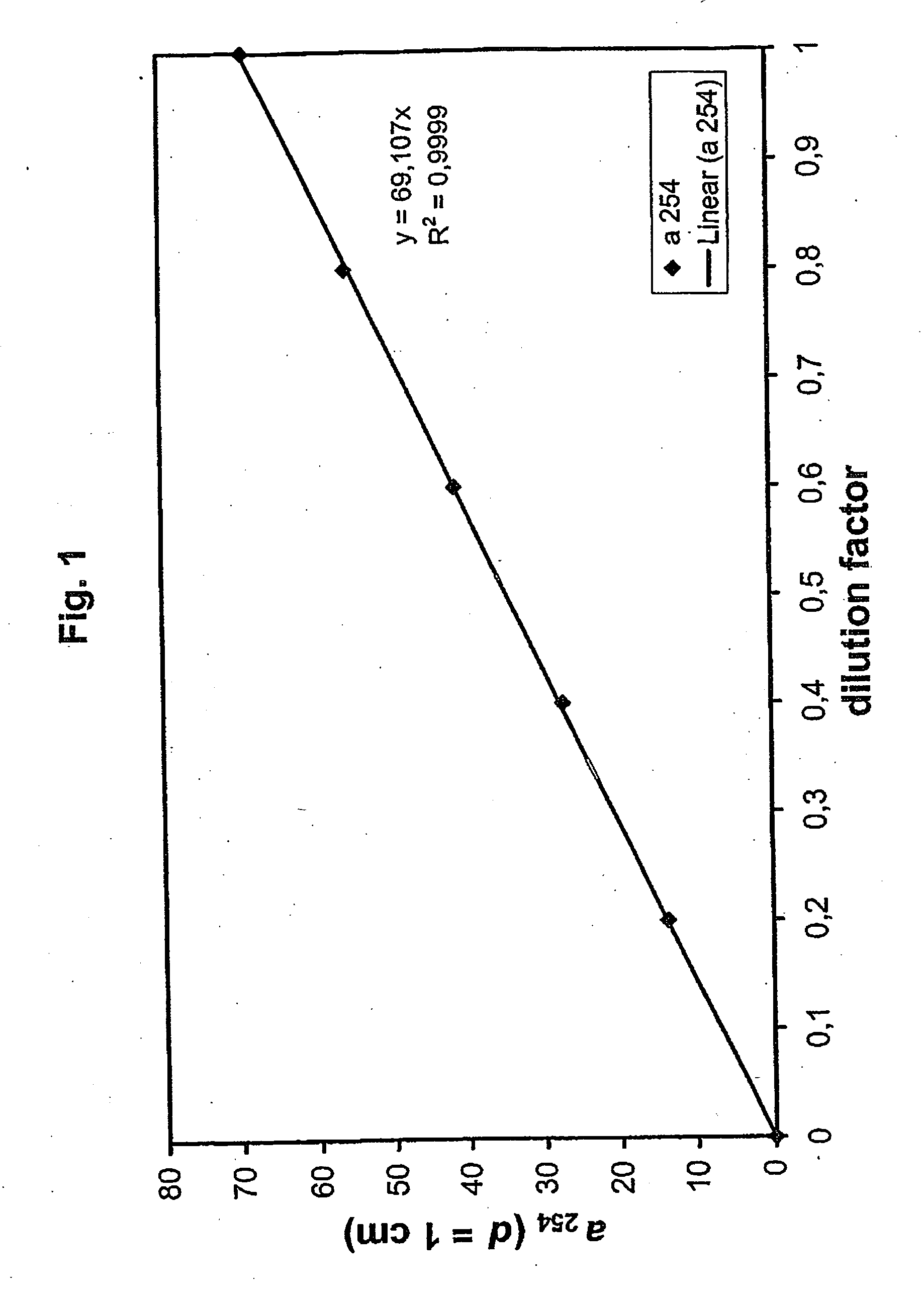
[0125] A solution containing 0.24 M KI and 0.04 M in 0.01 M borate buffer, pH=9.25, was diluted 0.2-, 0.4-, 0.6 and 0.8-fold with 0.01 M borate buffer and the absorption measured in a 0.1 mm cuvette. FIG. 1 shows that the absorption coefficient depends in a linear-ratio (a253.7=69 / cm×dilution factor) on the concentration.

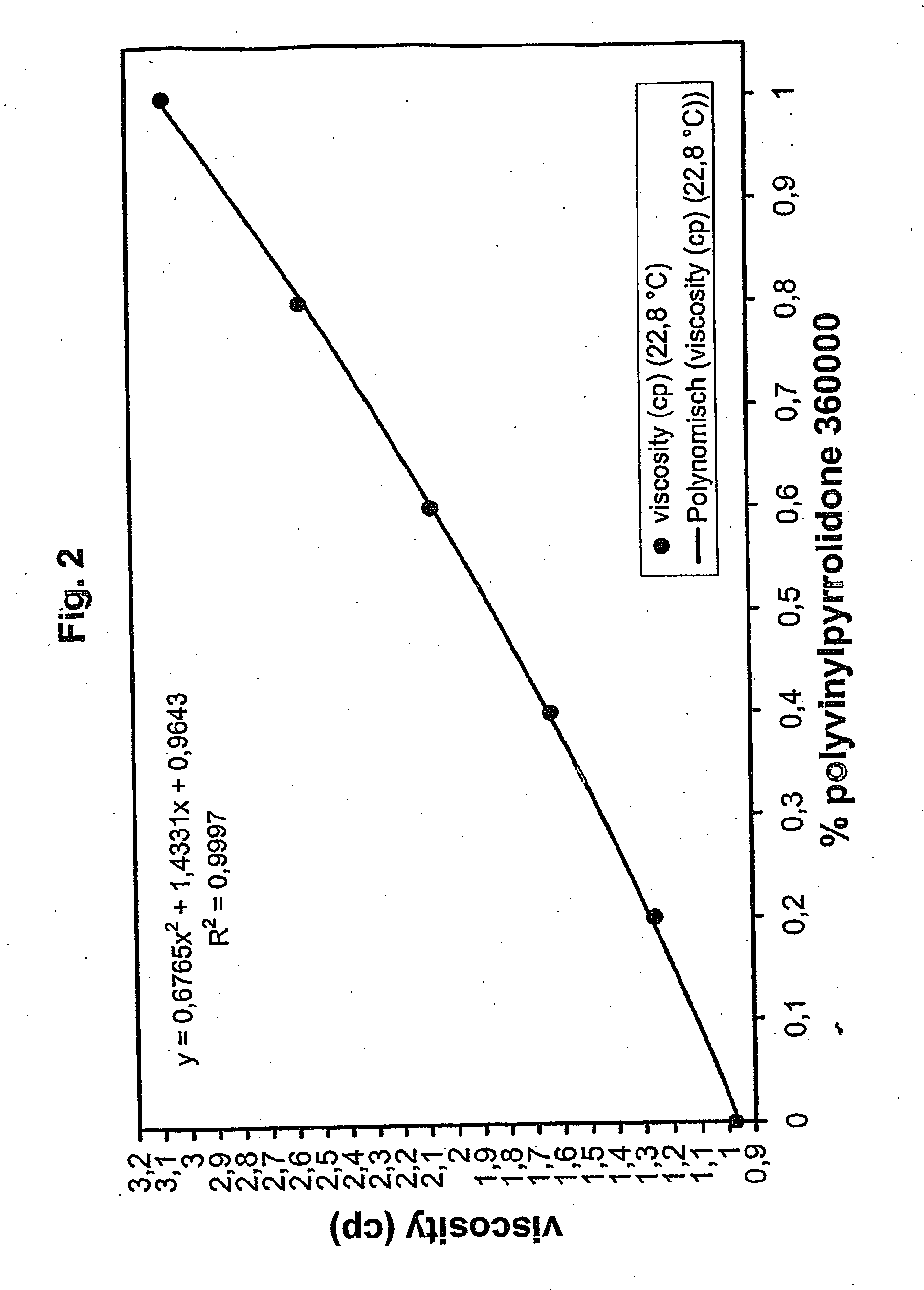
[0126] A solution containing 10 g polyvinylpyrrolidone K90 (PVP K90; average molar mass 360000 Da) in 1 L 0.01 M borate buffer was diluted 0.2-, 0.4-, 0.6-, 0.8-fold and the viscosity of the dilutions and the buffer were measured in a Schott 0.40 mm Ubbelohde capillary viscosimeter thermostated at 22.8° C. It can be seen from FIG. 2 that the viscosity increases with the concentration in a non-linear ratio.

[0127] Dilutions of the stock solution c...

example 2

Determination of the Irradiation Time in a Batch Photoreactor with Fixed Geometry with Actinometric Dosimetry Solutions

[0129] Dosimetry solutions with decadic absorption coefficients a253.7 of 2.1 / cm (0.0072 M KI+0.0012 M KIO3+0.5 g PVP K90 / L, for FVIII concentrate), 6.1 / cm (see Example 1), 10 / cm (0.0346 M KI+0.0058 M KIO3+1.21 g PVP K90 / L; for a 2% (w / v) immunoglobulin G solution), and 16 / cm (see Example 1) were prepared and calibration plots recorded as described in Example 1. A quartz test tube 4 cm in diameter was filled with 100 mL dosimetry solution, stirred with a magnetic stirrer bar on a magnetic stirrer, and irradiated between 2 low-pressure mercury vapor lamps equipped with polished stainless steel reflectors. At predefined intervals, samples were drawn and filled into the 0.2 mm cuvettes for spectrophotometric measurement, and the dose was read out from the calibration plot. The dose-rate was then calculated as (mJ / cm2) / min, and the inverse dose rate, i.e. the specific ...

example 3

Inactivation of MMV and Phi-X 174 in Thin Layer- and Batch Irradiation

[0131] MMV stock solution from cell culture supernatant (˜108 tissue-culture infectious doses (TCID50) / mL) and bacteriophage Phi-X 174 lysate (˜1×10⁹ plaque-forming units (PFU) / mL) from propagation in Escherichia coil were diluted in 20 mM phosphate-buffered 0.15 M NaCl (phosphate-buffered saline, PBS) and 2.5 mL were irradiated with different UV-C doses in 32 mm polystyrene petri dishes shaken horizontally under a CAMAG TLC lamp. The irradiance at the sample surface distance was determined with a Dr. Groebel RM21-radiometer with a daylight-blind UV-C sensor, and the self-absorbance of the solution measured in a spectrophotometer to correct for the effective fluence (UV dose) (Morowitz 1950). MMV and Phi-X 174 titers were determined by titration on host cell culture and host bacteria respectively.

log10 pfu Phi-XUV doselog10 TCID50UV dose (mJ / cm2)174 / mL(mJ / cm2)MMV / mL0.006.4106.542.255.7925.314.504.3744.426.753.4...


PUM

PropertyMeasurementUnit
penetration depthaaaaaaaaaa
wavelengthsaaaaaaaaaa

Abstract

A validatable method for determining a photochemically effective dose for inactivating pathogens in a fluid sample is described herein. In particular, the instant invention covers methods for determining a photochemically effective dose sufficient to inactivate pathogens in a biological sample while leaving biologically active substances of interest unaffected. A batch irradiation reactor effective for inactivating pathogens in biological samples is also described.

Description

FIELD OF THE INVENTION [0001] The present invention relates to a method and apparatus for photochemical pathogen inactivation by batch irradiation, which prior to the present invention was considered too complex and unworkable. In particular, the invention advantageously permits those skilled in the art to determine an effective light dose for photochemical pathogen inactivation. BACKGROUND OF THE INVENTION [0002] Treatment of blood, blood cells, plasma, serum, plasma fractions, other biological fluids and protein solutions by irradiation has been a widely investigated approach to inactivate pathogenic viruses, in particular, small single-stranded non-enveloped DNA viruses. Examples of irradiation treatment currently under study include short-wave ultraviolet light, long-wave ultraviolet light, visible light with photosensitizng compounds, and broad-spectrum high-intensity flash light. [0003] In the 1930's, small volumes of autologous whole blood were exposed to UV irradiation from ...

Claims


Application Information

Patent Timeline
Patent Type & Authority Applications(United States)
IPC IPC(8): C12Q1/68A61L2/00A61L2/08A61L2/10A61L2/28G01J1/00G01J1/08G01J1/42G01J1/48
CPCA61L2/0011A61L2/0017A61L2/0052A61L2/10A61L2/28G01J1/48A61M1/3681C02F1/32C02F2201/326G01J1/08G01J1/42A61L2202/22A61M2205/053A61M1/3683A61P31/12A61P7/08
Inventor ANDERLE, HEINZMATTHIESSEN, PETERSCHWARZ, HANS-PETERTURECEK, PETERKREIL, THOMASBOGGS, DANIEL R.
Owner TAKEDA PHARMA CO LTD
Why Eureka
Try Eureka