
Methods for modulating osteochondral development using bioelectrical stimulation

a bioelectric stimulation and osteochondral technology, applied in biochemical equipment and processes, specific use bioreactors/fermenters, therapy, etc., can solve the problems of total knee joint replacement, loss of productivity and independence, and deterioration of joint cartilage, so as to maximize the utility and application of desired pemf waveforms, optimize osteoblast development, and improve bone mass

Active Publication Date: 2006-12-28
MEDRELIEF
View PDF58 Cites 45 Cited by
  • Summary
  • Abstract
  • Description
  • Claims
  • Application Information

AI Technical Summary

Benefits of technology

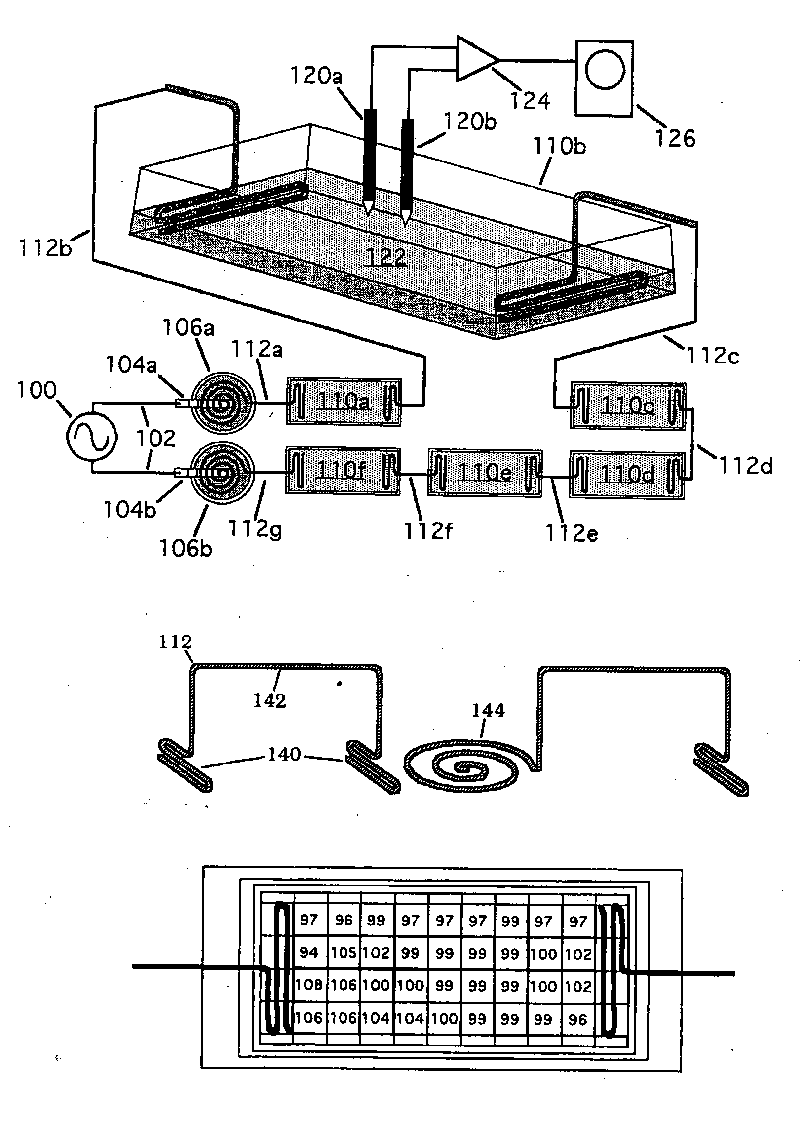
[0012] The present invention overcomes the shortcomings of prior art devices and methods by enabling the delivery of bioelectrical signals optimized to correspond to selected features of natural body signals resulting in accelerated and more permanent healing. The signals described herein conform to selected features of natural signals and consequently tissues subjected to electrostimulation according to the present invention undergo minimal physiological stress. In addition, the present invention is non-invasive and cost-effective making it desirable for multiple applications for personal and individual use. Furthermore, the present methods provide electrical stimulation where the electrical signals closely mimic selected characteristics of natural body signals. The stimulated tissue is therefore subjected to minimal stress and growth and repair is greatly facilitated.
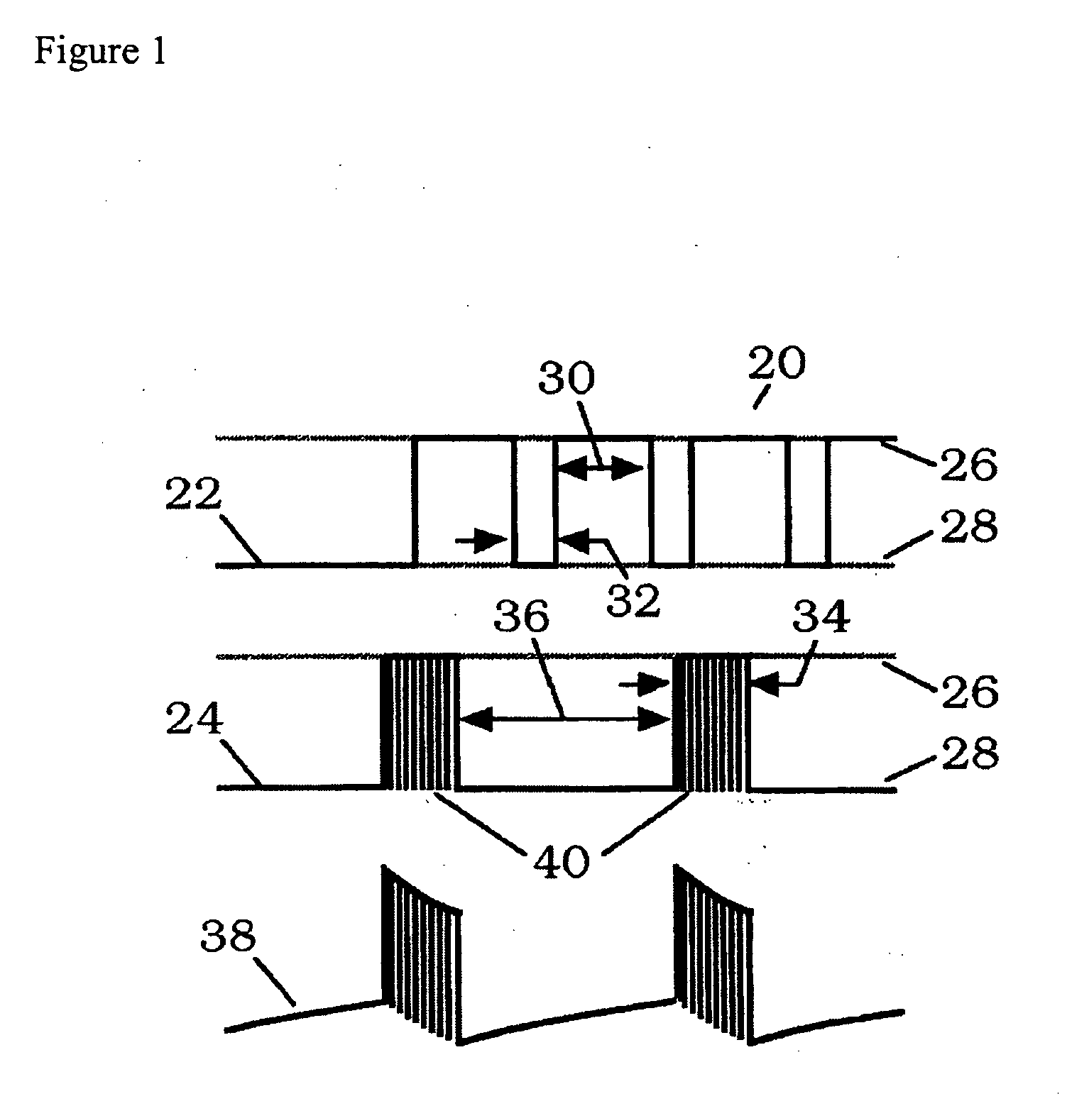
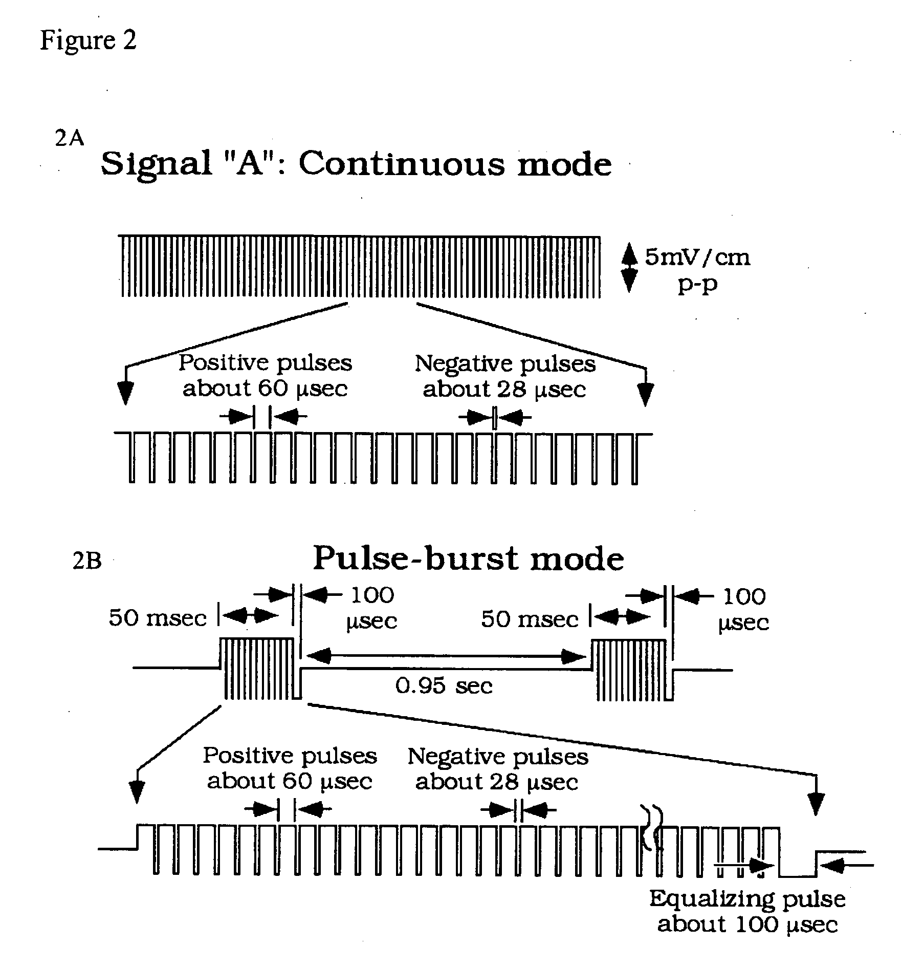
[0015] Although no commercial bioelectrical devices are currently approved for osteoporosis therapy, the present invention provides a promising candidate. As demonstrated herein, unique pulsed electromagnetic field (PEMF) wave patterns may be advantageously applied at both a macroscopic level (i.e. common bone fractures) as well as at microscopic levels (i.e. osteoblast development). Certain embodiments of the invention maximize the utility and application of desired PEMF waveforms: for example, the spine, hip and / or wrist are the most common sites of osteoporotic fracture, for such types of fractures the inventors provide simple, self-adhesive, skin contact electrode pads as electrotherapeutic delivery vehicles. The use of such electrode pads results in the improvement of bone mass at such key anatomical sites. At a microscopic level, the present inventors have identified specific PEMF waveforms and frequencies that optimize osteoblast development. As described in greater detail in the Examples (see Example 1) the inventors demonstrate that PEMF signals enhance osteoblast mineralization and matrix production, and that the signal confers structural features as well. The inventors also show that other PEMF signals enhanced cell proliferation and accompanying increases in bone morphogenetic proteins (BMPs). While both pulse-burst and continuous electrical signals may be used in the present invention, the administration of continuous rather than pulse-burst signals provided the more pronounced effects on proliferation and mineralization.
[0016] The electrical signals of the present invention may be used to promote the repair and growth of structural tissues such as bone and cartilage. However, such systems and methods need not be confined to use with intact organisms, since isolated cells or tissue cultures can also be affected by electrotherapeutic waveforms (appropriate electrical stimuli have been observed to modify the rates of cell metabolism, secretion, and replication). Electrical signals are generally applicable to other connective tissues such as skin, ligaments, tendons, and the like. The electrical signals described herein may be used to stimulate other tissues to increase repair of the tissues and promote growth of tissues for transplantation purposes. Isolated skin cells, for example, might be treated with the devices and waveforms of the present invention in an appropriate growth medium to increase cell proliferation and differentiation in the preparation of tissue-cultured, autogenous skin-graft material. In a like manner, these bioelectric signals can be applied directly to injured or diseased skin tissue to enhance healing.
[0017] Exogenous delivery of bioelectrical signals and progenitor cells such as bone marrow stromal cells-BMSCs to a fracture can lead to enhanced healing and repair of recalcitrant fractures. Both of these factors (bioelectricity and cell recruitment) are, in fact, parts of the natural healing process. For these applications, electrical stimulation using the waveforms described herein can be applied immediately after injury with an electrotherapy system. The electrotherapy system may be lightweight, compact and portable. Both electrical stimulation and universal cell-based therapy can be applied within a few days after injury. Autologous cells may be added at time further after injury. The present invention also provides methods to induce bone repair or development that regenerates natural tissues rather than scar tissue.

Problems solved by technology

About 10% of these have delayed healing and of these, 150,000 to 200,000 nonunion fractures occur accompanied by loss of productivity and independence.
In the case of cartilage, severe and chronic forms of knee joint cartilage damage can lead to greater deterioration of the joint cartilage and may eventually lead to a total knee joint replacement.
Approximately 200,000 total knee replacement operations are performed annually and the artificial joint generally lasts only 10 to 15 years leading to similar losses in productivity and independence.
Furthermore, the incidence of bone fractures is also expected to remain high in view of the incidence of osteoporosis as a major public health threat for an estimated 44 million Americans.
Such approaches also hold potential for cartilage injuries.
Unfortunately, most electrotherapeutic devices now available rely on direct implantation of electrodes or entire electronic packages, or on inductive coupling through the skin using coils which generate time-varying magnetic fields, thereby inducing weak eddy currents within body tissues which inefficiently provides the signal to tissues and thus in addition to bulky coils requires relatively large signal generators and battery packs.
The need for surgery and biocompatible materials in the one case, and excessive circuit complexity and input power in the other, has kept the price of most such apparatus relatively high, and has also restricted the application of such devices to highly trained personnel.

Method used


Image

  • Methods for modulating osteochondral development using bioelectrical stimulation
  • Methods for modulating osteochondral development using bioelectrical stimulation
  • Methods for modulating osteochondral development using bioelectrical stimulation

Examples


example 1

Effect of PEMF Signal Configuration on Mineralization and Morphology in a Primary Osteoblast Culture

[0127] The goal of this study was to compare two PEMF waveform configurations delivered with capacitative coupling by evaluating biochemical and morphologic variations in a primary bone cell culture.

Methods

[0128] Osteoblast cell culture: Primary human osteoblasts (CAMBREX®, Walkersville, Md.) were expanded to 75% confluence, and plated at a density of 50,000 cells / ml directly into the LAB-TEK™ (NALGE NUNC INTERNATIONAL™, Rochester, N.Y.) chambers described previously. Cultures were supported initially with basic osteoblast media without differentiation factors. When the cultures reached 70% confluence within the chambers, media was supplemented with hydrocortisone-21-hemisuccinate (200 mM final concentration), β-glycerophosphate (10 mM final concentration), and ascorbic acid. Osteoblasts were incubated in humidified air at 37° C., 5% CO2, 95% air for up 21 days. Media was changed ...

example 2

Use of a Niobium “Salt” Bridge for In Vitro PEMF Stimulation

Introduction

[0141] A passive electrode system using anodized niobium wire was developed to couple time-varying electric signals into culture chambers. The intent of the design was to reduce complexity and improve reproducibility by replacing conventional electrolyte bridge technology for delivery of PEMF-type signals, such as those induced in tissue by the EBI repetitive pulse burst bone grown stimulator, capacitively rather than inductively, in vitro for cellular, tissue studies. Anodized niobium wire is readily available and requires only simple hand tools to form the electrode bridge. At usable frequencies, typically between 5 Hz and 3 MHz, DC current passage is negligible.

Background

[0142] Capacitively-coupled electric fields have typically been introduced to culture media with conventional electrolyte salt bridges which have limited frequency response and are difficult to use without risk of contamination for exte...

example 3

Stimulation of Cartilage Cells Using a Capactively Coupled PEMF Signal

Introduction

[0147] A PEMF signal similar to that used clinically for bone repair is currently being tested for its ability to reduce pain in joints of arthritic patients. Of interest is whether this pain relief signal can also improve the underlying problem of impaired cartilage.

Background

[0148] Compared to drug therapies and biologics, PEMF based therapeutics offer a treatment that is easy to use, non-invasive, involves no foreign agent with potential side effects, and has zero clearance time. Issues with PEMF therapeutics include identifying responsive cells, elucidating a physical transduction site on a cell, and determining the biological mechanism of action that results in a cell response. The purpose of this study was to determine whether a specific PEMF signal currently being tested for pain relief (MEDRELIEF®, Healthonics, Inc, Ga.) could stimulate cartilage cells in vitro and whether a biological me...


PUM


Abstract

Compositions and methods are provided for modulating the growth, development and repair of bone, cartilage or other connective tissue. Devices and stimulus waveforms are provided to differentially modulate the behavior of osteoblasts, chondrocytes and other connective tissue cells to promote proliferation, differentiation, matrix formation or mineralization for in vitro or in vivo applications. Continuous-mode and pulse-burst-mode stimulation of cells with charge-balanced signals may be used. Bone, cartilage and other connective tissue growth is stimulated in part by nitric oxide release through electrical stimulation and may be modulated through co-administration of NO donors and NO synthase inhibitors. Bone, cartilage and other connective tissue growth is stimulated in part by release of BMP-2 and BMP-7 in response to electrical stimulation to promote differentiation of cells. The methods and devices described are useful in promoting repair of bone fractures, cartilage and connective tissue repair as well as for engineering tissue for transplantation.

Description

CROSS REFERENCE TO RELATED APPLICATIONS [0001] This application claims the benefit of U.S. provisional patent application 60 / 687,430 filed Jun. 3, 2005, U.S. provisional patent application 60 / 693,490 filed Jun. 23, 2005, U.S. provisional patent application 60 / 782,462 filed Mar. 15, 2006 and U.S. provisional patent application 60 / 790,128 filed Apr. 7, 2006.BACKGROUND [0002] Diseases and injuries associated with bone and cartilage have a significant impact on the population. Approximately five million bone fractures occur annually in the United States alone. About 10% of these have delayed healing and of these, 150,000 to 200,000 nonunion fractures occur accompanied by loss of productivity and independence. In the case of cartilage, severe and chronic forms of knee joint cartilage damage can lead to greater deterioration of the joint cartilage and may eventually lead to a total knee joint replacement. Approximately 200,000 total knee replacement operations are performed annually and t...

Claims


Application Information

Patent Timeline
Patent Type & Authority Applications(United States)
IPC IPC(8): A61N1/00
CPCA61N1/40A61N1/326C12M21/08C12M35/02
Inventor KRONBERG, JAMES W.GANEY, TIMOTHYGORDON, STEPHEN L.
Owner MEDRELIEF
Learn More