

Method of inducing fenestrae
a fenestrae and endothelial cell technology, applied in the field of induction of fenestrae in an endothelial cell line, can solve the problems of limited descriptive and morphological analysis of fenestrae, inherently difficult study of fenestrae, etc., and achieves the effect of confirming the non-discriminatory effect of drugs, reducing the amount of stress fibers, and increasing relative induction
- Summary
- Abstract
- Description
- Claims
- Application Information
AI Technical Summary
Benefits of technology
Problems solved by technology
Method used
Image

Examples
example 1
Mammalian Tissue Culture
Isolation of Mouse Embryonic Fibroblasts
[0082] 14.5 day old embryos were dissected from the extraembryonic membranes in Dulbeccos Modified Eagle's Medium (DMEM; Invitrogen). The liver was discarded, the head was removed for genotyping and the remainder of the embryo was trypsinised by incubating with 1 ml of trypsin / versene for 45 minutes on a shaker set at 37° C. Using a plastic Pasteur pipette, the embryo was dissociated by pipetting up and down five times, and split between two 60 mm dishes.
[0083] Maintenance of Mammalian Cell Lines
Cell LineSpeciesOriginPassage NoCulture conditionsbEND5mousebrain13-25DMEM high glucose with sodium pyruvate,endothelioma10% FBS, 4 mM L-glutamate, penicilin / streptomycin,5 μM β-mercaptoethanol, non-essential amino acids.37° C. incubator with 10% CO2Py4.1mouseear and tailDMEM high glucose with sodium pyruvate, 2% FBS,hemangiomaspenicilin / streptomycin. 37° C. incubator with10% CO2NIH 3T3mouseembryoDMEM high glucose with so...
example 2
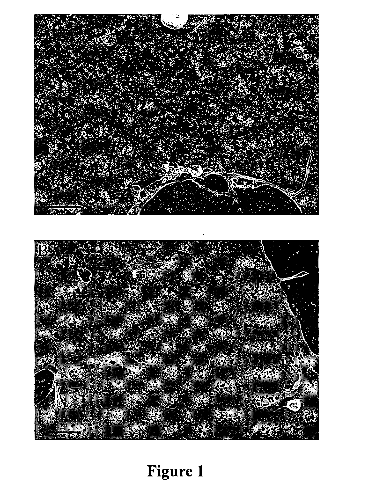
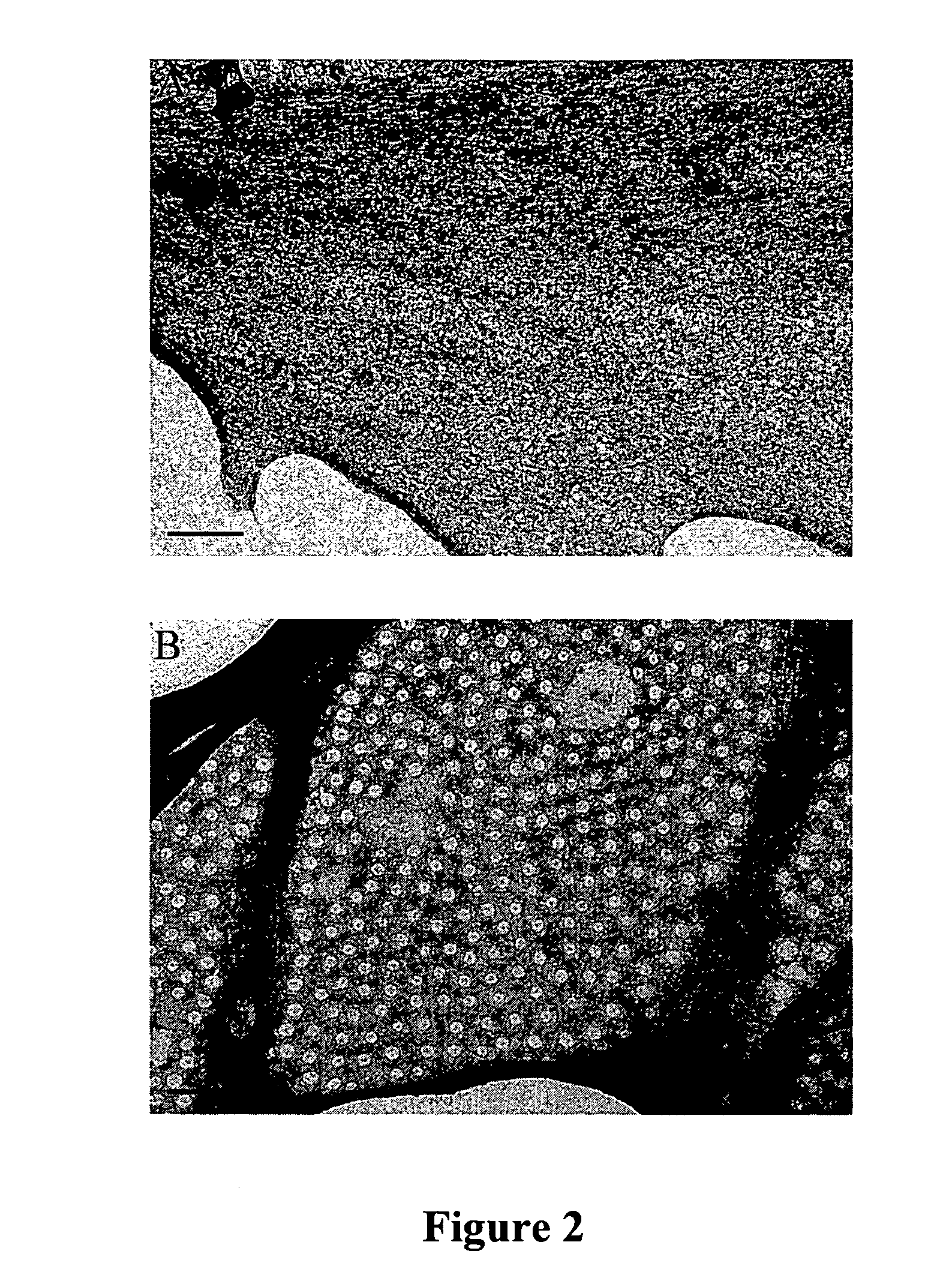
Fenestrae Induction in Endothelial Cells
[0090] Coverslips and dishes were coated with 1% gelatin (Sigma) solution in PBS for 30 minutes at room temperature. Endothelial cells were seeded overnight at a density equivalent to 1.5×10⁶ cells per 100 mm dish. Cultures were induced with Cytochalasin B (Sigma) at 10 μM for 2 hours, with Latrunculin A (Molecular Probes) at 2.5 μM for 3 hours, or with a combination of recombinant mouse 75 ng / ml VEGF (R&D systems) for 6-72 hours and 10 μM Cytochalasin B for 2 hours. Cells were processed for biochemistry or morphology immediately after the end of the induction.
[0091] To inhibit protein synthesis during fenestrae formation, cells were incubated with 10 μg / ml Cycloheximide (Sigma) for 30 minutes, and then induced with VEGF (75 ng / ml) for 6 hours and Cytochalasin B (10 μM) for the last 2 hours.
example 3
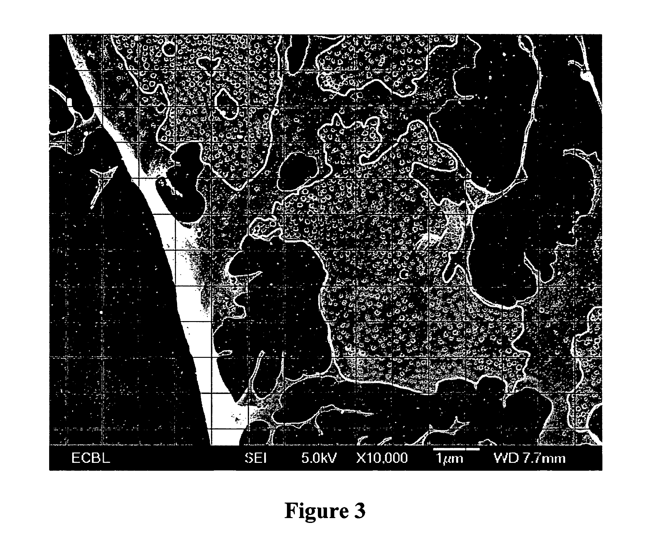
Light Microscopy
[0092] Images were captured using the following instruments and software packages:
[0093] 1) LSM510 laser scanning confocal microscope (Zeiss); 63×1.40 NA Plan-Achromat oil immersion objective
[0094] 2) TCS SP2 spectral confocal microscope (Leica); 40×1.25 NA Plan-Achromat oil immersion objective; 63×1.4 NA Plan-Achromat oil immersion objective; 100×; Leica confocal software version 2.5
[0095] 3) Widefield DMRA4 microscope (Leica); orca ER2 camera (Hamamatsu); Metamorph Software (Universal Imaging Corporation)
[0096] 4) MZFL III Fluorescence Stereomicroscope (Leica); Retiga Camera (Q-Imaging); OpenLab 3.1.7 (Improvision, Inc.)
[0097] Digital images were processed using Adobe Photoshop 7.0 (Adobe Systems Inc.)
PUM
| Property | Measurement | Unit |
|---|---|---|
| Molar density | aaaaa | aaaaa |
| Molar density | aaaaa | aaaaa |
| Concentration | aaaaa | aaaaa |
Abstract
Description
Claims
Application Information