
Quantitative molecular probes

a molecular probe and quantification technology, applied in the field of molecular probes, can solve the problems of difficult amplification steps, particularly difficult quantification of mirna, and inability to provide complete spatial-temporal profiles of gene expression at the single cell level, so as to improve signal-to-background, improve spatial quantification, and improve quantification

Inactive Publication Date: 2009-04-23
THE TRUSTEES OF THE UNIV OF PENNSYLVANIA
View PDF2 Cites 23 Cited by
  • Summary
  • Abstract
  • Description
  • Claims
  • Application Information

AI Technical Summary

Benefits of technology

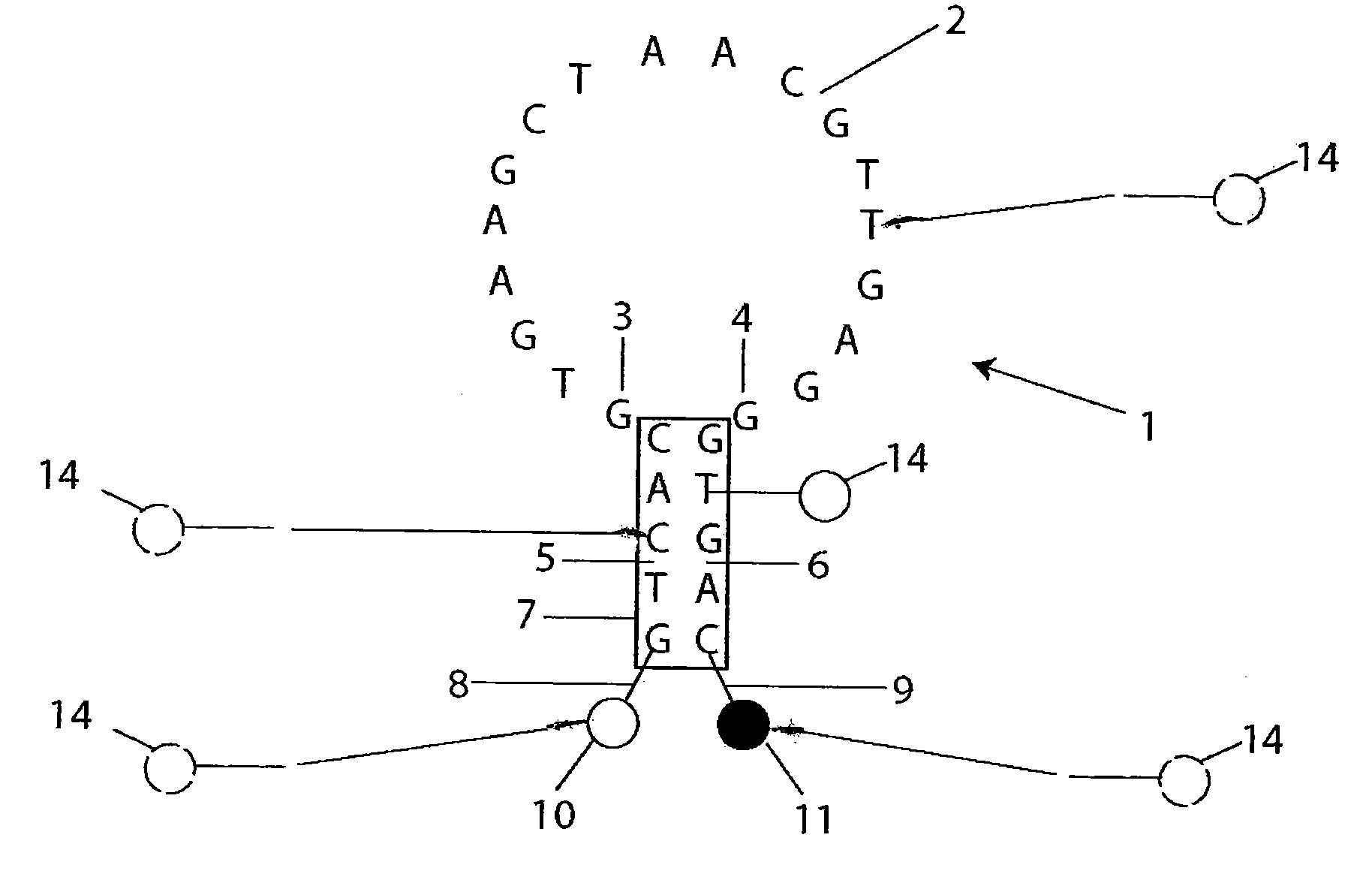
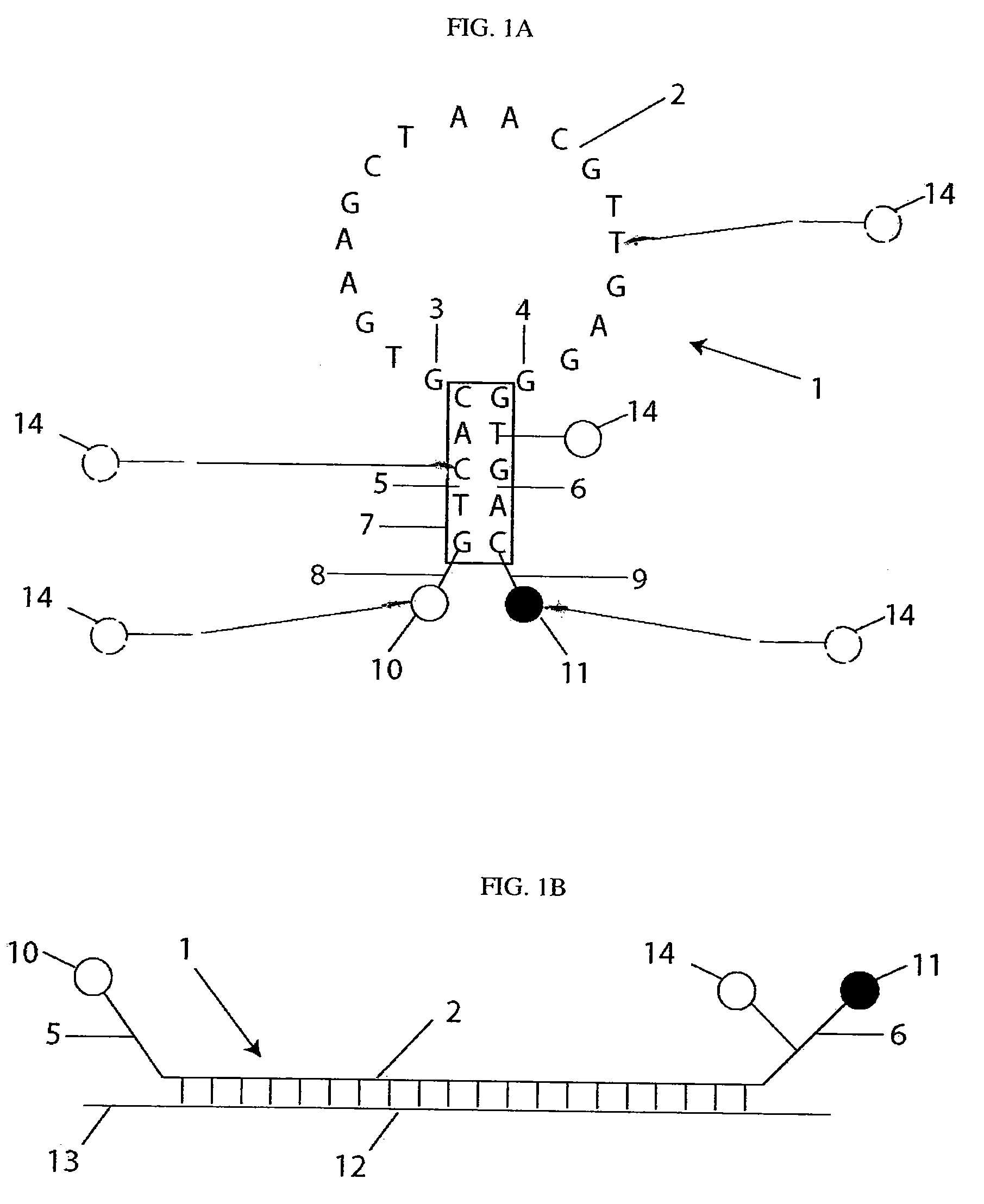
[0028]The attachment of a reference dye to a molecular beacon (or dual-labeled oligonucleotide) provides significant advantages as compared to current methods. These advantages include the following:
[0037]The unquenched reference dye / nanoparticle expands upon the versatility of the molecular beacon by not only improving the ability to accurately and sensitively detect RNA expression and localization (for the reasons described above), but it also provides a means for mRNA quantification. Specifically, the reference dye signal can be quantified to determine the number of probes in the cell and ratiometric imaging, comparing the emission of the ‘report’ dye to the ‘reference’ dye, provides a simple means to quantify the extent of probe hybridization to target RNA. These quantitative measurements can subsequently be used to calculate the exact copy number of RNA within single cells. An advantage of using a nanoparticle / macromolecule as the reference dye is it prevents nuclear localization. Thus, the construct is not sequestered in the nucleus and no non-specific signal is observed in the cell (e.g. nucleus), which is often the case with conventional molecular beacons.

Problems solved by technology

However, many commonly used approaches such as fluorescence in situ hybridization (FISH), real-time PCR, and microarrays do not provide complete spatial-temporal profiles of gene expression at the single cell level that are vital for understanding the role of genetic processing in cellular function.
However, these methods generally provide only the relative change in gene expression for a population of cells and not an absolute measure of RNA copies at the single cell level.
Quantifying miRNA is particularly challenging because the short nature of miRNAs makes enrichment, ligation, and / or amplification steps technically challenging.
Further, cross-hybridization is problematic for microarrays due to the sequence similarity between some miRNAs or large variations in G-C content of others (i.e., require very different hybridization temperatures).
However, these oligonucleotide-based probes still require PCR-amplification of the miRNA2.
A single-molecule method for the quantification of miRNA gene expression is available; but the technology is not widely accessible like flow cytometry1.
Although single cell microdissection techniques can potentially be combined with any of these approaches to provide single cell miRNA measures, large numbers of cells could not be analyzed in a high-throughput fashion.
Although molecular beacons obviously have several advantageous features for imaging RNA in living cells, they also possess several limitations.
Specifically, conventional molecular beacons only provide a qualitative / relative measure of RNA expression and do not yield a rigorous quantification.
Further, there is a high potential for false-negatives and false-positives.
Consequently, inhomogeneous transfections in each cell can make interpretation of fluorescence very difficult.
This generally limits the use of molecular beacons to the study of RNAs that undergo dramatic changes in expression level.
Although many of these problems can be avoided with microinjection only, then only a few cells can be studied at a time and therefore this approach does not scale well for high-throughout screening.
Another problem faced by conventional molecular beacons is they immediately localize to the nucleus, where they emit a non-specific signal.
Also, the non-specific fluorescence that emanates from the nucleus makes image analysis and RNA quantification difficult.
Perhaps the most significant limitation is that molecular beacon signals from individual cells are difficult to compare directly to each other because of the potential bias arising from the different number of beacons present across cells.
Normalization of fluorescence by ratiometric imaging not potentially allows for differentiating RNA expression levels from cell-to-cell, and for more accurate / precise monitoring of RNA expression trends.
A major drawback of using two unlinked reporters is the variance in intracellular lifetime that exists between hybridized and unhybridized oligonucleotides.
A second drawback of the dual oligonucleotide approach is that varying ratios of each oligonucleotide in each cell, considered common in liposome / dendrimer-based transfection methods, can also cause errors in quantification of RNA.
Microinjection is invasive and only allows for the study of a small number of cells.
A third drawback is that the two oligonucleotide probes used for ratiometric imaging do not necessarily co-localize making it difficult to perform true ratiometric imaging.
However, this reference does not describe utilizing polymersomes for calibration or encapsulating molecular probes.

Method used


Image

  • Quantitative molecular probes
  • Quantitative molecular probes
  • Quantitative molecular probes

Examples


example 1

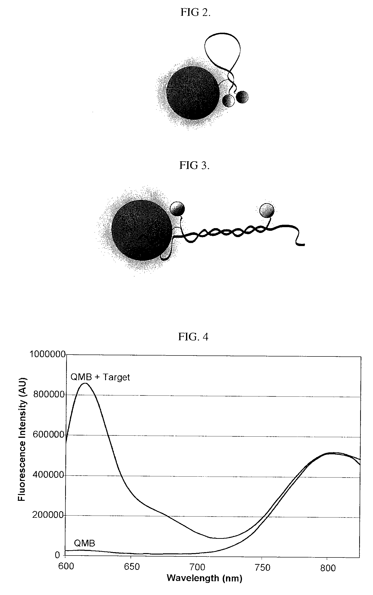
[0172]Synthesis of quantitative molecular probes: As a precursor to the ‘quantitative molecular probe’ (QMP), an antisense molecular beacon possessing an amino-linker within its stem was synthesized using standard phosphoramidite chemistry (FAM-GTCACCTCAGCGTAAGTGATGTCG / aminoC6T / GAC-Dabcyl) (SEQ ID NO: 15). To cross-link the aminated molecular beacons to quantum dots (Qdot 655, Quantum Dot Corp.), a 10 μM sample of the beacon was reacted with a 50-fold excess of Disuccinimidyl Suberate (DSS, Pierce) in DMSO at 40° C. for 2 hours. The activated molecular beacon was then acetone precipitated, and subsequently reacted with 6 μM Amino (PEG) quantum dots (QDs) at molar ratios of 25:1, 10:1, or 5:1 molecular beacons to QD in 50 mM Sodium Borate, pH 8.2 at 37° C. for 2 hours. QD-molecular beacon conjugates (QD-QMPs) were purified from unbound molecular beacons by gel chromatography (Superdex, Amersham). The number of molecular beacons per QD was quantified spectrophotometrically. The QMPs p...

example 2

[0174]Synthesis of quantitative molecular probes: As an alternative to QD-QMPs, another embodiment consists of neutravidin-based QMPs, where fluorescently-labeled neutravidin serves as the reference dye. Cy5.5-neutravidin was synthesized by first reacting 33 μM neutravidin with Cy5.5 N-hydroxysuccinimide (Amersham) at molar ratios of 3:1, 5:1, 10:1, and 20:1 in PBS, pH 7.4 for 2 hours. The Cy5.5-neutravidin conjugate was purified from free Cy5.5 by gel chromatography (PD-10, Amersham) and the labeling ratio was determined spectrophotometrically. The number of Cy5.5 dyes per neutravidin varied from 0.5 to 2. To attach the aminated-molecular beacons to the Cy5.5-neutravidin conjugate, 33 μM molecular beacons were reacted with a 10-fold excess of NHS-biotin (Pierce) in DMSO at 40° C. for 2 hours. The molecular beacon-biotin conjugate was purified by gel chromatography (NAP-5, Amersham). Alternatively, molecular beacons were synthesized with a biotin label directly incorporated into the...

example 3

[0175]Synthesis of quantitative molecular probes: Antisense Firefly luciferase (pGL3-Luc 235-252, promega) and antisense c-myc (564-581, GenBank Accession V00568) molecular beacons were labeled at the 5′-end with a CAL Fluor® Red 610 (Biosearch Technologies) fluorophore, Cal610, and at the 3′-end with Iowa Black RQ quencher, IAbRQ. In addition, a biotin linker was inserted within the 3′-stem. The luciferase antisense molecular beacon sequence and labeling scheme was / Cal610 / GTC ACC TCA GCG TAA GTG ATG TCG / ibiodT / GA C / 3IabRQ. The c-myc antisense molecular beacon sequence and labeling scheme was / Cal610 / GTC ACG TGA AGC TAA CGT TGA GGG / ibiodT / GA C / 3IabRQ. Luciferase and c-myc target oligonucleotides were synthesized with the sequences, GTC ACG ACA TCA CTT ACG CTG AGT TT and GTC ACC CTC AAC GTT AGC TTC ACT TT, respectively. Antisense c-myc 2′-O-methyl oligonucleotides were synthesized with the sequence GTG AAG CTA ACG TTG AGG (SEQ ID NO: 27).

[0176]Biotinylated molecular beacons were cro...


PUM

PropertyMeasurementUnit
Forceaaaaaaaaaa
Amphiphilicaaaaaaaaaa
Lengthaaaaaaaaaa

Abstract

In accordance with this invention, a molecular probe for detection of a nucleic acid target containing a preselected target sequence is constructed and has at least two sources of a signal: a conventional reporter source and a reference source in a form of a luminescent material, e.g., a fluorophore, quantum dot, fluorescent nanoparticle, or other fluorescent reference dye / nanoparticle / microparticle conjugated to the molecular probe.

Description

BACKGROUND OF THE INVENTION[0001]1. Field of Invention[0002]This invention relates to molecular probes for detecting and quantifying nucleic acid sequences.[0003]2. Description of Related Art[0004]Over the past several decades, numerous analysis tools have been developed to help identify the genetic variations that lead to the onset and progression of various diseases such as cancer. Ability to detect and quantify nucleic acids is invaluable in providing solutions to researchers and doctors.[0005]Nucleic acids such as RNAs (e.g., microRNA, non-coding RNAs and mRNA) have been identified as potential targets for gene therapy, imaging, cell manipulation, diagnostics, etc. RNA is often the ideal target for imaging and therapeutic treatments because many disease states stem from the deregulation of RNA expression and / or defects in RNA splicing. For example, BRCA1 mRNA is often spliced incorrectly in breast cancer, a mutant SMN2 gene is often associated with spinal muscular atrophy, incre...

Claims


Application Information

Patent Timeline
IPC IPC(8): C12Q1/68C07H21/04G01N33/00
CPCC12Q1/6818Y10T436/143333C12Q2565/1015C12Q2525/301
Inventor TSOURKAS, ANDREWCHEN, ANTONY
Owner THE TRUSTEES OF THE UNIV OF PENNSYLVANIA
Learn More