
Device and method for determining the concentration of a substance

a technology of substance concentration and measurement device, applied in the field of medicine, can solve the problems of mechanical disturbance, limited pre-clinical application, and unsuitable clinical settings, and achieve the effects of reducing the number of patients

Active Publication Date: 2010-10-07
ACADEMISCH MEDISCH CENT BIJ DE UNIV VAN
View PDF4 Cites 4 Cited by
  • Summary
  • Abstract
  • Description
  • Claims
  • Application Information

AI Technical Summary

Benefits of technology

[0031]Preferably, a method of the invention is provided wherein the endogenous compound comprises a compound capable of being excited to a triplet state since molecular oxygen is a molecule of which the ground state is a triplet state. Oxygen is, therefore, capable of quenching an excited triplet state. Hence, a compound capable of being excited to a triplet state is particularly suitable for determining an oxygen concentration with a method of the present invention. As used herein, “quenching an excited triplet state” means causing relaxation of an excited triplet state to occur at a rate that is higher than the rate of spontaneous relaxation. “Spontaneous relaxation” means relaxation without the presence of a substance capable of accelerating relaxation. For instance, in the presence of oxygen, the lifetime of an excited triplet state is shortened as compared to the lifetime of an excited triplet state in the absence of oxygen.
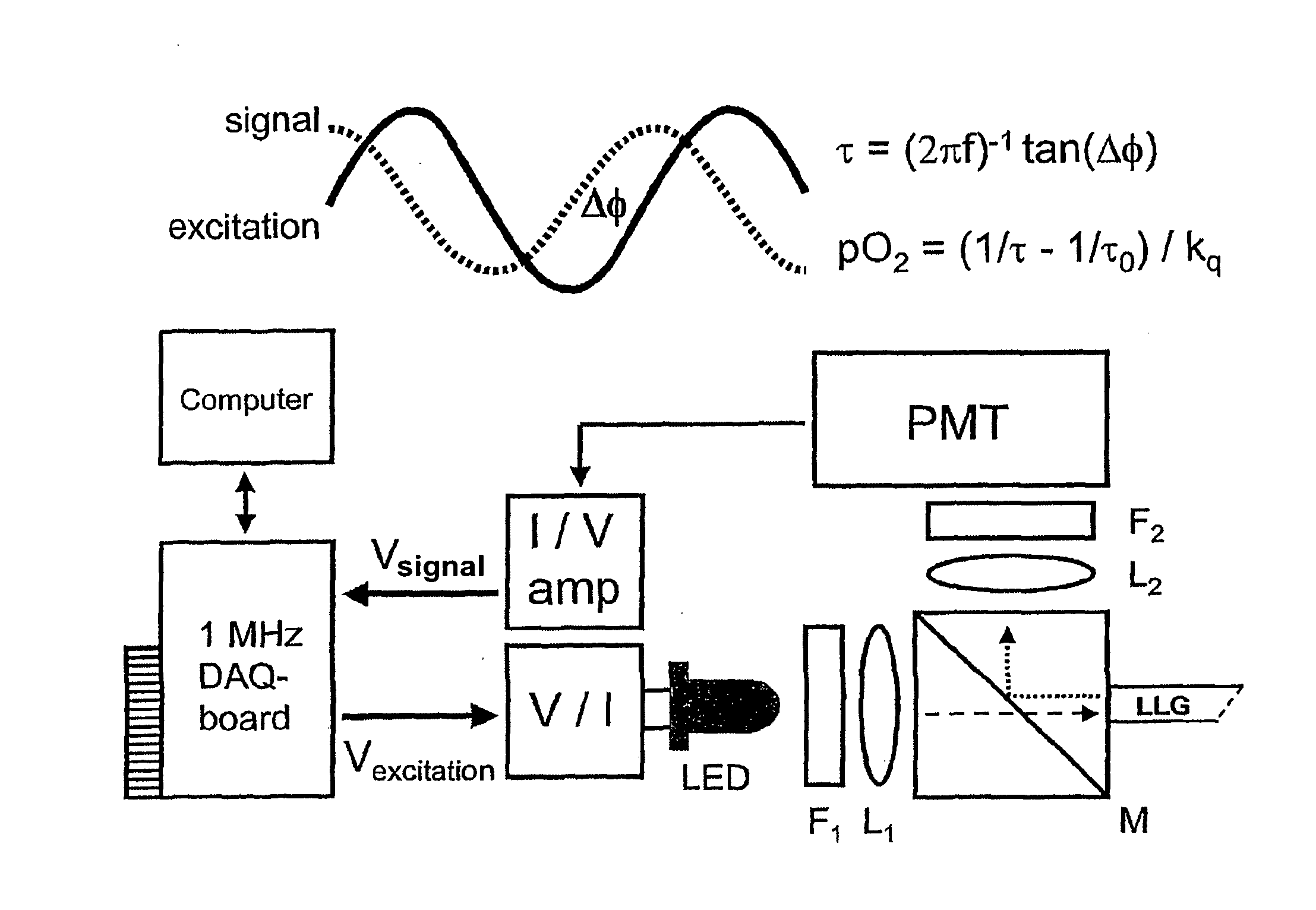
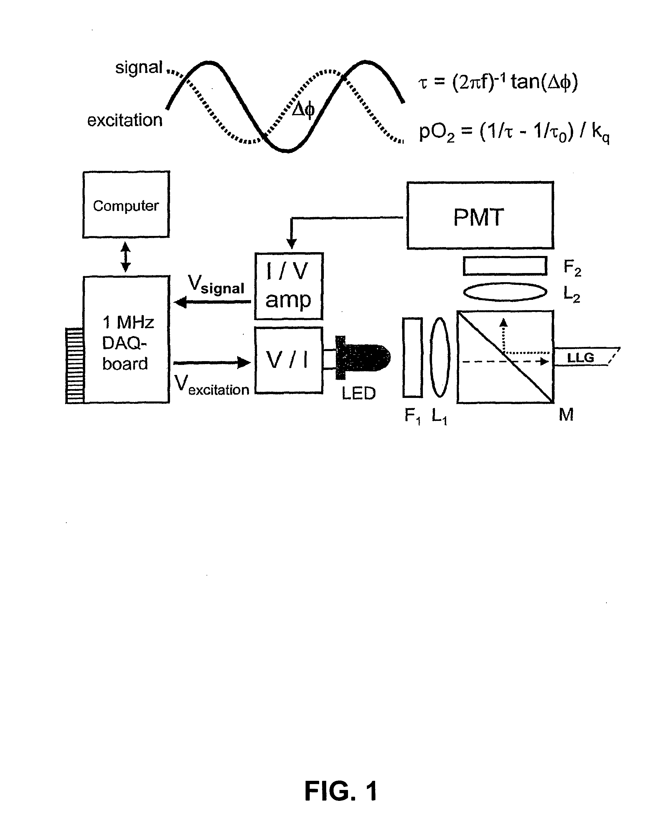
[0062]Preferably, equipment for optical spectroscopy comprises an illumination light source, an optical system (for instance, comprising filters, mirrors and lenses) and a detection unit. The detector, for instance, comprises a sensitive CCD camera, photomultiplier tube and / or spectrophotometer. Several descriptions of optical systems are described in the literature (Carlsen et al., 2002; Baxter et al., 1997; Green et al., 1988). An example of a frequency domain phosphorescence lifetime measurement device is described in Shonat et al., 1997, incorporated herein by reference. Non-limiting examples of a device of the invention are outlined in the Examples. In a preferred embodiment, a combination of a prism and a bandpass filter is used, at least partly preventing a high amount of excitation light to reach the filters in order to avoid possible disturbance of a delayed fluorescence signal as a result of fluorescence and / or phosphorescence of the filters themselves. In a further preferred embodiment, a device of the invention comprises a fast shutter in front of a PMT, preferably a pockel cell, in order to prevent distortion of the first 20 to 30 μs of a signal, which would otherwise occur if a PMT is gated by switching the voltages of the second and third dynodes during the laser pulse. Alternatively, a semiconductor device, preferably an avalanche-photodiode, is used, which is cheaper.

Problems solved by technology

The drawback of these conventional techniques is that they are either mechanically disruptive (insertion of oxygen electrodes) or qualitative (spectrophotometry).
These constraints have led to the development of alternative methods.
An important drawback of this technique is, however, that it relies on injection of palladium-porphyrin into the circulation, making this technique unsuitable for clinical settings because of long-term toxicity.
The use is limited to pre-clinical applications.
Moreover, this technique is only suitable for measuring oxygen levels in the microcirculation.
Since the molecules are large and cell-impermeable, this technique cannot be applied for intracellular oxygen measurements without disrupting the intracellular compartment by micro-injection (Hogan, 1999).
Unfortunately, this phosphorescence cannot be used for quantitative oxygen measurements because of the complex decay kinetics arising from the different tryptophan-containing proteins (Vanderkooi et al., 1987b).
The use of tryptophan phosphorescence for in vivo applications is furthermore limited because of the excitation in the UV region (283 nm), resulting in extremely shallow penetration depths in tissue, besides the well-known photo-toxicity of this high energetic light.
Although both oxygen-dependent quenching of phosphorescence and hemoglobin saturation measurements give information about the microvascular oxygenation status, they do not provide a direct measurement of the adequacy of tissue oxygenation.
Since for the conversion of mitochondrial NADH to NAD+ the availability of molecular oxygen is mandatory, lack of oxygen results in accumulation of NADH and subsequent increase in fluorescence intensity.
However, even with proper calibration, exact quantification of the NADH levels remains impossible (Masters, 1993).
Hence, although oxygen is one of the most important biological molecules, concentration measurements in vivo remain cumbersome.
The same kinds of problems arise when the concentration of another substance is measured.

Method used


Image

  • Device and method for determining the concentration of a substance
  • Device and method for determining the concentration of a substance
  • Device and method for determining the concentration of a substance

Examples


example 1

[0080]The spectra of prompt and delayed luminescence were recorded using a LS50B luminescence spectrometer (Perkin-Elmer, Wellesley, Mass., USA). Prompt fluorescence was measured using the fluorescence mode with excitation source correction. Delayed luminescence was recorded in the phosphorescence mode, using varying delay times with respect to the excitation flash and a gate width of 100 μs. The measurements were made at room temperature. Excitation and emission wavelengths and slit widths will be specified in the results section. Spectra were recorded with either air-saturated samples or samples containing zero oxygen. Adding a sufficient amount of ascorbic acid (20 μl of 200 mM solution) to the already ascorbate oxidase-containing samples (1 unit ascorbate oxidase per ml, 3 ml total sample volume) created the zero-oxygen conditions. This method of reducing oxygen levels is explained in more detail below. The amounts of ascorbate oxidase and ascorbic acid used did not interfere wi...

example 2

[0094]In this example, we demonstrate the feasibility of the proposed method for measuring intramitochondrial oxygen levels in living cells.

[0095]Equipment

[0096]In this Example, a method of the invention is, for instance, performed in the time domain using pulsed excitation from an experimental high-power tunable laser. The laser of this Example consists of a doubled flash-lamp pumped Nd-YAG laser pumping an optical parametric oscillator (OPO). This results in a tunable laser providing 10 mJ pulses of 6 ns duration. The laser is coupled to a quart cuvette containing the studied samples using a glass fiber. Perpendicular to the laser beam is a detector consisting of coupling lens, monochromator and photomultiplier tube (PMT). The photomultiplier (Hamamatsu R928) is working in photon-counting mode and is gated during laser excitation by reversing the polarities of the second and third dynode. The current from the PMT is voltage converted using a fast-switching integrator (integration ...

example 3

[0101]An example of an experimental two-photon set-up is given in FIG. 13. In this example, excitation is achieved using a Q-switched laser operating at 1064 nm (Laser 1-2-3, Schartz Electro-Optics Inc., Orlando, Fla., USA). The laser provides pulses of approximately 10 ns duration and an energy ranging from 10 mJ per pulse for in vitro experiments to 100 mJ per pulse in in vivo experiments. The bundle diameter of the laser beam is slightly expanded to a final diameter of 5 mm by a beam expander, before being directed to the focusing lens by an optical mirror with an enhanced silver reflection surface (Opto Sigma, Santa Anna, Calif., USA). The focusing lens is a single plan-convex lens with a focal length of 2.0 cm. Based on Gaussian beam optics, the bundle diameter of 5 mm combined with a lens with a focal length of 2.0 cm results in a focal spot size of 8 μM and a focus length of 94 μm (in air). Assuming a refractive index in tissue of 1.4, the measurement volume is approximately ...


PUM

PropertyMeasurementUnit
Timeaaaaaaaaaa
Timeaaaaaaaaaa
Lifetime stabilityaaaaaaaaaa

Abstract

Described are methods for determining a concentration of a substance in a compartment comprising exciting an endogenous compound, or a functional part, derivative, analogue or precursor thereof, measuring the lifetime of the luminescence and / or transient absorption exhibited by the compound, functional part, derivative, analogue and / or precursor, and correlating the lifetime with the concentration of the substance.

Description

CROSS-REFERENCE TO RELATED APPLICATIONS[0001]This is a continuation of co-pending Ser. No. 11 / 988,535 filed on Jan. 7, 2008 and accepted on Nov. 13, 2008, which is a national phase entry under 35 U.S.C. §371 of International Patent Application PCT / NL2006 / 000341, filed Jul. 6, 2006, published in English as International Patent Publication WO 2007 / 004873 A1 on Jan. 11, 2007, which claims the benefit under 35 U.S.C. §119 of European Patent Application 05076565.0 filed Jul. 6, 2005, the contents of the entirety of each of which are incorporated herein by this reference.TECHNICAL FIELD[0002]The invention relates to the field of medicine. More specifically, the invention relates to monitoring the concentration of a substance.BACKGROUND[0003]Health control, diagnosis of disease and / or monitoring of treatment of disease often involves measurement of various parameters. One parameter is the concentration of a certain substance, such as oxygen, within at least part of an organism. Local tissu...

Claims


Application Information

Patent Timeline
IPC IPC(8): C12Q1/02C12M1/34
CPCA61K49/0021A61K49/0036Y10T436/207497G01N21/6408A61K49/0056
Inventor MIK, EGBERT G.SINAASAPPEL, MICHIEL
Owner ACADEMISCH MEDISCH CENT BIJ DE UNIV VAN
Learn More