
Novel molecular assembly, molecular probe for molecular imaging and molecular probe for drug delivery system using the same, and molecular imaging system and drug delivery system

a molecular imaging and molecular probe technology, applied in the field of new molecular assemblies, can solve the problems of limited life of the indicator, high diagnostic equipment cost, and inability to accurately diagnose optically to a living body, and achieves simple and safe method, high safety for a living body, and easy control

Inactive Publication Date: 2011-05-05
SHIMADZU CORP
View PDF4 Cites 35 Cited by
  • Summary
  • Abstract
  • Description
  • Claims
  • Application Information

AI Technical Summary

Benefits of technology

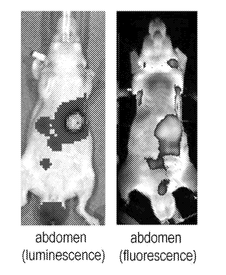
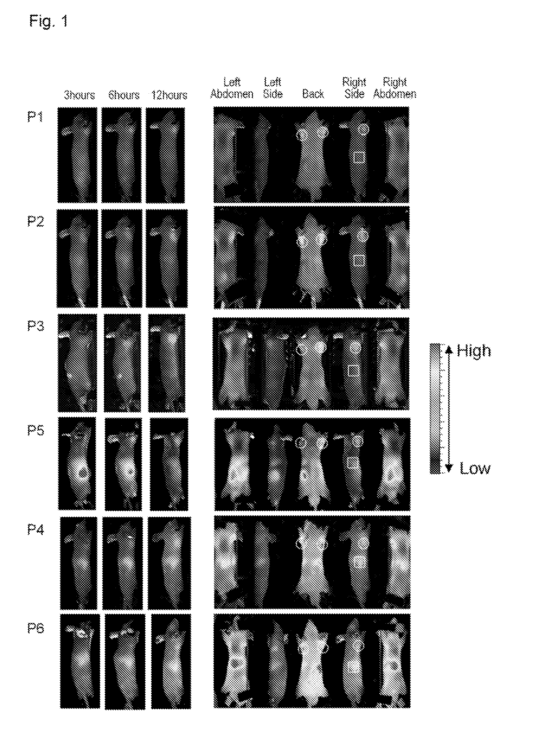
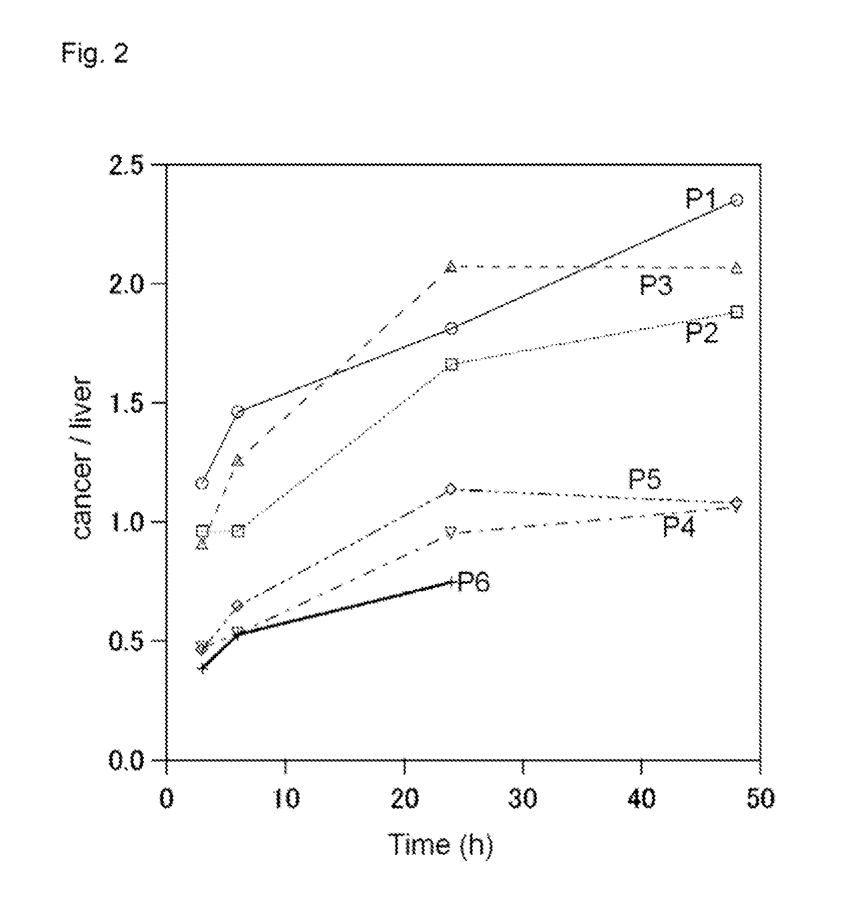
[0121]According to the present invention, it is possible to provide a molecular assembly which is less likely to accumulate in tissue other than cancer tissue, is highly safe for a living body, and can be prepared by a simple and safe method and whose particle size can be easily controlled. Therefore, according to the present invention, it is possible to provide a molecular imaging system and a molecular probe useful for the system and to provide a drug delivery system and a molecular probe useful for the system.
[0122]More specifically, the molecular assembly according to the present invention uses a polylactic acid-type labeled polymer to reduce accumulation in tissue other than cancer tissue. This makes it possible to allow the molecular assembly to specifically accumulate in cancer tissue. Further, the molecular assembly according to the present invention is highly safe for a living body, can be easily applied to a molecular probe, can be prepared by a safe method, and has excellent biocompatibility and biodegradability. Further, the shape and size of the molecular assembly itself can be controlled by controlling the amount of lactic acid as a monomer in the process of its preparation.
[0123]The molecular assembly according to the present invention selectively accumulates in a cancer and enables imaging in a short period of time, and is therefore particularly useful for imaging of liver cancers and cancers of organs near the liver. Further, the molecular assembly is also useful as a molecular probe for PET or DDS targeting liver cancers or cancers of organs near the liver and the like.
[0124]Further, when performing diagnostic imaging using chemiluminescence, the molecular assembly makes it possible to safely perform cancer diagnostic imaging without the need for genetic modification, and is therefore applicable to diagnostic imaging of tumors in human body.
[0125]Further, according to the present invention, it is possible to provide a method for preparing molecular assembly capable of arbitrarily controlling the particle size of molecular assembly. This makes it possible to allow molecular assembly having a signal group or a drug to effectively accumulate in cancer tissue by utilizing EPR effect.

Problems solved by technology

In a case where a radioactive indicator is used, the lifetime of the indicator is limited due to its half-life.
In addition, a diagnostic apparatus is very expensive.
In addition, such optical diagnosis is noninvasive to a living body.
It is known that these neovessels are fragile and therefore molecules leak from the vessels even when the molecules are somewhat large.
Further, the excretory system of a substance in cancer tissue is underdeveloped, and therefore molecules leaking from the vessels are retained in cancer tissue for a certain period of time.

Method used


Image

  • Novel molecular assembly, molecular probe for molecular imaging and molecular probe for drug delivery system using the same, and molecular imaging system and drug delivery system
  • Novel molecular assembly, molecular probe for molecular imaging and molecular probe for drug delivery system using the same, and molecular imaging system and drug delivery system
  • Novel molecular assembly, molecular probe for molecular imaging and molecular probe for drug delivery system using the same, and molecular imaging system and drug delivery system

Examples


experimental example 1

Synthesis of Aminated Poly-L-Lactic Acid (a-PLA)

[0377]In this experimental example, aminated poly-L-lactic acid (a-PLA) was synthesized using L-lactide (compound 1) and N-carbobenzoxy-1,2-diaminoethane hydrochloride (compound 2) (Scheme 1).

[0378]To N-carbobenzoxy-1,2-diaminoethane hydrochloride (compound 2) (310 mg, 1.60 mmol) served as a polymerization initiator, a dispersion liquid obtained by dispersing tin octanoate (6.91 mg) in toluene (1.0 mL) was added. The toluene was distilled away under reduced pressure, and then L-lactide (compound 1) (3.45 g, 24 mmol) was added to perform polymerization reaction at 120° C. under Ar gas atmosphere. After 12 hours, the reaction container was air-cooled to room temperature to obtain a yellowish-white solid. The yellowish-white solid was dissolved in a small amount of chloroform (about 10 mL). The chloroform was dropped into cold methanol (100 mL) to obtain a white precipitate. The white precipitate was collected by centrifugation and dried ...

experimental example 2

Synthesis of Amphiphilic Block Polymer A1 (Polysarcosine-Poly-L-Lactic Acid; PSL1)

[0380]In this experimental example, an amphiphilic substance, polysarcosine-poly-L-lactic acid (PSL1) was synthesized from sarcosine-NCA (Sar-NCA) and aminated poly-L-lactic acid (a-PLA) (Scheme 2)

[0381]Dimethylformamide (DMF) (140 mL) was added to a-PLA (383 mg, 0.17 mmol) and sarcosine-NCA (Sar-NCA) (3.21 g, 27.9 mmol) under Ar gas atmosphere, and the mixture was stirred at room temperature for 12 hours to obtain a reaction solution. Then, the reaction solution was cooled to 0° C., and then glycolic acid (72 mg, 0.95 mmol), O-(benzotriazol-1-yl)-N,N,N′,N′-tetramethyluronium hexafluorophosphate (HATU) (357 mg, 0.94 mmol), and N,N-diisopropylethylamine (DIEA) (245 μL, 1.4 mmol) were added thereto to perform reaction at room temperature for 18 hours.

[0382]The DMF was distilled away under reduced pressure using a rotary evaporator, and then purification was performed using an LH2O column. Fractions showi...

experimental example 3

Synthesis of Sarcosine-Poly(Leucine-Aminoisobutyric Acid) (SLA)

[0383]In this experimental example, an amphiphilic substance, sarcosine-poly(leucine-aminoisobutyric acid) (SLA) was synthesized from sarcosine-NCA (Sar-NCA) and poly(leucine-aminoisobutyric acid) (LAI) (Scheme 3).

[0384]Boc-(Leu-Aib)8-OMe (600 mg, 0.349 mmol) was added to a mixed solution of 6.0 mL of trifluoroacetic acid (TFA) and 0.6 mL of anisole to remove a Boc group to obtain a trifluoroacetate derivative. The trifluoroacetate derivative was washed with isopropyl ether and dried under vacuum for 2 hours to obtain a dry product. The dry product was dissolved in chloroform and neutralized with a 4 wt % aqueous sodium hydrogen carbonate solution to remove a TFA group. The chloroform solution was concentrated to obtain 420 mg (0.259 mmol) of poly(leucine-aminoisobutyric acid) (LAI) (H-(Leu-Aib)8-OMe; LAI).

[0385]The thus obtained LAI was dissolved in 8.0 mL of a 1:1 (V / V) mixed solution of DMF and HCl3, and the mixed sol...


PUM

PropertyMeasurementUnit
particle sizeaaaaaaaaaa
optical purityaaaaaaaaaa
particle sizeaaaaaaaaaa

Abstract

The present invention provides a molecular assembly which is less likely to accumulate in tissue other than cancer tissue, is highly safe for a living body, and can be prepared by a simple and safe method and whose particle size can be easily controlled. The present invention provides a molecular imaging system and a molecular probe useful for the system, and a drug delivery system and a molecular probe useful for the system. The present invention provides a method for preparing molecular assembly, by which the particle size of molecular assembly having a signal group or a drug can be arbitrarily controlled in order to allow the molecular assembly to effectively accumulate in cancer tissue by utilizing EPR effect. A molecular assembly comprising: an amphiphilic block polymer A comprising a hydrophilic block chain and a hydrophobic block chain having 10 or more lactic acid units; a hydrophobic polymer A2 having at least 10 or more lactic acid units; and / or a labeled polymer B comprising at least 10 or more lactic acid units and a labeling group.

Description

TECHNICAL FIELD[0001]The present invention relates to a novel molecular assembly made of a biocompatible amphiphilic substance and a molecular imaging system or a drug delivery system using the same.BACKGROUND ART[0002]As described in JP-A-2005-172522 (Patent Document 1), in recent years, there has been a growing interest in nanotechnology, and new functional materials utilizing the characteristics inherent in nanosized substances have been developed. These new functional materials can be used in various fields such as energy, electronics, and medical and pharmaceutical fields. Among such various fields, nanotechnology has been attracting attention in detection of substances in biological samples and in-vivo imaging. Particularly, in medical and pharmaceutical fields, liposomes, which are nanoparticles composed of phospholipid, and the like are used as carriers for drug delivery system (DDS).[0003]In medical and pharmaceutical fields, as described in JP-A-2005-220045 (Patent Documen...

Claims


Application Information

Patent Timeline
Patent Type & Authority Applications(United States)
IPC IPC(8): A61K51/06A61K49/00A61K49/04
CPCA61K9/5153A61K49/0032A61K49/0034A61K51/1234A61K49/0082A61K51/06A61K49/0054A61P35/00
Inventor HARA, ISAOYAMAHARA, RYOOZEKI, EIICHITAKEUCHI, ERIKIMURA, SHUNSAKUKONDOH, SHINAEMAKINO, AKIRAYAMAMOTO, FUMIHIKO
Owner SHIMADZU CORP
Learn More