Method For The Treatment Of Proliferative Disorders Of The Eye

a proliferative disorder and eye technology, applied in the field of eye proliferative disorders, can solve the problems of retinal detachment and blindness, retinal detachment, and compromising the success of retinal re-attachment surgery, and achieve the effects of inhibiting the potential for cell proliferation, and preventing the formation of cytotoxic substances

Inactive Publication Date: 2011-08-18
ONCOTX
View PDF4 Cites 9 Cited by
  • Summary
  • Abstract
  • Description
  • Claims
  • Application Information

AI Technical Summary

Benefits of technology

[0074]A particular advantage of the present invention is the intraocular delivery of a drug (or set of drugs) that specifically and irreversibly inhibits the potential for cell proliferation without killing nonproliferating cells. The present method has the following major advantages: a) a single brief exposure to the drug will generally be sufficient to inhibit the potential for cell proliferation and treat the proliferative eye disorder; by contrast current therapies require multiple and prolonged dosing regimens; b) broad-spectrum activity; the ability to inhibit essentially all processes that require proliferation including, angiogenesis, fibrosis, gliosis, scar tissue formation, RPE cell proliferation, and the evolution and progression of cancer; c) therapeutic activity regardless of the growth factors that drive cell proliferation in the eye; d) the inhibition of the potential for cell proliferation is independent of the cell cycle; (This is important because only a fraction of malignant cells actively proliferate at a given time); e) absence of cytotoxicity to nonproliferating cells and tissue; (This is in sharp contrast to the prior art, which involves the use of cytotoxic agents) and f) a large therapeutic index, the ratio of LD50 to TGI is large.
[0075]Conventional anti-proliferative drugs such as adriamycin, cisplatin, taxol, vincristine, mitomycin, busulfan, Actinomycin-D, and phosphoramide mustards are highly cytotoxic, kill proliferating as well as nonproliferating cells, and cause tissue damage after local administration. In addition such agents damage cells and can impair cell function. Cytostatic agents generally require continuous drug exposure for continuous or inhibition of cell proliferation. For typical antiproliferative agents the concentration required to inhibit cell proliferation is comparable to that which is cytotoxic to cells. (See FIG. 1).
[0076]In a preferred embodiment the drug is bizelesin. In another preferred embodiment the drug is adozelesin. In a preferred embodiment one or more drugs is selected from the following group: adozelesin; bizelesin; U77809, U78779, carzelesin; Brostallicin; tallimustine, or derivatives, prodrugs, analogues, and active metabolites thereof. These drugs covalently bind to the minor groove of DNA in adenine-thymine rich regions and inhibit cell proliferation at sub-nanomolar concentrations. Bizelesin; and its active metabolite U77809 cross-link adjacent DNA strands. One skilled in the art can readily identify using routine and well-known experimental techniques additional drugs that can irreversibly and permanently arrest the potential for cell replication without causing extensive cell death or tissue necrosis upon local injection. Such drugs are to be within the scope of the present methods and invention.
[0077]The following references relate to this matter: Li, L. H. et al., Invest New Drugs, 9(2): 137-48 (May 1991); Liu, J. S. et al., Mol Cancer Ther, 2(1): 41-7 (January 2003); Liu, J. S. et al., Mutat Res, 532(1-2): 215-26 (Nov. 27, 2003); Liu, J. S. et al., J Biol Chem, 275(2): 1391-7 (Jan. 14, 2000); Cao, P. R. et al., Mol Cancer Ther, 2(7): 651-9 (July 2003); Hess, M. T. et al., Nucleic Acids Res, 24(5): 824-8 (Mar. 1, 1996); Cristofanilli, M. et al., Anticancer Drugs, 9(9): 779-82 (October 1998); Burris, H. A. et al., Anticancer Drugs, 8(6): 588-96 (July 1997); Liu, J. S. et al., J Biol Chem, 275(2): 1391-...

Problems solved by technology

PDR is characterized by new blood vessel growth anterior to the retina, bleeding, proliferation of inflammatory cells and fibrosis, which can ultimately lead to retinal detachment and blindness.
The pre-retinal membrane can cause retinal detachment and also compromise the success of retinal re-attachment surgery.
However, these drugs do not inhibit the proliferation of fibroblasts, glial cells, and RPE cells and do not effectively prevent fibrosis and scar tissue formation.
While these drugs are beneficial, vision loss can still occur from proliferative processes.
POCO can result in vision loss requiring operative treatment.
Nonetheless PCO remains a significant clinical problem.
Scarring and fibrosis due to...

Method used


Image

  • Method For The Treatment Of Proliferative Disorders Of The Eye
  • Method For The Treatment Of Proliferative Disorders Of The Eye
  • Method For The Treatment Of Proliferative Disorders Of The Eye

Examples


example 1

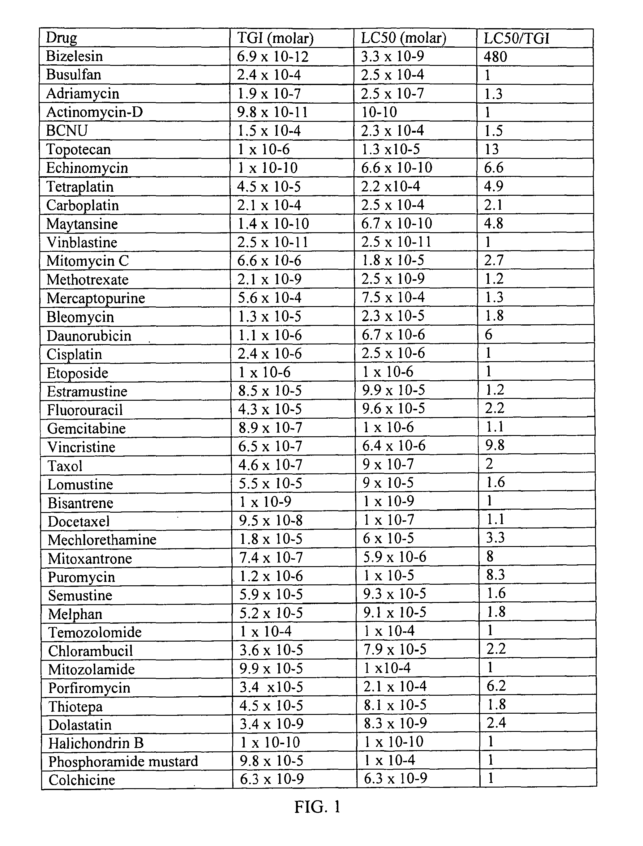
[0107]Intravitreal bizelesin was evaluated in the mouse model of ischemic proliferative retinopathy. Details of the model are provided in Xie, B. et al., J Cell Physiol, 218(1): 192-8 (January 2009). The animal work was done in accordance with the Association for Research in Vision and Ophthalmology Statement for the Use of Animals in Ophthalmic and Vision Research and the guidelines of the Animal Care and Use Committee.

[0108]Bizelesin was dissolved in DMSO PharmaSolvent (Gaylord Chemical, Inc.) at 20 microgram / ml and filtered sterilized with a 0.2 micron Milliex-LG Millipore filter. HPLC analysis of the DMSO drug solution post filtration revealed that the bizelesin concentration was 11 microgram / ml. The bizelesin solution was stored at −65 C or below. Immediately prior to administration the drug solution was thawed and diluted with sterile phosphate buffered saline (PBS).

[0109]In brief, for the ischemic proliferative retinopathy model, liters of C57BL / 6 mice were exposed to 75% oxy...

example 2

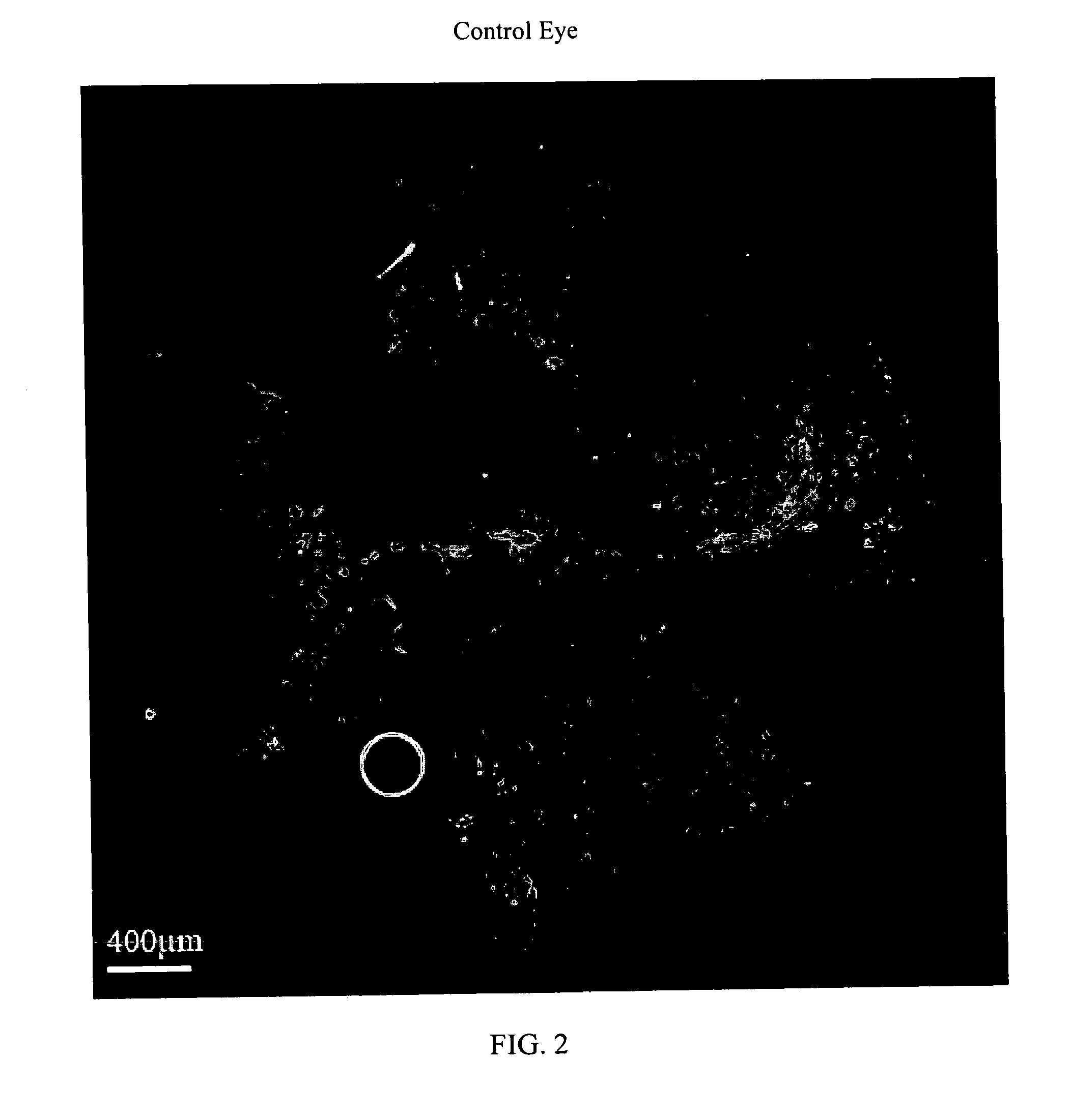
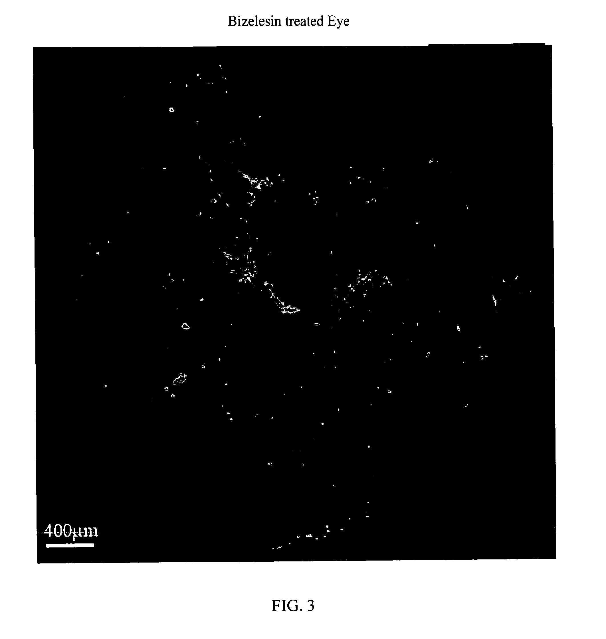
[0111]Intravitreal bizelesin was evaluated in the laser induced mouse model of choroidal neovascularization. Details of the model are provided in Xie, B. et al., “Blockade of Sphingosine-1-phosphate Reduces Macrophage Influx and Retinal and Choroidal Neovascularization,”J Cell Physiol, 218(1): 192-8 (January 2009 Bizelesin was formulated in PBS as described in Example 1. In brief, 5-6 week old C57BL / 6 mice on day 0 were anesthetized and choroidal neovascularization was induced by laser photocoagulation-induced rupture of Bruch's membrane. The mice were then given an intravitreal injection of 1 microliter of control diluent in one eye and in the contralateral fellow eye 1 microliter of solution containing 6 ng to 0.006 ng of Bizelesin. Ten mice were employed per dose level. On day 14 the mice were perfused with fluorescein-labeled dextran, the retinas were isolated, flat mounts prepared, and the area of neovascularization was determined with fluorescent microscopy and computerized im...

example 3

[0112]This is an example of a single dose kit for intravitreal injection of bizelesin for the treatment of proliferative eye disorders including but not limited to diabetic proliferative retinopathy, age related macular degeneration associated proliferative retinopathy, proliferative vitreoretinopathy, sub-retinal fibrosis, polypoidal choroidal vasculopathy, proliferative vitreoretinopathy, epimacular membranes choroidal neovascularization, neovascularization of the retina, retinopathy of prematurity, neovascularization related to ocular histoplasmosis, retinal hemagioblastoma in von Hippel-Landau syndrome, uveal melanoma, ocular nevi, retinoblastoma, ocular lymphoma, and metastatic cancers to the eye.

[0113]The kit consists of:

[0114]A sterile solution of bizelesin (0.05 ng to 100 ng) dissolved in 10 to 50 microliters of pharmaceutical grade, anhydrous, dimethylsulfoxide in a labeled, amber glass vial, with a Teflon coated rubber septum, filled with dry nitrogen. The vial is stored a...


PUM

PropertyMeasurementUnit
Massaaaaaaaaaa
Massaaaaaaaaaa
Timeaaaaaaaaaa

Abstract

The present invention relates to method for the treatment or prevention and of proliferative eye diseases including but not limited to: age related macular degeneration associated proliferative retinopathy, proliferative diabetic retinopathy, proliferative vitreoretinopathy, posterior capsular opacification, scaring and fibrosis after glaucoma filtration surgery, uveal melanoma, and retinoblastoma. The method comprises contacting cells in the eye by means of intra-ocular injection or infusion, with a drug that irreversibly inhibits cellular proliferation without causing extensive tissue necrosis or cytotoxicity. In a preferred embodiment the drug is bizelesin.

Description

RELATED APPLICATION[0001]This application claims the benefit of U.S. Provisional Application No. 61 / 196,894, filed on Oct. 22, 2008. The entire teachings of the above application are incorporated herein by reference.BACKGROUND OF THE INVENTION[0002]Proliferative diseases of the eye are the leading cause of blindness and vision loss in the United States. Proliferation of blood vessels and scar tissue within the eye plays a major role in a number of diseases that result in vision loss, including age-related macular degeneration (ARMD), proliferative diabetic retinopathy (PDR), and proliferative vitreoretinopathy (PVR). Approximately 1.8 million Americans have severe ARMD and 4 million people in the U.S. have diabetic retinopathy. These numbers are expected to nearly double by the year 2020. PVR develops in approximately 8% of patients who have surgery for retinal detachment.[0003]ARMD proliferative retinopathy is characterized by choroidal neovascularization, vascular leakage, fibrosi...

Claims


Application Information

Patent Timeline
IPC IPC(8): A61K31/409A61K31/407A61K9/00A61P27/02
CPCA61K9/0048A61K47/40A61K31/404A61K9/0051A61P27/02
Inventor GLAZIER, ARNOLD
Owner ONCOTX
Why Eureka
Try Eureka