Methods for detection or measurement of viruses

a technology for applied in the field of methods of detecting or measuring viruses, can solve the problems of not always highly sensitive or specific methods, require long procedures, and still present risk of secondary infection, and achieve the effect of simple and sensitive detection and quantitation of virus antigens

Inactive Publication Date: 2011-10-27
AOYAGI KATSUMI +4
View PDF0 Cites 20 Cited by
  • Summary
  • Abstract
  • Description
  • Claims
  • Application Information

AI Technical Summary

Benefits of technology

[0014]It is an object of the present invention to provide a method for detecting various virus antigens, including a method for detecting HCV antigen, that is suitable for treating a large number of samples as in screening in the blood industry and health checkups. In other words, the object of the present invention is to provide the detection system for various virus antigens including a method of detecting HCV antigen that has a sensitivity and specificity equivalent to those of the PCR method, that permit simple pretreatment, or that can be easily automated without pretreatment. Preferred embodiments of the present invention will now be explained hereinbelow with a main reference to HCV.
[0028]According to the second embodiment of the present invention, there is provided a method to detect or determine a virus antigen during the window period in which antibodies against said virus have not yet been generated. In this method, the disruption of the virus particle to expose the virus antigen is sufficient and there is no need to disrupt antibodies against the virus antigen in the blood.

Problems solved by technology

However, these methods are not always highly sensitive or specific though the sensitivity and the specificity may vary with the type of virus to be detected.
Even when they are sensitive and specific enough, they are often expensive and require lengthy procedures as in the culture and isolation of a virus.
However, as for other common viral infections such as by the human immunodeficiency virus (HIV), there is a period of time until the appearance of antibodies after infection, or the so-called window period in which a virus is unidentifiable by existing testing methods.
This means that the risk of secondary infection is still present, due to blood-borne components that cannot be identified by antibody testing methods, in areas where blood-selling is legal or in some regions of Japan.
The antibody testing method also has a drawback in that it cannot distinguish a person who has recuperated from an infection and a person who is in the active stage of infection because of its principle of testing.
However, since the antibody titer starts to decline only after the reduction of antigen stimulation or several months after the elimination of antigen, it is impossible to determine whether IFN administration resulted in the elimination of HCV, at a desired timing and accuracy, by the antibody testing alone.
It was difficult to establish a method of directly detecting the virus particle (virus antigen) of HCV because blood levels of the virus are very low as compared to other viruses such as hepatitis B virus (HBV) and because the virus cannot be propagated in vitro or using an animal etc. as a host.
But, the method of detecting viral genomes have several problems when compared to the method of detecting virus antigens.
First, it has been pointed out that since the substance to be detected is RNA that is not very stable during storage, the procedure of freezing and thawing of serum may cause a reduction in the measured value.
Although the testing methods that involve the use of a PCR method are the most sensitive for detecting gene fragments, they have problems in that : reverse transcription from a genomic RNA to a template DNA is often accompanied by losses, which therefor requires great skills to obtain an accurate quantitative value, and: since amplification is an important principle in the methods, a high incidence of false-positives may occur in case of contamination, and thus the processing of a large volume of samples at one time is impossible.
Furthermore, even those methods which are postulated to be a simple procedure take 2 hours or more for pretreatment of samples and are complicated since repeated procedures of centrifugation and the like are required.
In addition, such complicated procedures lead to increased chances of contamination and thereby increased chances of obtaining false-positive results.
On the other hand, the branched-DNA probe method is low in detection sensitivity and besides takes about 20 hours before obtaining test results (Igaku to Yakugaku [Medicine and Pharmacology] 31: 961-970, 1994), and hence the method leaves much to be desired in terms of sensitivity and processing time.
However, there are still several major problems that need be solved as in the methods of detecting the viral genome.
One such problem is that the sensitivity, compared to the PCR method, is so low that it cannot be used as a final test method of serum screening.
Furthermore, the complicated procedure of treating samples for measurement, and the long time it takes, pose problems when it is used in screening.
In addition, the process of dispersing, with urea, the precipitate having an increased viscosity due to the PEG treatment requires great skill.
Furthermore, such processes as centrifugation, supernatant removal, etc. are not amenable to automation and render the simultaneous treatment of a large number of samples very difficult.
Thus, from a viewpoint of ease of handling as well, the method is not suited for applications that require the treatment of a large volume of samples as in screening tests.
However, because the disclosed method of detecting the core antigen, as indicated above, is not amenable to automation and is low in sensitivity so that it cannot be a gold standard in applications that require high sensitivity such as in the blood industry, it cannot be applied to tests that handle a large number of samples such as screening, and cannot make the best use of its advantageous features over the PCR method.
Furthermore, clinically useful assay methods must always face the challenges of sensitivity, specificity, reproducibility, ease of handling, and low cost, and sustained efforts are needed to satisfy these challenges as much as possible.
With regard to detection of virus antigens other than HCV, especially for use in screening handling a large number of samples, there are many methods that are not put into practical use because they are low in sensitivity, as compared to the PCR method, or the desired antigen could not be fully exposed.

Method used


Image

  • Methods for detection or measurement of viruses
  • Methods for detection or measurement of viruses
  • Methods for detection or measurement of viruses

Examples


example 1

Expression and Purification of a HCV-Derived Polypeptide

(A) Construction of an Expression Plasmid

[0122]A plasmid corresponding to the core region of HCV was constructed as follows: one microgram each of DNA of plasmids pUC·C11-C21 and pUC·C10-E12 obtained by integrating the C11-C21 clone and the C10-E12 clone (Japanese Unexamined Patent Publication (Kokai) No. 6 (1994)-38765) respectively, into pUC119 was digested in 20 μl of a restriction enzyme reaction solution [50 mM Tris-HCl, pH 7.5, 10 mM MgCl2, 1 mM dithiothreitol, 100 mM NaCl, 15 units of EcoRI and 15 units of ClaI enzyme] and the restriction enzyme reaction solution [10 mM Tris-HCl, pH 7.5, 10 mM MgCl2, 1 mM dithiothreitol, 50 mM NaCl, 15 units of ClaI and 15 units of KpnI enzyme] at 37° C. for one hour each, and then was subjected to 0.8% agarose gel electrophoresis to purify about 380 by of EcoRI-ClaI fragment and about 920 by of ClaI-KpnI fragment.

[0123]To the two DNA fragments and a vector obtained by digesting pUC119 ...

example 2

Method of Constructing a Hybridoma

[0132]The fusion polypeptide (TrpCll) prepared by the method described above was dissolved in 6 M urea, and then diluted in 10 mM phosphate buffer, pH 7.3, containing 0.15 M NaCl to a final concentration of 0.2 to 1.0 mg / ml, and mixed with an equal amount of adjuvant (Titermax) to make a TrpCll suspension. This suspension prepared at 0.1 to 0.5 mg / mi of TrpCll was intraperitoneally given to 4 to 6 week old BALB / c mice. Similar immunization was conducted every two weeks and after about two more weeks 10 μg of TrpCll dissolved in physiological saline was administered through the tail vein.

[0133]Three days after the last booster, the spleen was aseptically isolated from the immunized animal and was cut into pieces using scissors, which were then crumbed into individual cells and washed three times with the RPMI-1640 medium. After washing, a mouse myeloma cell line SP2 / 0Ag14 at the logarithmic growth phase as described above, 2.56×10⁷ of said cells and ...

example 3

Construction of Monoclonal Antibody

[0136]The hybridomas obtained in the method of Example 2 were inoculated to the abdominal cavity of mice treated with pristane etc., and the monoclonal antibodies produced in the ascites fluid was collected. The monoclonal antibodies were purified using the Protein A-bound Sepharose column to separate IgG fractions.

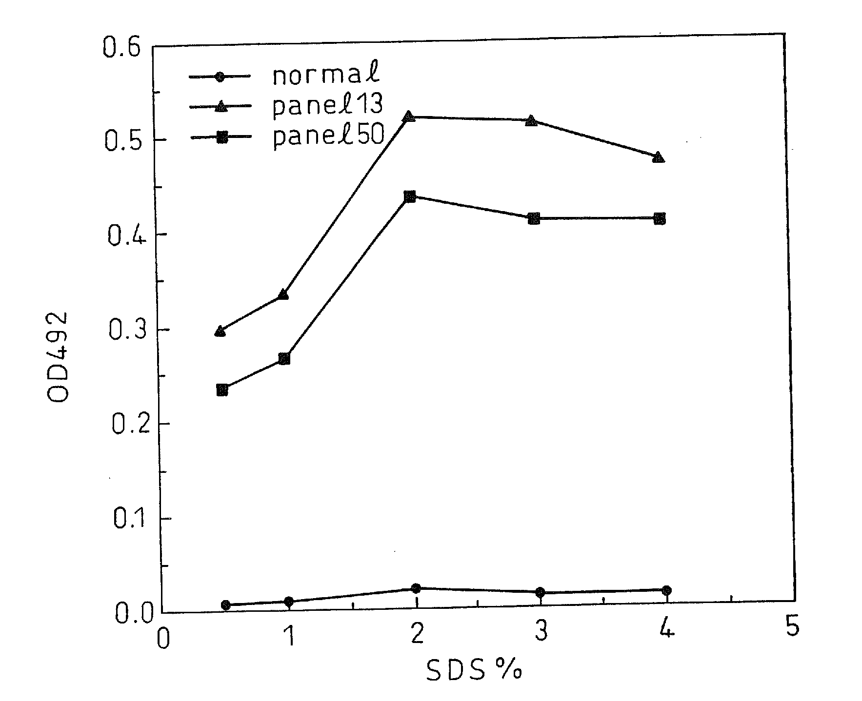
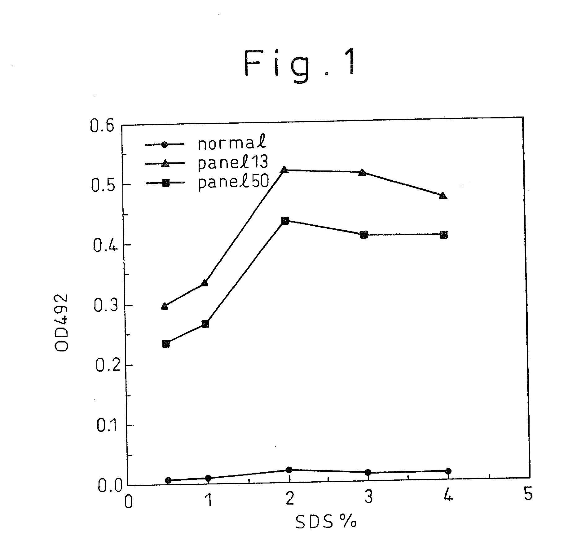
[0137]By an immunoassay using rabbit anti-mouse Ig isotype antibody (manufactured by Zymed), the isotype of each of the monoclonal antibodies C11-14, C11-11, C11-10, C11-7; and C11-3 produced from the above five hybridomas, respectively, was found to be IgG2 for C11-10 and C11-7; and IgGl for CH11-11, C11-14, and C11-3. For the five monoclonal antibodies obtained, epitope analysis was conducted using the synthetic peptides composed of 20 amino acids synthesized according to the sequence derived from the HCV core region. The result indicated, as shown in Table 1, that they were the monoclonal antibodies that specifically recognize part of...


PUM

PropertyMeasurementUnit
temperatureaaaaaaaaaa

Abstract

A method for treating a virus-containing sample, characterized by treatment of a virus-containing sample with a treatment solution containing (1) an anionic surfactant and (2) an amphoteric surfactant, nonionic surfactant or protein denaturant; a virus assay method using said treating method; a method for treating a virus-containing sample, characterized by treatment of a virus-containing sample with a treatment solution containing (1) a chaotropic ion and (2) an acidifying agent; a virus assay method using said treating method; a virus assay method, characterized in that a virus antigen and a virus antibody are measured based on their binding to their probe in the presence of a surfactant with an alkyl group of 10 or more carbon atoms and a secondary, tertiary or quaternary amine, or a nonionic surfactant, or of both of them; and a monoclonal antibody and a hybridoma producing the same for carrying out said method.

Description

TECHNICAL FIELD[0001]The present invention relates to methods of detecting or measuring viruses and reagents therefor.BACKGROUND ART[0002]Currently, various methods of detecting viruses have been used to detect the presence of infectious viruses in blood or blood products, and to identify the presence of viruses in patients with diseases. However, these methods are not always highly sensitive or specific though the sensitivity and the specificity may vary with the type of virus to be detected. Even when they are sensitive and specific enough, they are often expensive and require lengthy procedures as in the culture and isolation of a virus. As a background to the present invention, type C hepatitis (hepatitis C) will be mentioned in detail below.[0003]The causative agent of hepatitis C had long been unknown, but when the gene of the virus was cloned (Science 244: 359-362, 1989) and a diagnostic method by antibody measurement using a recombinant antigen generated based on said gene w...

Claims


Application Information

Patent Timeline
Patent Type & Authority Applications(United States)
IPC IPC(8): C12Q1/70C07K16/08G01N33/569G01N33/576
CPCG01N33/56983G01N2333/18G01N2333/02G01N33/5767G01N33/569
Inventor AOYAGI, KATSUMIOHUE, CHIHARUIIDA, KUMIKOKIMURA, TATSUJIYAGI, SHINTARO
Owner AOYAGI KATSUMI
Why Eureka
Try Eureka