
Kits for quantitative detection of k-ras mutations

a technology of kras and kras fragments, applied in the field of kras mutation quantitative detection kits, can solve the problems of not producing quantitative, requiring a lot of human labor, and the restriction fragment length polymorphism method, and achieve the effect of accurately and quantitatively determining the ratio of kras mutations

Inactive Publication Date: 2012-11-15
BEIJING ACCB BIOTECH
View PDF5 Cites 2 Cited by
  • Summary
  • Abstract
  • Description
  • Claims
  • Application Information

AI Technical Summary

Benefits of technology

[0004]The detecting method of the present invention has the following advantages: easy manipulation, and easy standardization. Other methods, such as allele specific oligonucleotide probe hybridization method, are very much dependent on hybridization conditions, and therefore require strict control of the experimental conditions. The restriction fragment length polymorphism method, on the other hand, needs a lot of human labor, and can not generate quantitative results. The method of the present invention has short experimental cycle, and can be completed with 2 hours. It doesn't need verification the results by sequencing, whereas the direct sequencing and high resolution melting analysis need 4 days to 2 weeks. Sensitivity of the method of the present invention is high, which, after optimizing experimental conditions, can reach 1% for detecting mutations, whereas sensitivity of direct sequencing is 20-50%. Specificity of the method of the present invention is also high. Immunohistochemistry (IHC) method can easily get pseudo-positive and pseudo-negative results, and can not determine the position and types of point mutations. The unique advantage of the present invention is accurate quantification. By using absolute quantification method to analyze data, draw standard curve, and accurately determine the content of wild-type gene and mutant gene in the samples, one can obtain ratio of the mutant gene in the samples, which will be helpful for clinical diagnosis and therapeutic selection. Furthermore, the present invention is safe and non-toxic, other methods such as chemical breaking method of mismatch base need isotope and toxic chemical agents.
[0005]The question that the present invention addresses is to provide an assay kit for quantitatively detecting an K-ras gene mutation, which can quantitatively detect the following mutations: GGT at position 2155 in K-ras Codon 12 (SEQ ID NO:1; SEQ ID NO:2) replaced with GTT, AGT, GAT or TGT; or GGC in Codon 13 (SEQ ID NO:1; SEQ ID NO:3) replaced with GAC.
[0006]To address the above question, the present invention provides quantitative detection kit containing a mixture comprising Taq enzyme, 10× Taq buffer, MgCl2, dNTP mixture, PCR primers which can specifically amplify the sequences at K-ras gene mutation positions, and probes which can specifically identify wild-type sequences and mutant sequences, together with method of the detection, as follows:
[0007](1) Separately design upstream and downstream primers around the mutation positions of Codon 12 and 13 of K-ras gene; and design specific probes according to each mutant site. Said probes can specifically bind wild-type sequences or the mutant sequences to be detected at specific K-ras sites, so as to determine whether the tested mutations occur at said sites.
[0008](2) To accurately and quantitatively determine the ratio of the K-ras mutations, standards were designed in the present invention.
[0009](3) Use fluorescent quantitative PCR to detect the samples and standards.

Problems solved by technology

The restriction fragment length polymorphism method, on the other hand, needs a lot of human labor, and can not generate quantitative results.
Immunohistochemistry (IHC) method can easily get pseudo-positive and pseudo-negative results, and can not determine the position and types of point mutations.

Method used


Image

  • Kits for quantitative detection of k-ras mutations
  • Kits for quantitative detection of k-ras mutations
  • Kits for quantitative detection of k-ras mutations

Examples


example 1

Extracting Genome DNA from Fresh Human Tumor Tissues, Paraffin Embedded Tissues, Peripheral Blood, Pleural Effusion, and Human Cell Lines

[0027]The tumor cell lines we tested included cell lines of: non-small-cell carcinoma (NSCLC; A549, H460, H838 and H1703), breast cancer (MCF-7, BT474 and HuL100), malignant mesothelioma (H513, H2052, H290, MS-1 and H28), colon cancer (SW480), head and neck cancer (U87), cervical carcinoma (Hela), sarcoma (Mes-SA, Saos-2 and A204).

[0028]The fresh human tumor tissues, peripheral blood, paraffin embedded tissues we tested included: NSCLC, mesothelioma, colon cancer, malignant melanoma, renal carcinoma, esophagus cancer, thyroid carcinoma, malignant cancer and ovarian cancer.

[0029]Extraction of Sample DNA

[0030]DNA extracting kit from Qiagen Inc., Promega Inc., or Roche Inc. can be used to extract genomic DNA from the samples. Content and purity of the extracted DNA can be determined by using Nanodrop ND1000 (Gene Inc.) (OD260 / OD280 is about 1.8, OD260...

example 2

Preparation of the Plasmid Standards Containing Mutant and Wild-Type Sequences

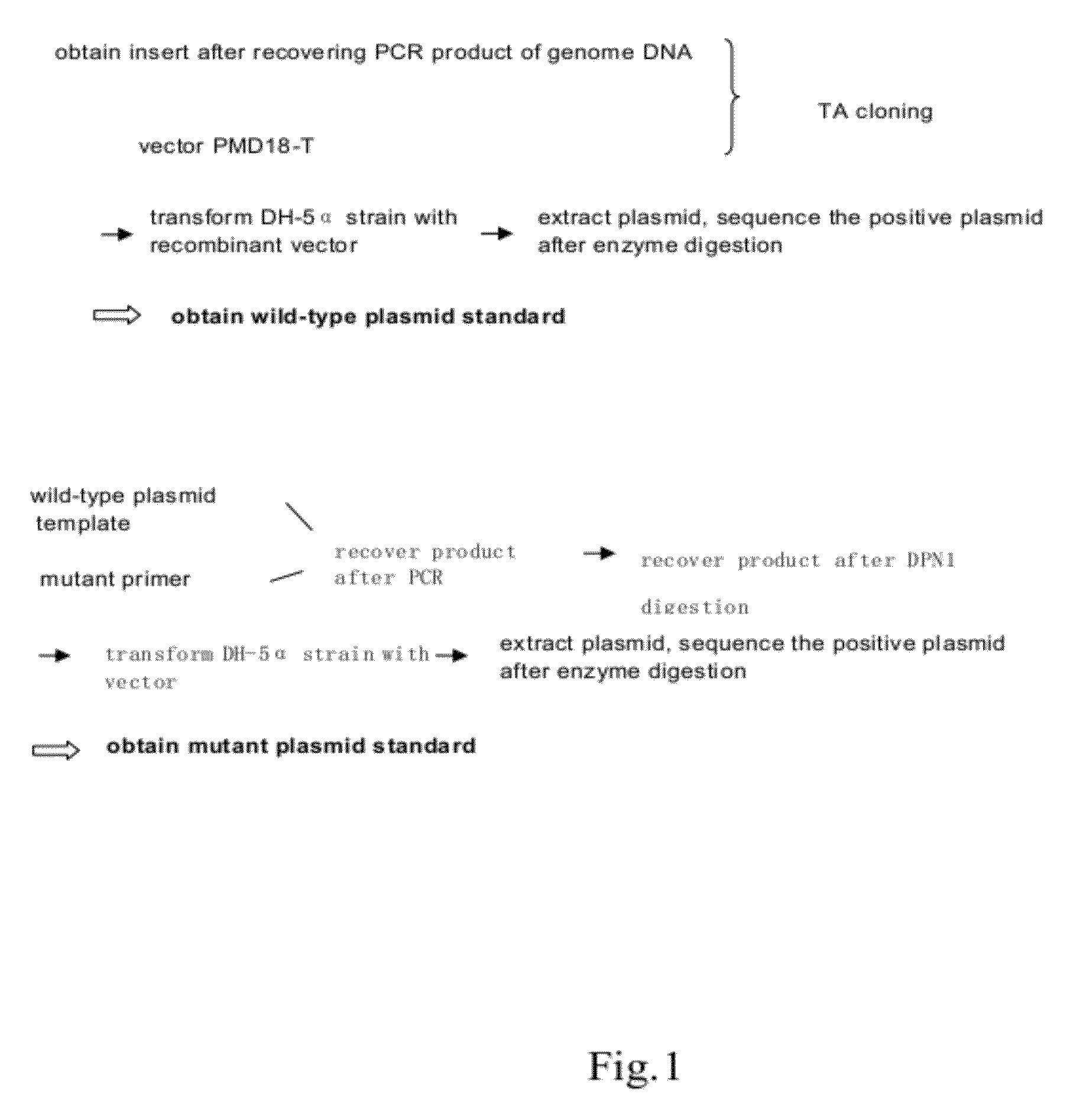
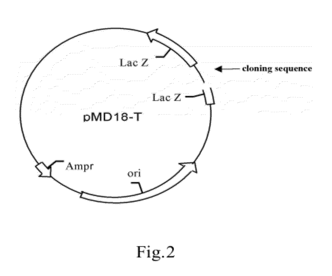
[0085]1. Construction of Wild-Type Plasmids (FIG. 1, FIG. 2)

[0086]1.1 Preparation of the carrier

[0087]TA cloning carrier pMD18-T was purchased from TAKARA Inc.

[0088]1.2 Preparation of the insert

[0089]The insert is prepared using PCR. The template of PCR is the sample genome DNA extracted in Step 1. The reaction system and amplification condition are shown in the following tables (Table 1, Table 2 and Table 3):

TABLE 1PCR reaction system (50 μl)reagentsamount(μl / tube)double-distilled water29.7510x buffer (free of Mg2+)5MgCl2 (25 mM)7.5dNTP (10 mM)1.25upstream primer (25 μM)1.25downstream primer (25 μM)1.25Taq enzyme1DNA template3total volume50

TABLE 2PCR primersnameSequenceK-ras-F1CCTCTATTGTTGGATCATATT(SEQ ID NO: 3)K-ras-F2AATGACTGAATATAAACTTGTGGTAGT(SEQ ID NO: 4)K-ras-R1TGACTGAATATAAACTTGTGGT(SEQ ID NO: 5)K-ras-R2AAATGATTCTGAATTAGCTGTATCGT(SEQ ID NO: 6)

TABLE 3PCR amplification conditionstepcyclestemperature ...

example 3

Detection of K-Ras Mutations from Genome DNA of Human Cell Lines, Human Fresh Tumor Tissues, Peripheral Blood, and Paraffin Embedded Tissues, Using Lung Cancer and Cervical Carcinoma as Examples

[0101]1. The templates for fluorescent quantitative PCR are the genome DNA of lung cancer and cervical carcinoma samples extracted in Example 1, and the standards prepared in Example 2. Double-distilled water is served as negative control. For drawing the standard curves, the standards are diluted as 1 ng / μl, 0.5 ng / μl, 0.25 ng / μl, 0.125 ng / μl. 0.0625 ng / μl, 0.03125 ng / μl.

[0102]2. The reaction system and condition are shown in Table 2, Table 5, Table 6 and Table 7, wherein the fluorescent emission group bound to the probe is selected from FAM, TET, HEX or ROX, the quench group is selected from BHQ or TAMARA.

TABLE 5Reaction system for fluorescent quantitative PCR (20 μl / tube)reagentamount (μl / tube )double-distilled water9.910 × buffer (free of Mg2+)2MgCl2 (25 mM)3dNTP (10 mM)0.5upstream primer...


PUM

PropertyMeasurementUnit
fluorescent quantitative PCRaaaaaaaaaa
fluorescenceaaaaaaaaaa
fluorescence emittingaaaaaaaaaa

Abstract

The present invention relates to an assay kit for quantitatively detecting k-ras gene mutations. Particularly, the present invention relates to detection method and a detection kit for K-ras gene mutations, which relates to the therapeutic efficacy of targeted molecular anti-cancer drugs. More particularly, the present invention relates to a fluorescent quantitative PCR method and kit for detecting mutations at hotspots of K-ras gene, together with the use thereof. The present invention detects the mutations at specific sites of K-ras gene, and can predict the therapeutic efficacy of anti-EGFR tyrosine kinase inhibitors. Therefore, it can provide a guidance to individualized treatments for cancer patients.

Description

CROSS REFERENCE TO RELATED APPLICATIONS[0001]This application claims the priority of Chinese Patent Application No. 200910215835.8, filed on Dec. 30, 2009, the disclosure of which is incorporated herein by reference.BACKGROUND OF THE INVENTION[0002]K-ras belongs to ras oncogene family, it is also referred as p21 gene because it encodes 21 kD ras protein. K-ras transduces an important signal cascade. The encoded product of K-ras gene locates in the downstream of K-ras signaling cascade, functioning as an important “switch” during tumor cell growth, proliferation and angiogenesis. When mutation occurs in K-ras gene, it keeps K-ras in a constitutively active state, leading to deregulation of cell growth and inhibition of apoptosis. The mutation rate of K-ras in different tumor tissues are various, wherein pancreas cancer is 82%, colon cancer is 43%, lung cancer is 30%, hypothyroid cancer is 29%, bladder, liver, kidney and cervical cancer are 10% or lower (Bos J. L. et al, Cancer Res, 1...

Claims


Application Information

Patent Timeline
Patent Type & Authority Applications(United States)
IPC IPC(8): G01N21/64C12N15/63
CPCC12Q1/6886C12Q2600/156C12Q2600/106
Inventor XU, JUNPUCHEN, ZHAO
Owner BEIJING ACCB BIOTECH
Learn More