

Method and Apparatus for Analyzing Biomolecules Using Raman Spectroscopy
a biomolecule and raman spectroscopy technology, applied in the field of raman spectroscopy analysis methods and apparatuses, can solve the problems of inability to search for the target molecule based on a predicted mass shift, inability to specify the binding site, and high time-consuming, etc., to achieve high accuracy and improve measurement sensitivity, the effect of shortening the processing tim
- Summary
- Abstract
- Description
- Claims
- Application Information
AI Technical Summary
Benefits of technology
Problems solved by technology
Method used
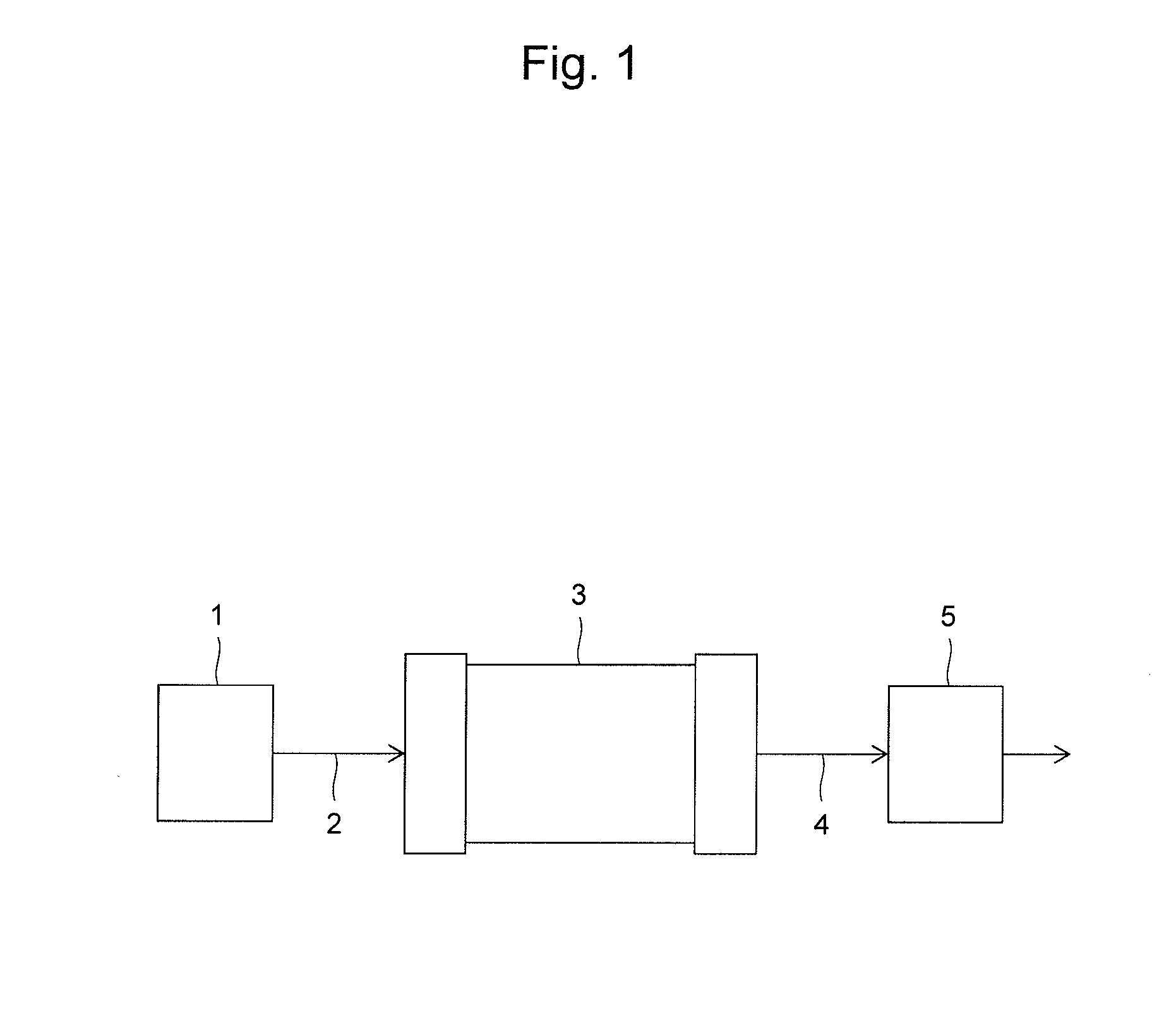
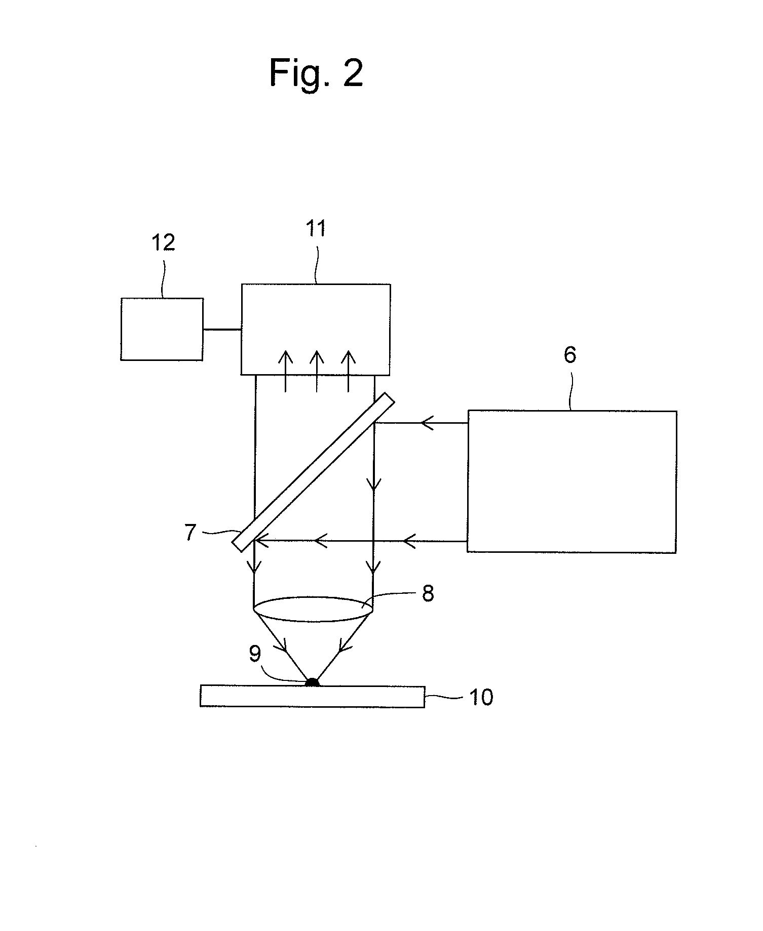
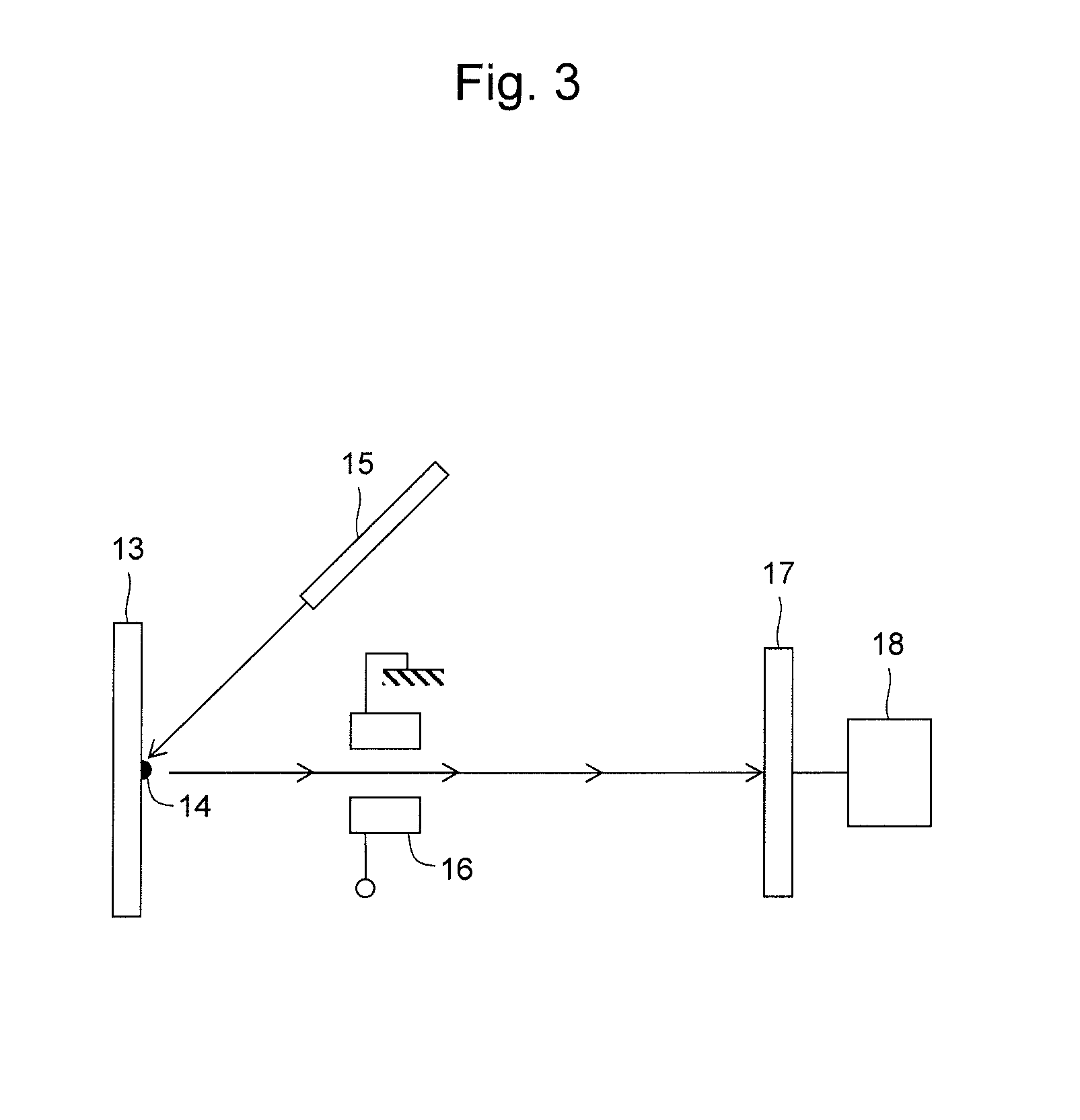
Image

Examples
example 1
Raman Spectroscopy of Low-Molecular-Weight Compounds Having Raman Labels
[0154]Low-molecular-weight peptides having the amino acid sequence of EQWPQCPTXK (SEQ ID NO: 4), and specifically a peptide in which X is isoleucine and a peptide in which X is propargyl glycine, were synthesized. Hereinafter, the former is referred to as peptide 1, and the latter is referred to as alkyne peptide 1 in this example. Peptide 1 was synthesized by the solid phase synthesis (Fmoc) method. Similarly, alkyne peptide 1 was also synthesized by the solid phase synthesis method (all peptides were synthesized at the RIKEN Brain Science Institute). A commercially available product was used for propargyl glycine. These structures are shown in the upper section of FIG. 6.
[0155]Alkyne peptide 1 was fractionated by liquid chromatography, and then subjected to Raman spectroscopy. The results are shown in FIG. 8-1 and FIG. 8-2. Liquid chromatography and Raman spectroscopy were performed by / under techniques and con...
example 2
Preparation of a Low-Molecular-Weight Compound, RAT8-AOMK
[0159](S)-3-(2-((((4-ethynylbenzyl)oxy)carbonyl)amino)-3-phenyl propane amide)-2-oxopropyl 2,6-dimethylbenzoate) (hereinafter, referred to as RAT8-AOMK) was prepared by the following procedure.
NBoc-AOMK Synthesis
[0160]19 ml of 10% sodium hydroxide was added to a solution of THF (29 ml) of methylethyl 2-(2-((tert-butoxycarbonyl)amino)-3-phenyl propane amide) acetate (2.0 g, 5.7 mmol) and methanol (29 ml), and then the mixture was stirred at 10° C. for 10 minutes. After reaction, the solution was neutralized with 7.5% hydrochloric acid, followed by 6 times of extraction with dichloromethane. The solvent was removed under reduced pressure to prepare a THF (27 ml) solution. N-methylmorpholine (970 μl, 8.8 mmol) and isobutyl chloroformate (1.05 ml, 8.1 mmol) were added. After stirring at 10° C. for 30 minutes, diazomethane / diethylether was added. The mixture was stirred for at least 3 hours at room temperature, 33% HBr in acetic ac...
example 3
Labeling of Cathepsin B with RAT8-AOMK
[0173]A sample lot, namely, FL-S10, containing cathepsin B labeled with RAT8-AOMK was prepared by the following procedure. Cathepsin B (6 μg, about 200 pmol, CALBIOCHEM Catalog No. 219362) was dissolved in 300 μl of a labeling buffer (50 mM acetic acid (pH 5.6), 5 mM MgCl2, and 2 mM dithiothreitol (DTT)). The solution was left to stand at room temperature for 15 minutes, and then 3 μl of 2 mM RAT8-AOMK dissolved in 3.0 μl dimethyl sulfoxide (DMSO) was added to the solution. The mixture was incubated at 37° C. for 3 hours, and then the protein (cathepsin B) was precipitated by TCA precipitation. The thus obtained precipitate was dissolved in 20 μl of a denaturation buffer (7 M guanidinium hydrochloride (GuHCl)), 1M Tris-HCl (pH 8.5)), followed by 1 hour of incubation at 37° C. After reduction and alkylation with DTT and iodacetamide (IAA), 1.5 μl of trypsin (100 ng / μl) was added to the sample, followed by several hours of incubation at 37° C. Her...
PUM
| Property | Measurement | Unit |
|---|---|---|
| diameter | aaaaa | aaaaa |
| Excitation wavelength | aaaaa | aaaaa |
| exposure time | aaaaa | aaaaa |
Abstract
Description
Claims
Application Information