Method for preparing fluorescent-encoded microspheres coated with metal nanoshells

a technology of metal nanoshells and microspheres, applied in the field of micronano materials preparation and application, can solve the problems of complex preparation processes, low surface coverage, poor uniformity, etc., and achieve the effects of simple preparation process, high surface coating rate and good uniformity

Active Publication Date: 2021-09-02
HANGZHOU SHINEDO BIOTECH CO LTD
View PDF0 Cites 6 Cited by
  • Summary
  • Abstract
  • Description
  • Claims
  • Application Information

AI Technical Summary

Benefits of technology

[0006]Therefore, technicians skilled in the art are committed to developing a method of coating metal nanoshells on the surface of polymer microspheres with simple preparation process, good uniformity (CV<10% supported by relevant data) and controllable LSPR peak (400-900 nm supported by relevant data), to obtain a liquid biochip detection technique with high stability, high reproducibility and high sensitivity and provide a powerful tool for diagnosis of diseases.
[0026]The preparation method has the advantages of simple process, high surface coating rate, good uniformity and controllable LSPR peaks, which can solve the problems of existing commonly used metal nanoshell coating methods such as low surface coating rate, poor uniformity, complex preparation process and uncontrollable LSPR peaks, etc. Using this kind of fluorescent-encoded microspheres coated with metal nanoshells on the surface as a carrier, it can achieve fast and high-throughput multiple quantitative detection of proteins and nucleic acids, etc., which can dramatically improve the stability of encoded microspheres, increase the detection sensitivity. Therefore, it has very important significance and extensive application prospects in the liquid phase biochip detection technologies.

Problems solved by technology

However, these preparation methods generally have the problems such as low surface coverage, poor uniformity, complex preparation processes and uncontrollable LSPR peaks, etc.

Method used


Image

  • Method for preparing fluorescent-encoded microspheres coated with metal nanoshells
  • Method for preparing fluorescent-encoded microspheres coated with metal nanoshells
  • Method for preparing fluorescent-encoded microspheres coated with metal nanoshells

Examples


example 1

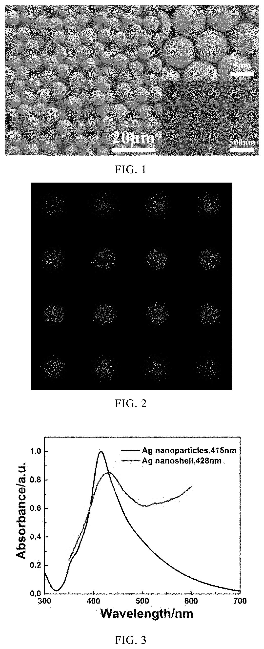
[0075]FIG. 1 showed a preferred embodiment of a method for preparing fluorescent-encoded microspheres coated with metal nanoshells provided by the present invention. The fluorescent-encoded microspheres coated with metal nanoshells prepared in this embodiment were CdSe / ZnS QDs / PSMA@Ag nanoshell (wherein precious metal nano-material was Ag nanoparticle, fluorescent-encoded material was CdSe / ZnS quantum dots, and polymer was styrene-maleic anhydride copolymer (PSMA). It included two groups, and the preparation methods were SPG membrane emulsion method and Pickering emulsion method respectively, and the specific preparation procedures were as follows.

[0076]The first group(SPG):

[0077]0.05 mg of CdSe / ZnS quantum dots (purchased from Suzhou Xingshuo Nanotech Co., Ltd.) and 0.5 g of styrene-maleic anhydride copolymer (PSMA) were dissolved in 8 mL of toluene to form a dispersed phase;

[0078]Nanoparticles (particle size of 100 nm) were resuspended in 60 mg of Ag in ethanol (90% ethanol) and d...

example 2

[0087]The fluorescent-encoded microspheres coated with metal nanoshells (the first group) prepared in this embodiment were CuInS2 / ZnS QDs / PSMA@Ag nanoshell, wherein the precious metal nano-material was Ag nanoparticle, and the fluorescent-encoded material was CuInS2 / ZnS quantum dots. The polymer was styrene-maleic anhydride copolymer (PSMA). In addition, in another group, no metal nanoshell was used and sodium lauryl sulfate (SDS) was used as a surfactant to prepare a fluorescent-encoded microsphere, and the group was the control group (the second group). The preparation method was SPG-Pickering emulsion method, and the specific preparation steps were as follows.

[0088]The first group:

[0089]1 mg of CuInS2 / ZnS quantum dots and 0.5 g of styrene-maleic anhydride copolymer (PSMA) were dissolved in 8 mL of toluene to form a dispersed phase;

[0090]Nanoparticles (particle size of 80 nm) were resuspended in 75 mg of Ag in ethanol and dispersed in 100 mL of ultrapure water uniformly under ultr...

example 3

[0100]The fluorescent-encoded microspheres coated with metal nanoshells prepared in this example were HPS / MOTAS@Au nanoshell, wherein the precious metal nano-material was Ag nanoparticle and the fluorescent-encoded material was 1,1,2,3,4,5-hexaphenylsilole (HPS), the polymer was styrene-acrylic acid copolymer (MOTAS). The preparation method was SPG-Pickering emulsion method, and the specific preparation procedures were as follows.

[0101]0.8 mg of HPS and 0.5 g of styrene-acrylic acid copolymer (MOTAS) were dissolved in 8 mL of toluene to form a dispersed phase;

[0102]Nanoparticles were resuspended in 100 mg of Ag in ethanol and dispersed in 100 mL of ultrapure water uniformly under ultrasound conditions, to form a continuous phase;

[0103]SPG membranes with the specification of Φ10×L20 mm (effective length 10 mm), with pore size of 3 μm and SPG membrane emulsifying device (purchased from Beijing Jiasheng Xingye Technology Co., Ltd., imported from Japan, model: MN-20) were used. The volu...


PUM

PropertyMeasurementUnit
concentrationaaaaaaaaaa

Abstract

A method for preparing fluorescent-encoded microspheres coated with metal nanoshells is disclosed herein. By using SPG method, metal nano-material modified with a certain ligand is used as a new surfactant in the emulsification process, and different kinds and different amounts of fluorescent materials are doped into polymer microspheres to prepare fluorescent-encoded microspheres with different fluorescent-encoded signals and uniformly coated metal nanoshells in one step. The prepared fluorescent-encoded microsphere comprises a metal nanoshell, a polymer, and a fluorescent-encoded material. The fluorescent-encoded microsphere has a particle size of 1 μm˜20 μm, CV of less than 10%, which can be used for protein / nucleic acid detection. The preparation method has the advantages of simple process, high surface coating rate, good uniformity and controllable LSPR peaks, which can solve the problems of existing commonly used metal nanoshell coating methods such as low surface coating rate, poor uniformity, complex preparation process and uncontrollable local surface plasmon resonance (LSPR) peaks, etc.

Description

CROSS REFERENCE TO RELATED APPLICATION[0001]The present application claims the priority from China Patent Application No. 202010135367.X, filed on Mar. 2, 2020. The entirety of this application including all tables, diagrams and claims are incorporated hereby as reference.FIELD OF THE INVENTION[0002]The present invention relates to preparation and application of micro-nano materials, in particular to fluorescent-encoded microspheres coated with metal nanoshells, a preparation method and applications thereof.BACKGROUND OF THE INVENTION[0003]Liquid-phase biochip technique uses encoded microspheres as a carrier for liquid-phase reactions and adopts fast and high-throughput flow cytometry as the analysis method based on the rules of protein-protein, nucleic acid-nucleic acid interactions, to perform fast and high-throughput multiplex quantitative detection of proteins and nucleic acids, with a wide range of applications in the protein expression, nucleic acid analysis, early diagnosis o...

Claims


Application Information

Patent Timeline
Patent Type & Authority Applications(United States)
IPC IPC(8): C09K11/02B01F13/08B01F3/08
CPCC09K11/025B01F13/08G01N2021/258B01F3/088B01F3/0853B01F3/0811C09K11/883C09K11/623C09K11/06C12Q1/689C12Q1/6837G01N33/582G01N33/585G01N33/57473C09K2211/1096C12Q2563/107C12Q2563/149C09K11/565C09K11/58C09K11/621C09K2211/1007C09K11/02Y02A50/30B82Y15/00B82Y20/00B82Y30/00B82Y40/00G01N21/25B01F23/43B01F23/49B01F23/4105B01F33/45B01F23/06B01F23/48B01F23/4146B01J13/04B01J13/02
Inventor FANG, JIANQIUZHONG, CHUNMEILI, WANWAN
Owner HANGZHOU SHINEDO BIOTECH CO LTD
Why Eureka
Try Eureka