

Bio-artificial organ containing a matrix having hollow fibers for supplying gaseous oxygen
a bio-artificial organ and fiber technology, applied in the field of bio-artificial organs containing fibers, can solve the problems of putting stringent demands on both oxygenation and oxygenation, severely hindering the development of so-called bio-artificial liver systems,
Inactive Publication Date: 2002-04-16
ACADEMISCH ZIEKENHUIS BIJ DE UNIV VAN AMSTERDAM ACADEMISCH MEDISCH CENT +1
View PDF16 Cites 48 Cited by
- Summary
- Abstract
- Description
- Claims
- Application Information
AI Technical Summary
Benefits of technology
It is a further object of the invention to provide an improved solid support and bioreactor enabling direct liquid contact between the cells and the liquid medium to be treated while at the same time maintaining a homogeneous flow of liquid medium to all parts of said support.
It is another object of the invention to provide a method for the culturing of liver cells, with which liver cells can be kept viable in an amount and during a period of time that are practical for use in a bio-artificial liver.
Problems solved by technology
A special problem in the cultivation of adherent cells--compared to the cultivation of cells in suspension or in confluent layers--is to provide sufficient nutrients and / or oxygen to the cells and / or provide for sufficient removal of waste products and / or carbon dioxide.
This is especially a problem with cells that put stringent demands on both oxygenation as well as the removal of waste products, such as liver cells.
The non-availability of suitable solid supports and methods for the in vitro cultivation of liver cells has over the last 40 years severely hindered the development of the so-called bio-artificial liver (BAL) systems, systems that could be used in patients with liver defects for the support and / or replacement of the natural liver function.
However, due to the abovementioned lack of suitable methods and / or materials for cultivating and / or maintaining liver cells in vitro, the bio-artificial liver systems from the prior art have so far proved insufficient, because they do not fully replace all the functions carried out by the liver of the patient in vivo, because they have insufficient capacity, and / or because the time during which they are therapeutically effective is too limited for practical use.
All these systems have been found to be insufficient.
The disadvantages of bio-artificial liver systems include ( .
. . ) the problem of maintaining normal hepatocyte viability and function at the high cell density necessary for clinical application.
However, the prior art hepatocyte systems also suffer from problems with regard to capacity and effective working time, see Sussman and Kelly:
. . ) the technology has severe limitations.
Coupled with the labour-intensive nature of cell preparation, this makes it almost impossible to scale up production to meet current needs in a cost-effective manner.
Moreover, freshly isolated cells do not appear to last very long during treatment.
A liver assist device that lasts for only 6 to 7 hours, as some have been reported to do, clearly falls short of allowing liver regeneration.
Finally, production of any such device using animal cells entails a number of problems, especially in areas of sterility and lot-to-lot variability.
However, a serious drawback of this system, besides the complexity of constructing and using a 200 glass plate-reactor, is that the monolayer culture of hepatocytes on said plates precludes the advantageous formation of hepatocyte aggregates.
Because of these limitations said hollow fiber reactor cannot easily be scaled up to a capacity required for practical therapeutic application.
Furthermore, this reactor is used with a very "closed path" column with a high density of the microcarriers, which leads to the formation of microcarrier pellets and to mass transfer problems with regard to the cells at the center of such a pellet.
Another disadvantage of this system is that the hepatocytes first have to be immobilized on the micro-carrier before the hepatocytes can be introduced into the hollow fiber reactor.
This involves further complicating processing steps that can lead to loss of cell viability.
According to this system, the liver cells are oxygenated by the patient's blood, because--as stated by the authors--"plasma does not provide the oxygen carrying capacity of whole blood".
Furthermore, perfusion with whole blood can lead to the fibers and / or the pores thereof within the bioreactor getting clogged, which problem could only be solved by totally replacing the hollow fiber module, requiring a fresh isolation / immobilization of the hepatocytes.
Other disadvantages of this and other hollow fiber systems using whole blood as the liquid medium are that "the hollow fiber membrane must first act as a plasma separator before any significant transport of nutrients and metabolites can take place across the fiber wall", and that it "requires systemic anticoagulation with heparin to prevent clotting in the module".
However, with regard to activity and function, the use of such tumor-derived cell lines is generally less preferred in the art than the use of isolated primary hepatocytes, also from a safety standpoint.
Furthermore, the C3A cell line used by Sussman lacks some very important functions of primary hepatocytes.
However, this system also requires a complicated and time consuming pre-immobilization of the hepatocytes.
In order to obtain sufficient attachment of the cells, the surfaces of the fibers must first be coated with a proteineous basement membrane product, such as Matrigel.RTM. or other collagen-based materials, requiring a separate and expensive pretreatment step.
Even so, as hollow fibers are not specifically designed and / or suited for use as a solid support in cell cultivation, the attachment and the speed thereof permitted by and / or obtainable with said reactors is limited and heavy inoculum charges are required when seeding the reactor.
Again, these large aggregates can lead to mass transfer problems with regard to the cells in the center of said aggregate.
Also, it is well known that hollow fibers are difficult to process, and in this respect the manufacture of the very complicated three-dimensional fiber network described by Gerlach et al., comprising four separate discrete capillary systems, suffers from a disadvantage from an economical point of view.
Also, this reactor is complicated to operate, requiring multiple separated inlet / outlet control systems.
A general problem of all the abovementioned hollow fiber bioreactors of the prior art is that the liquid medium (blood, plasma) to be treated is separated from the hepatocytes by the hollow fiber membrane; in other words, that there is no direct contact between the liquid medium and the hepatocytes in the reactor.
The passage through the membrane can lead to transport phenomena that can limit the achievable mass transfer, and therefore the efficiency of the BAL-system.
Also, the membranes can get clogged, especially when perfusion with whole blood is used.
In that case the BAL system or parts thereof have to be replaced, which means that therapy has to be interrupted or even stopped.
However, these proteins are of comparable sizes to antibodies, which could have an adverse effect when directed against nonautologous hepatocytes in the bioreactor.
In many bioreactors this gives rise to the formation of non-physiological hepatocyte pellets.
Hepatocytes in the center of these large aggregates show poor metabolic activity and even possible necrosis due to high gradients as a result of hindered transport of nutrients and oxygen to and carbon dioxide, toxins and cell products from these cells.
Besides, in most systems substrate exchange depends on diffusion which further limits mass transfer compared to the in vivo situation where hepatocytes function under perfusion conditions with low gradients.
Also, the bioreactors of the prior art are limited with respect to the amount of liquid medium that can be withdrawn from the hollow fiber lumen, as in general the fusion transport will be too slow.
Therefore, an active withdrawal of liquid medium from within the hollow fibers will be required, even so, the total flow through the hollow fiber membrane will be very slow and / or lead to the undesired formation of gradients, even with a high flow of liquid medium through the hollow fibers themselves.
Another general problem with the bio-artificial liver systems of the prior art is that they require the use of liver cell preparations with a high viability (>80%) and a high attachment.
As already acknowledged by Sussman and Kelly hereinabove, the production of such cells is a very costly, complex and time-consuming process requiring isolation and subsequent cultivation of suitable liver cells in sufficient viability and quantity which involves complicated procedures that do not reliably afford the required results, even when carried out by qualified experts.
Furthermore, known hepatocyte-containing BAL-systems cannot be stored before use for a prolonged period of time because the viability and function of the liver cells in the reactor cannot be maintained at a therapeutically acceptable level.
Also, the only technique available for preserving isolated liver cells over a longer period of time, i.e., cryopreservation, does not afford cells that are suitable for use with known BAL-systems, see Rozga et al., mentioned hereinabove:
However, [cryopreservation] may result in a significant loss of cell viability [.
Because of these problems, the known BAL-systems cannot be used as "off the shelf" units that can be kept and / or maintained in hospitals until their use is required, as is the case with other artificial systems for organ support such as for instance dialysis machines or artificial heart or lung systems.
Also, replacement during therapy of a spent primary liver cell based BAL system of the prior art with insufficient function with a fresh BAL system is usually not economically feasible over a prolonged period of time.
Better results have been obtained by using immortalized cells or the C3A cell line reported by Sussman et al., however, the use of this hepatoblast derived cell line has other disadvantages as already mentioned hereinabove.
This matrix material has some specific advantages over micro-capsules, which are costly and delicate to produce and give problems at high cell density growth, because frequently cells at the center of the capsule die.
Also, the microcapsules may burst prematurely losing their contents and each new inoculation requires a fresh encapsulating procedure.
With microcarriers, these cells are immobilized on the outside of the carrier particles, making them susceptible to shear stress and particle collisions, for instance during preparation or packing of the reactor.
In fact, the use of the "spiral wound" reactor according to GB-A-2,178,447 for culturing and / or maintaining hepatocytes would in practice lead to insufficient oxygenation, because the oxygen is supplied by means of just one conduit covering the entire length of the matrix mat.
The use of such a single conduit would lead to the generation of an undesired oxygen gradient along its length or even to local oxygen depletion, especially when the reactor is scaled up by increasing the number of matrix windings.
Furthermore, the use of a singular spiral wound conduit for liquid transport can lead to an inhomogeneous supply of liquid medium to all the parts of the bioreactor, for instance by the generation of undesired gradients.
It will be clear that this will make the 3D hollow fiber network according to Gerlach et al. very difficult and expensive to produce.
Again, this was not possible with the prior art BAL-systems.
This embodiment will usually not be suited for liver cells that are unable to divide and grow after they have been isolated, such as primary liver cells.
This method of distributing the cells throughout the reactor prevents the formation of a cell pellet at the bottom in the bioreactor, which would lead to mass transfer problems during use, especially with regard to the cells at the center of the pellet, which can lead to loss of functional activity and / or viability.
This favorable removal of dead cells cannot be achieved with the prior art hollow fiber bioreactors, because in these reactors, the liver cells essentially are present in an enclosed "compartment" between the hollow fibers, without means for perfusing said space, or captured within a (hydro)gel.
Method used
View moreImage
Smart ImageViewing Examples

Examples
example ii
In Vivo Results
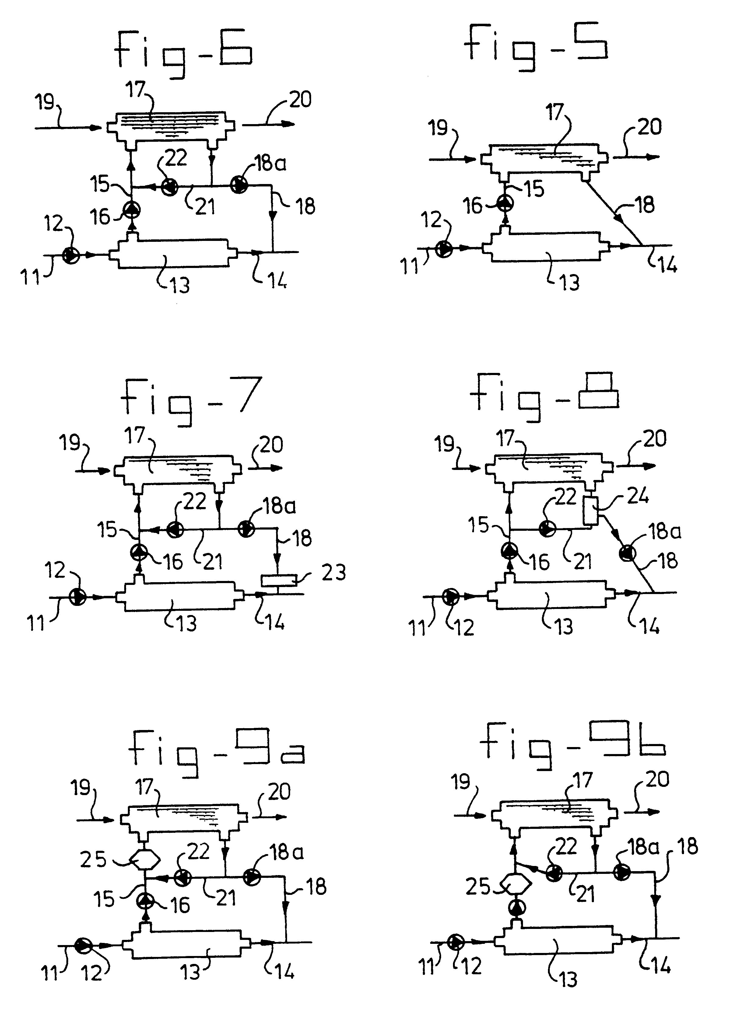
The abovementioned BAL was tested with an in vivo rat model.
The used population of rats was divided in three groups.
a. Reference group 1: liver ischemia (LIS) rats given only an infuse.
b. Reference group 2: LIS rats connected to the entire BAL system, but without hepatocytes. This reference is carried out to study the influence of plasmapheresis (possible negative effect) and the large volume of the extra-corporeal circulation (possible positive effect through dilution of toxins) on the survival.
c. LIS rats connected to the BAL with pig hepatocytes.
Login to View More PUM
 Abstract
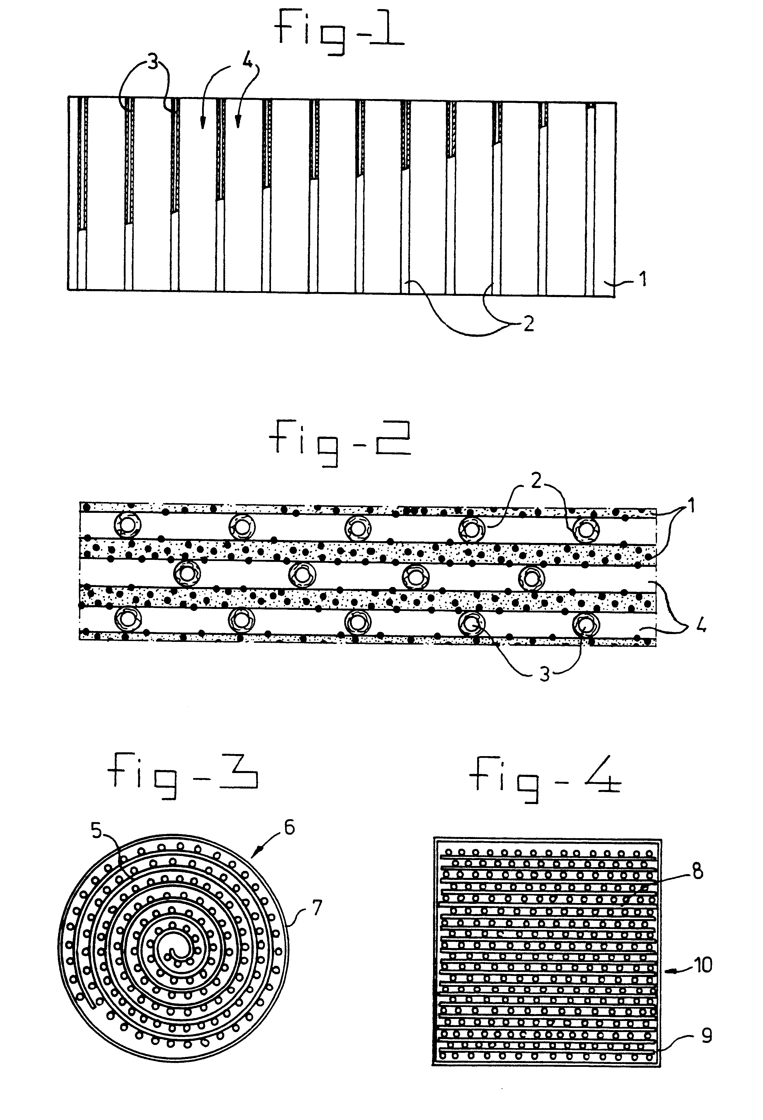
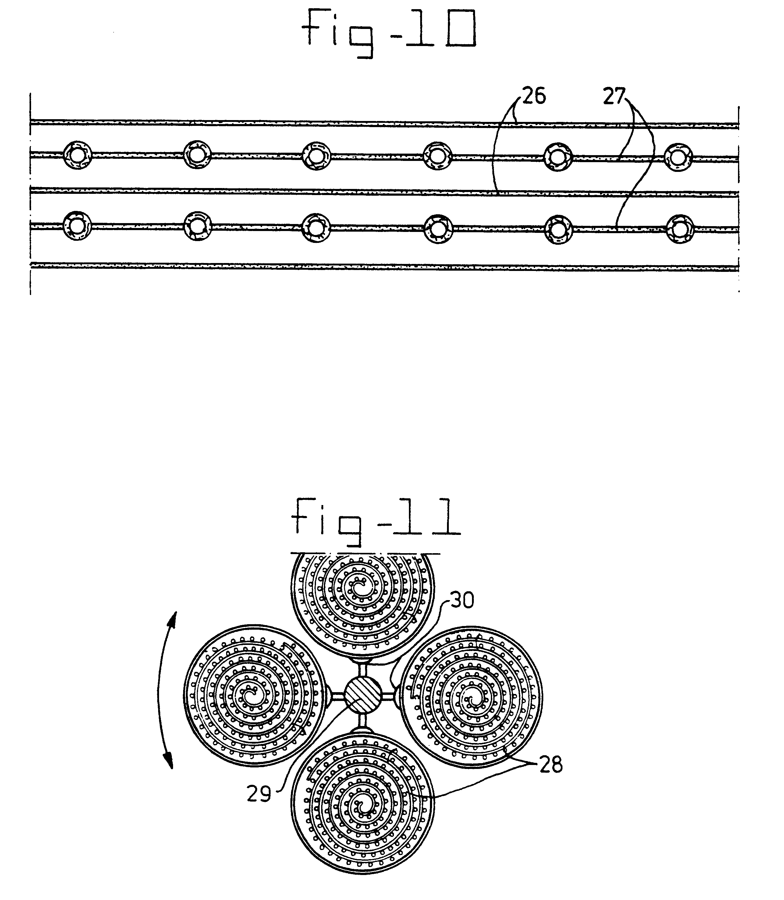
A bio-artificial organ system is provided comprising a wall surrounding a space which has a solid support for cell cultivation. The space includes a there dimensional matrix in the form of a highly porous sheet or mat and including a physiologically acceptable network of fibers or an open-pore foam structure; hydrophobic hollow fibers permeable to gaseous oxygen or gaseous carbon dioxide evenly distributed through the three dimensional matrix material and arranged in parallel running from one end of the matrix material to the other end of the matrix material. Cells obtained from organs are present in the extra fiber space for culture wherein the cells are provided with sufficient oxygenation and are maintained as small aggregates with three dimensional attachment.
Description
The present invention relates to the field of the cultivation of cells, especially of adherent tissue cells such as liver cells. More in particular, the invention relates to the field of biological methods and reactors for the cultivation and / or maintenance of cells, especially liver cells, and to the use of such methods in a bio-artificial liver system (BAL).BRIEF DESCRIPTION OF THE PRIOR ARTIt is generally known that most tissue cells require a solid support on which to grow and divide.Although it is possible to culture adherent tissue cells in ordinary vessels, such as glass bottles or Petri dishes, during which the cells adhere to the wall of the vessel, usually special reaction vessels or bottles with a high surface area are used so as to provide increased capacity for cell attachment. One way to improve said surface area is to use a solid support for cell adherence. Such solid supports are known in the art; examples include glass beads, microcarriers and cellulose fibers.A spe...
Claims
Login to View More Application Information
Patent Timeline
 IPC IPC(8): A61M1/34C12N5/00C12M1/40A61K35/12A61K35/407C12M1/36C12M3/00C12N5/06C12N5/08C12N5/10
CPCA61M1/3472C12M21/08C12M23/06C12M25/02A61M1/3489C12N5/0068C12M23/24C12M25/10C12M25/14C12N2533/30
InventorFLENDRIG, LEONARDUS MARCUS
OwnerACADEMISCH ZIEKENHUIS BIJ DE UNIV VAN AMSTERDAM ACADEMISCH MEDISCH CENT