
Assay of micro-organisms in cell cultures

a cell culture and microorganism technology, applied in the field of assay of microorganisms, can solve the problems of low level of microorganisms being accidentally introduced into cultures during routine feeding, invalidity and significance of research and the safety of biologicals produced from contaminated cell cultures,

Inactive Publication Date: 2005-10-04
LUMITECH UK
View PDF1 Cites 8 Cited by
  • Summary
  • Abstract
  • Description
  • Claims
  • Application Information

AI Technical Summary

Benefits of technology

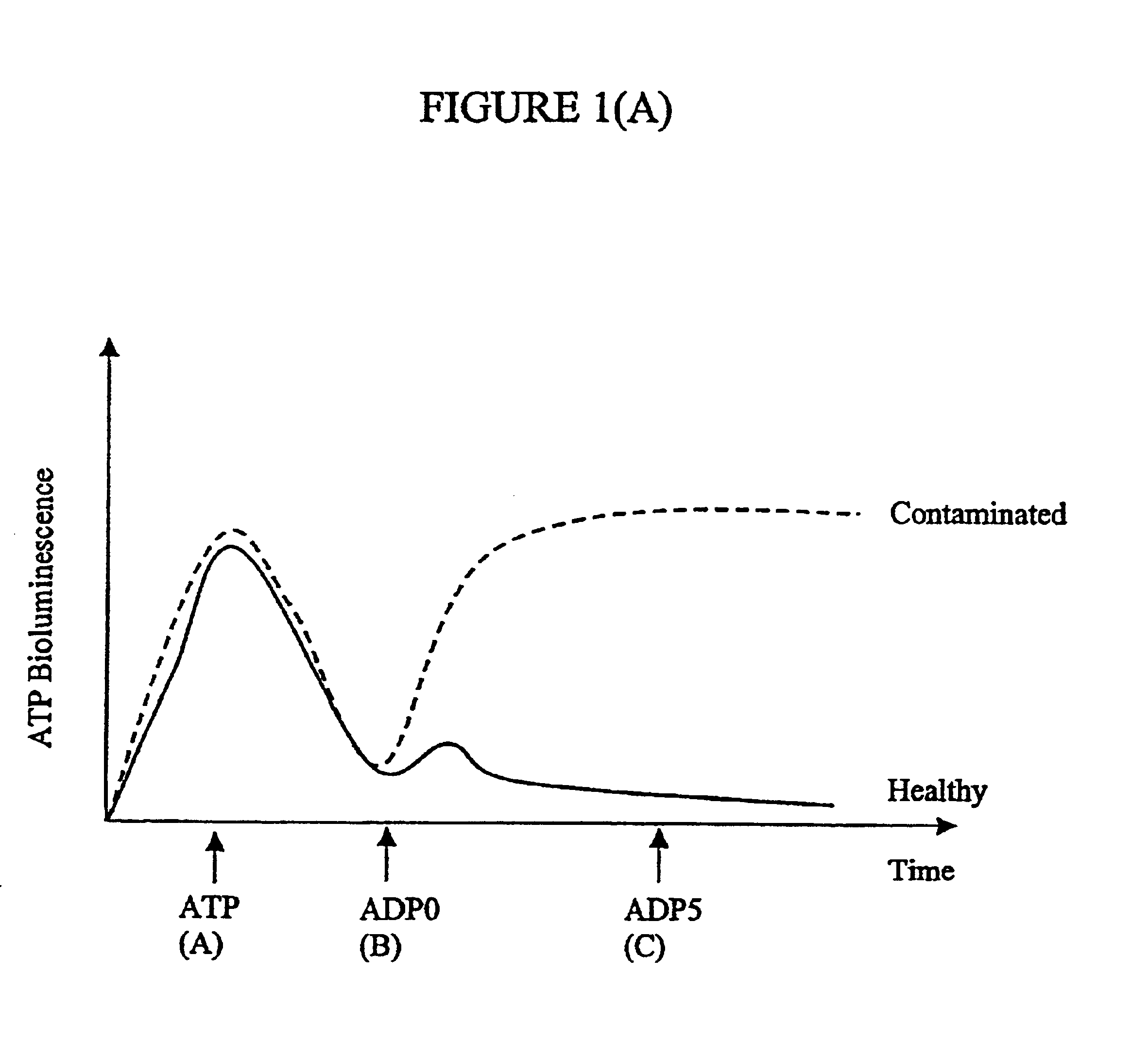
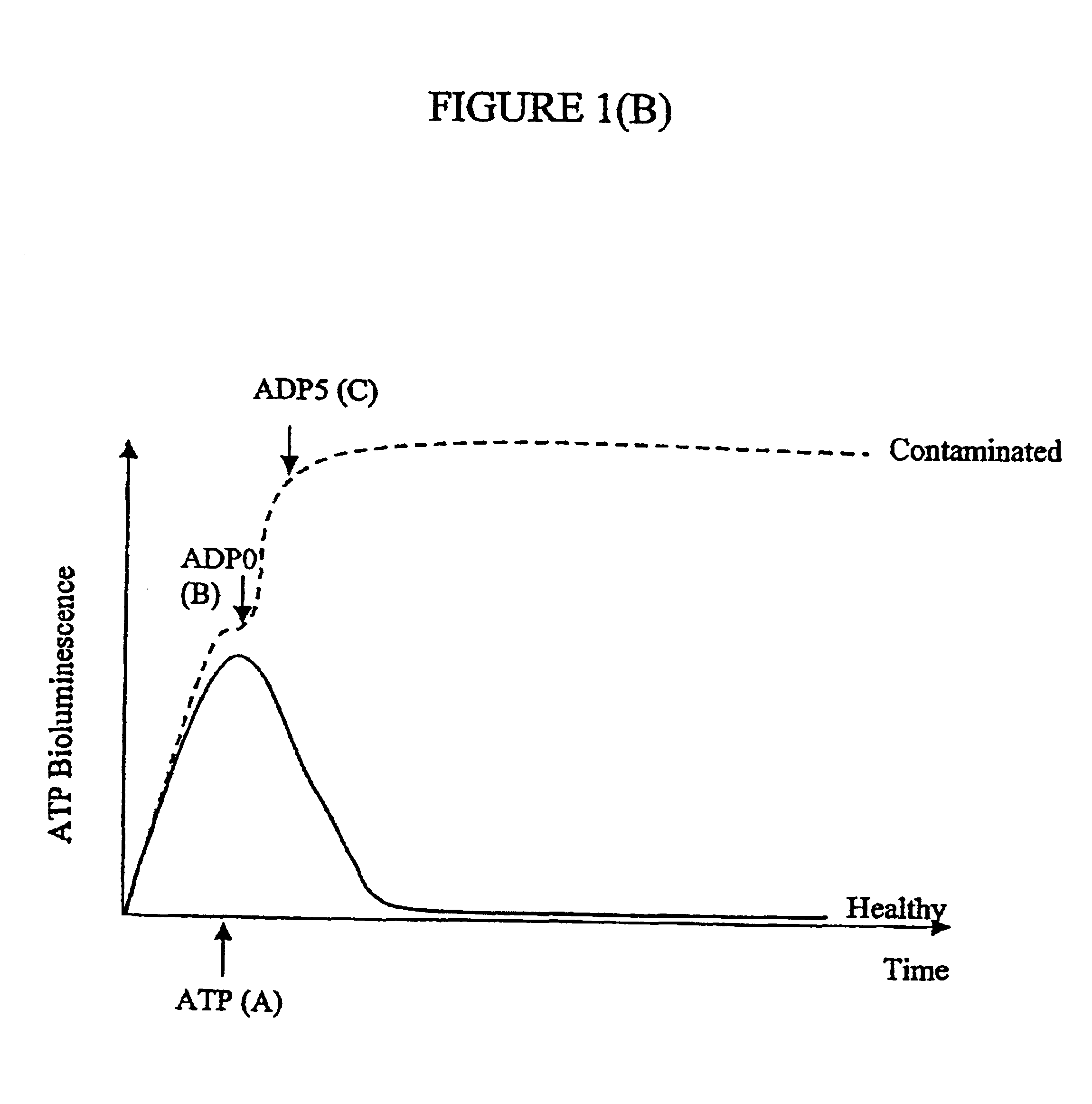
[0037]However, in the presence of the high levels of ADP associated with mycoplasma contamination, the conversion reaction results in a marked and rapid rise in bioluminescence, which can be detected much earlier than 5 minute delay typically needed prior to measuring reading ‘C’ (in fact, the bioluminescence signal will increase in a linear fashion for as long as 60-120 minutes in contaminated cells). In this case, the C-B value is large and positive, leading to a large and positive ADP:ATP ratio for such cells. The positive versus negative ratio in contaminated and healthy cells, respectively, provides a convenient and easy screening test for mycoplasma contamination of a culture.
[0045]For intracellular parasites, for example, infection by Plasmodium falciparum, red blood cells, or other suitable means of disrupting the host-cell membrane, the common feature is that the host cell will appear essentially healthy and therefore the ATP readings should be similar to an uninfected control cell. If the cells are treated with a Triton-based detergent, or other suitable means of disrupting the host-cell membrane, this will release all the adenylate nucleotides, the ATP results should not deviate significantly from uninfected cells. However, the presence of a parasite with ATPase activity will dramatically increase the levels of ADP. If the cell has an intact membrane and ADP is not being pumped out then there will be little ADP in the culture medium / sample. If the nature of the infection results in ADP being pumped out then there would be ADP present in the culture medium. There could therefore be two measurements done, one prior to lysis and one post disruption of the host cells.

Problems solved by technology

As a result of this widespread problem, research and production of biological materials is often unknowingly done using mycoplasmal contaminated cell cultures.
Thus, the validity and significance of research and the safety of the biologicals produced from contaminated cell cultures are jeopardised.
Because of their small size and lack of cell wall, mycoplasmas often pass through the 450 or even 200 nm filters used to “sterilise” cell culture media and sera, resulting in low levels of these organisms being accidentally introduced into cultures during routine feeding.
Even then, some mycoplasmal strains are difficult to grow in culture without cells.
Properly done, with appropriate positive and negative controls, direct culture testing offers the greatest security, but is rather slow, usually requiring up to 28 days for completion.
While faster than direct culture methods, usually taking only 1-5 days to complete, indirect tests are not yet as sensitive and usually require higher levels of contamination (e.g. 104 or more organisms / mL) for detection.
The PCR-based kit of Stratagene is widely used, but is expensive, additional PCR chemicals are required and the running of PCRs requires technical expertise not always conveniently available.
Also, competition between the internal control template DNA and the mycoplasma DNA frequently results in the control not being visible on the gel when the sample is contaminated with mycoplasma.
It is a problem to find an alternative rapid, indirect method which does not require PCR skills.

Method used


Image

  • Assay of micro-organisms in cell cultures
  • Assay of micro-organisms in cell cultures
  • Assay of micro-organisms in cell cultures

Examples


example 1

Reagents

Bioluminescent (Luciferin / Luciferase) Reagent

[0057]This was supplied by Labsystems, Helsinki, Finland as a freeze dried powder to be reconstituted prior to use. The powder contains luciferase from Photinus pyralis (40 μg), Luciferin (42 μg: believed to contain 96% D-Luciferin; 4% L-Luciferin), bovine serum albumin (50 mg), magnesium sulphate (1.23 mg) and Inorganic pyrophosphate (0.446 μg). It was reconstituted to 10 ml of 0.1 M Tris-acetate buffer, pH 7.75 containing 2 mM EDTA (dipotassium salt).

ATP-releasing Agent

[0058]0.1 M Tris acetate buffer pH 7.35 containing 2 mM EDTA (dipotassum salt), 0.25% v / v Triton X-100 and 1 μM dithiothreitol.

ADP-converting Agent

[0059]This was prepared by mixing equal volumes of 2 M potassium acetate, 500 units / ml pyruvate kinase (PK) and 100 mM phosphoenol pyruvate (PEP). An equal volume of Tris-acetate buffer pH 7.75 was then added. The PK and PEP were thus diluted 1:6 when the converting reagent was made up from the stock solutions. Since 20...

example 2

[0076]The procedure of Example I was followed.

[0077]The cell lines tested were U937, Jurkat, HL60, K562, CEM-7, L-929 and A549 (as in Example 1) plus:[0078]L-929 (freshly thawed) and[0079]BCP-1 (lymphoma cell line)

[0080]The DAPI stained cytospins were viewed under fluorescence microscopy, and all cell lines appeared positive. The PCR test showed that all cell lines except Jurkat were positive for M. fermentans. The Jurkat cells tested were positive for M. hyorhinis. The ADP / ATP ratios appeared to increase at a very early stage of infection.

[0081]The L-929 cells held as frozen stock seemed to be the source of the infection, as they were positive in the PCR assay before they had even been set up in culture. On thawing of these cells and culturing them for 48 hours, there was already an increase in the ADP:ATP ratio from around 0.1:1, normally observed in a healthy cell population, to 0.37:1 (average of 2 experiments).

[0082]The cells were regularly passaged by trypsinisation during a f...

example 3

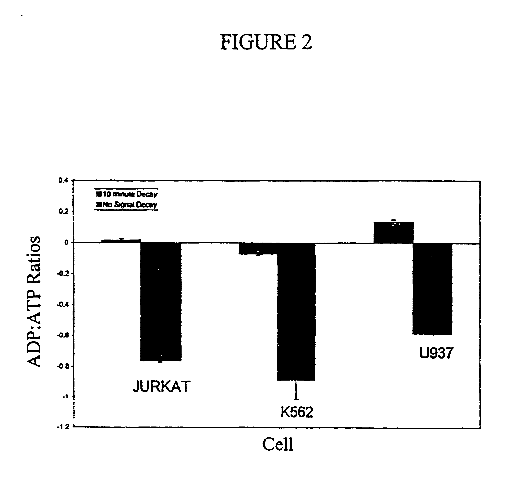
[0083]This Example demonstrates the effect of heat-treating the culture supernatants at 56° C. for 30 minutes. This would normally inactivate any ATPase activity present as a result of lysing the cells with the nucleotide releasing agent. The cells were incubated at 1×10⁵ ml−1 and ATP was measured immediately after removal from the water bath (controls were left at room temperature for 30 minutes) and 10 minutes later. It can be seen that the heat treatment reduced the light signal decay, by inactivating cellular ATPases and other degradative enzymes. ADP would have accumulated in the culture due to the presence of mycoplasma, and, as the ADP is beat stable, it was still detectable.

[0084]

TABLE 2ATPATP afterCell Typeinitially10 minutesADPRatioL-929-56° C.-RT26122189161555.3472693 348 96033.437U937-56° C.-RT56194378323204.98652441406 92131.466Jurkat-56° C.-RT94297242455894.06794441265118601.112K-562-56° C.-RT71866673343733.85565121578107951.415Cem-7-56° C.-RT72835752371724.31474541106...


PUM

PropertyMeasurementUnit
diameteraaaaaaaaaa
sizeaaaaaaaaaa
pHaaaaaaaaaa

Abstract

Micro-organisms which over-produce ATPase, especially mycoplasmas in a sample of a cell culture, are detected by making use of the ATPase activity to convert cellular or externally added ATP to ADP. The decrease in ATP externally added or the increase or absolute level of ADP produced from cellular ATP can then be detected or measured. For example, if a reagent such as phosphenol pyruvate / pyruvate kinase is added to convert that ADP back to ATP, the resulting high level of ATP can be detected bioluminescently using the reaction: Luciferin+luciferase enzyme+ATP→hv+products.

Description

BACKGROUND OF THE INVENTION[0001]1. Field of the Invention[0002]The present invention relates to a method of assay of micro-organisms, especially small wall-less bacteria such as mycoplasmas, in cell cultures.[0003]2. Description of the Related Art[0004]Mycoplasma are a large and widespread group of prokaryotes. They are essentially bacteria with the smallest known genomes and are found amongst members of mollicutes. This class presently comprises six eubacterial genera Acholeplasma, Anaeroplasma, Asteroplasma, Mycoplasma, Spiroplasma and Ureaplasma.[0005]The Mycoplasmas and Ureaplasmas are parasites in different vertebrates, from which they obtain their essential compounds such as fatty acids, amino acids, precursors for nucleic acid synthesis and cholesterol. Only Acholeplasma and Asteroplasma do not require cholesterol for growth.[0006]These flexible, pleomorphic organisms can be as small as 200-300 nm in diameter and can achieve very high densities in cell cultures, (107-108 org...

Claims


Application Information

Patent Timeline
Patent Type & Authority Patents(United States)
IPC IPC(8): C12Q1/66C12Q1/04C12Q1/00G01N21/78C12Q1/02C12Q1/48C12R1/35
CPCC12Q1/008C12Q1/04C12Q1/66G01N2333/30
Inventor CROUCH, SHARON PATRICIA MARYBRADBURY, DAWN ANNSLATER, KEVIN JOHN
Owner LUMITECH UK
Learn More