
Methods of protein destabilization and uses thereof

a protein destabilization and protein technology, applied in the field of protein analysis, can solve the problems of less robust methods, less facile or advanced reciprocal abilities for studying rna and protein molecules, and high cost and time-consuming and expensive genetic analysis of vertebrate organisms

Inactive Publication Date: 2007-08-28
AURORA BIOSCI CORP
View PDF30 Cites 7 Cited by
  • Summary
  • Abstract
  • Description
  • Claims
  • Application Information

AI Technical Summary

Benefits of technology

[0132]An important advantage of the present invention is the ability to titrate the degree of destabilization, and therefore the steady state concentration, of the target protein in the cell. Since the destabilized, unmodified sensor represents the substrate for the target activity, it is preferable to provide the substrate at a physiologically relevant concentration within the cell while retaining the appropriate turnover characteristics for each individual reporter molecule.
[0133]For assays measuring protease activity, the linker generally comprises a protease recognition motif within its sequence. The protease recognition motif may be placed anywhere within the linker moiety, but is conveniently placed close to the center of the linker unless there are steric, or other reasons, to position the recognition motif at a specific location. Typically, the recognition motif will provide for relatively specific recognition of the sequence by the target protease. In some cases it may be preferable for the linker to contain a second “control” protease site for a known protease for use as a positive control.
[0134]The expression vector will normally direct expression of the sensor to the cytosol of the cell, although other cellular compartments, such as the plasma membrane are also practical. Once the expression vector is introduced in a population of cells, the cells are typically screened for reporter moiety expression level in the absence of the target protease. This can be achieved by FACS™, after addition of appropriate substrates for the reporter moieties (if required). While cells may be selected for varying levels of expression of the reporter moiety within the population of cells, observations to date suggest that cells exhibiting somewhat lower levels of reporter moiety are superior to those that initially exhibit high levels of reporter moieties under these conditions. Cells may also be selected via antibiotic resistance to provide for stable cell lines.
[0135]Once isolated and characterized, the resulting cell line represents a living sensor for the activation or expression of the target protease that enables the identification and screening of compounds that modulate the activation of the target protease. Importantly these determinations can be completed within the living cell where other issues such as membrane permeability, specificity and toxicity may be directly assessed.
[0136]In most cases, it will be preferable to start with a cell line that does not normally express high levels of the active target protease. However if this is not possible, then the initial evaluation of the cell lines may be modified in order to screen for cells initially exhibiting high levels of reporter moiety expression. For example, by using an inhibitor of the reporter moiety to inhibit basal reporter gene activity, (as discussed below). In general any types of cells may be used with the present invention, including animal, plant, insect, yeast and other eukaryotic cells or prokaryotic cells.
[0137]In whole cell studies it may be desirable to add an inhibitor of protein synthesis such as cycloheximide in order to reduce the steady state level of the destabilized reporter moiety in the cell immediately prior to the measurement of reporter activity. This approach has the advantage of improving the dynamic range of the assay because in the absence of new protein synthesis, uncleaved and therefore destabilized reporter moieties are destroyed by targeting to the proteasome leaving the cleaved and stabilized reporters intact within the cell. (i.e. the background is reduced). This results in a larger net difference in reporter moiety activity in cells containing a suitable protease compared with those lacking a suitable protease. Typically for such uses, cycloheximide is added to cell in the range of 10 to 150 μg / ml cycloheximide, preferably 50 to 100 μg / ml. Generally cells are pretreated with an appropriate stimulus to activate the target protease, and then cycloheximide is added one to two hours prior to the addition of suitable substrates for the reporter moiety.

Problems solved by technology

Although we have made significant improvements in our ability to clone, sequence and analyze DNA sequences, our reciprocal abilities for studying RNA and protein molecules are significantly less facile or advanced.
A central challenge facing workers in the field today is to understand out how a protein's activity and function within a cell are regulated and coordinated within the native physiological context.
Unfortunately genetic analysis in vertebrate organisms is extremely time consuming and expensive.
Although there is tremendous interest in understanding the regulation and interactions of proteins within cells there are relatively few methods that are robust, simple to use, amenable to high throughput screening or can be used effectively within living cells.
Furthermore in many cases where specific assays do exist these are restricted in scope to individual enzymatic steps or to one or two defined pathways.
A key challenge in this area has been to provide a predictable way of creating graded levels of destabilization for a given protein that that can be utilized in manipulating the steady state levels or dynamic temporal regulation of that protein.

Method used


Image

  • Methods of protein destabilization and uses thereof
  • Methods of protein destabilization and uses thereof
  • Methods of protein destabilization and uses thereof

Examples


example 1

Generation of Multimerized Destabilization Domains

[0194]The cDNA encoding human ubiquitin was isolated from a human genomic DNA preparation obtained from Jurkat cells by polymerase chain reaction (PCR) using the PCR primers Ubi5 (SEQ. ID. NO. 15) and Ubi3 (SEQ. ID. NO. 16) and cloned into pBluescript H vector (Stratagene). The C-terminal residue of ubiquitin was altered from glycine to valine by site-directed mutagenesis (Kunkel) in order to generate a mutant form of ubiquitin that cannot be cleaved by cellular α-NH-ubiquitin endopeptidases. This mutant is hereafter referred to as ubiquitinG76V (SEQ. ID. NO. 17). The ubiquitinG76V (SEQ. ID. NO. 17) mutant was then amplified by PCR using the oligonucleotide primers Ub5′ (SEQ. ID. NO. 18) and Ub3′, (SEQ. ID. NO. 19). These primers introduce a Bgl II restriction site at the 5′ end of the coding sequence and a BamH I site at the 3′ end of the coding sequence. The PCR fragment from the reaction was digested with Bgl II and BamH I and lig...

example 2

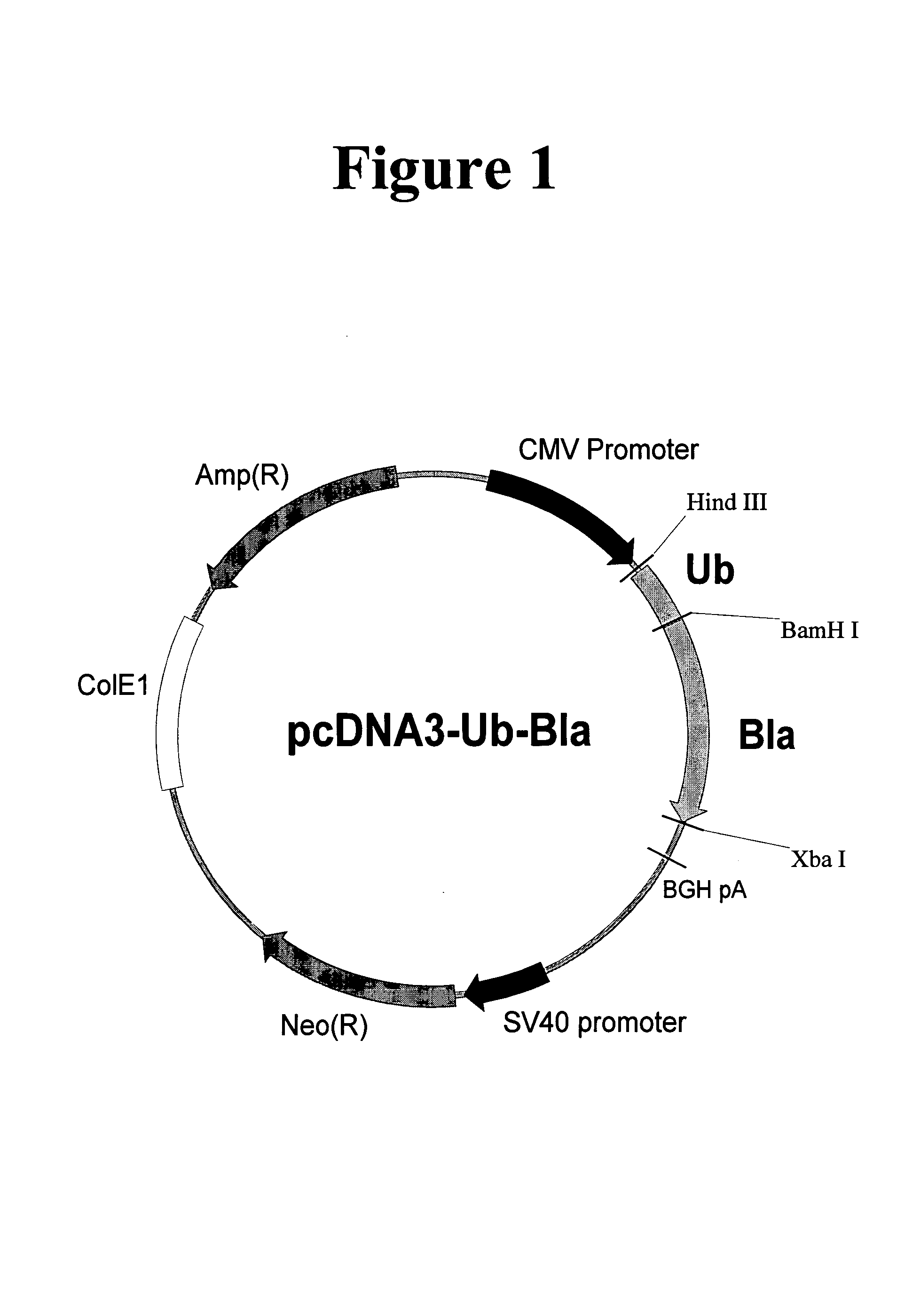
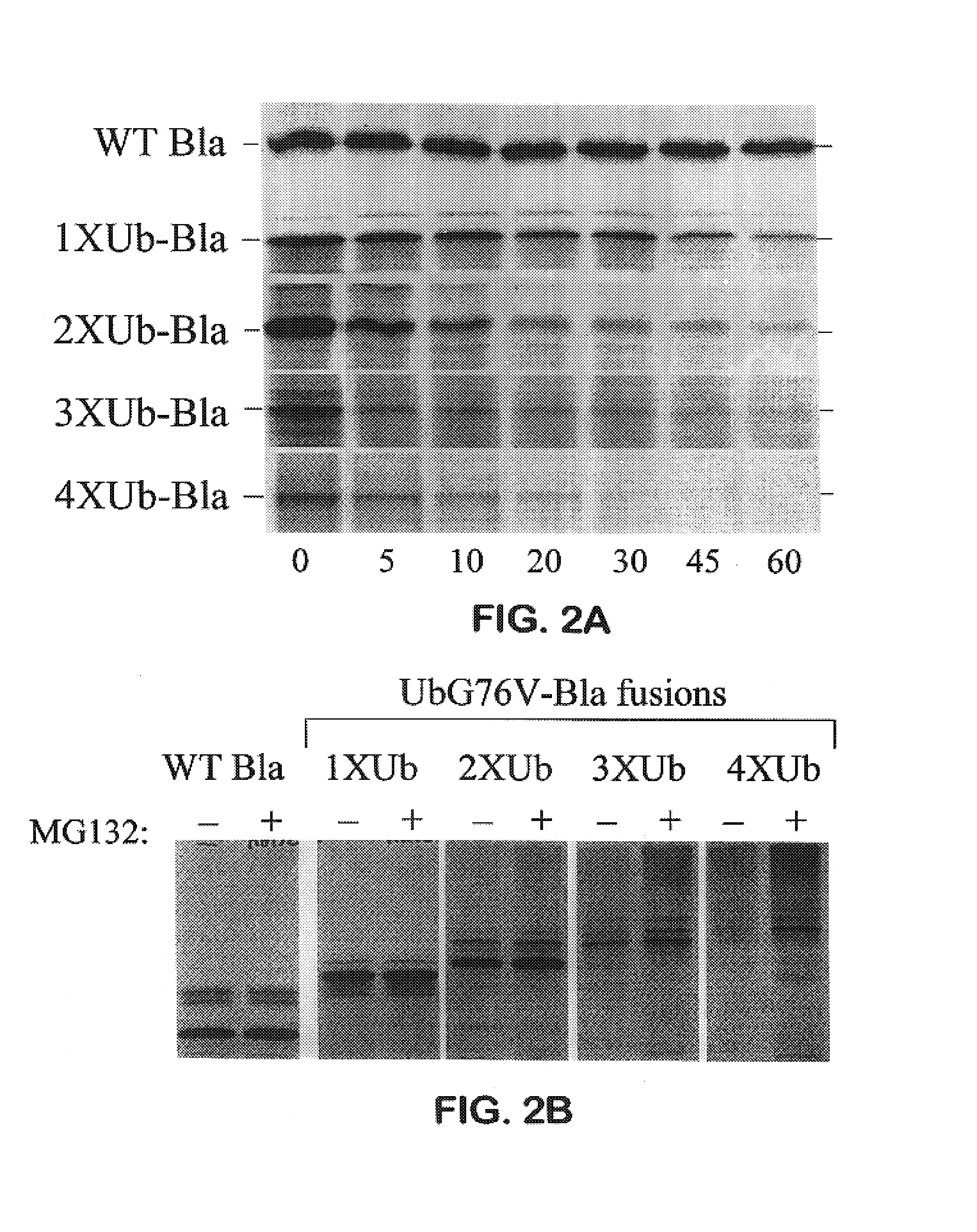
Creation of Multimerized Destabilization Domain-β-Lactamase Fusion Proteins

[0195]The gene encoding the E. coli TEM-1 β-lactamase was isolated from the plasmid pBluescript (Stratagene) by polymerase chain reaction (PCR) amplification using the PCR primers BLA5 (SEQ. ID. NO. 20) and ABSC107, (SEQ. ID. NO. 21) resulting in the deletion of the signal sequence and introduction of a BamH I restriction site and the amino acids below at the 5′ end of the coding sequence.[0196]BamH1 HHSGAWLHPETLVKYK (SEQ. ID. NO:73)

[0197]Amino acids in bold represent original β-lactamase coding sequence, underlined amino acids represent the BamH I restriction site. An Xba I site was inserted at the 3′ end of the coding sequence. The PCR fragments from these reactions were digested with BamH I and Xba I and ligated into pcDNA3 (Invitrogen) via the same sites. The resulting construct, pcDNA3-B1a (SEQ. ID. NO. 22), was then used to create in-frame fusions with the multimerized ubiquitinG76V constructs above. Th...

example 3

Creation of Multimerized Destabilization Domain-Naturally Fluorescent Protein Fusions

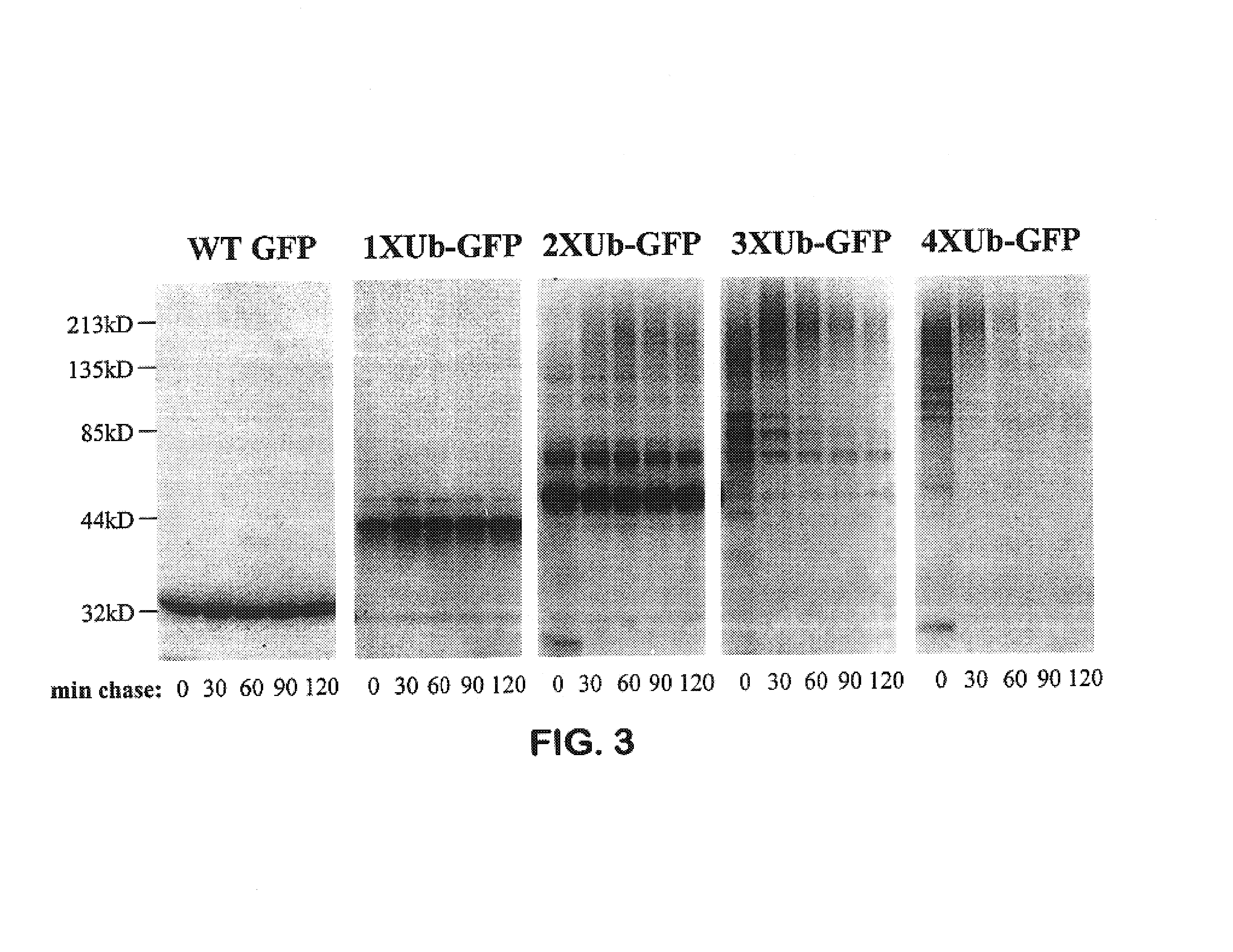
[0198]The gene encoding the GFP mutant Emerald (S65T, S72A, N149K, M153T, I167T) (SEQ. ID. NO. 28) was amplified by PCR using the oligonucleotides GFP5′ (SEQ. ID. NO. 29) and GFP3′, (SEQ. ID. NO. 30). The resulting PCR product had a BamH I restriction site at the 5′ end of the coding sequence and a Xba I site at the 3′ end of the coding sequence. The PCR fragment from this reaction was digested with BamH I and Xba I and ligated into pcDNA3 via the same sites. The resulting construct, pcDNA3-GFP was then used to create in-frame fusions with the multimerized ubiquitinG76V constructs described above. This was achieved by digesting the pcDNA3-1-4XUb-B1a constructs (SEQ. ID. NOs. 23 to 26) with the restriction enzymes BamH I and Hind III, and then ligating the fragment encoding the various multiUb destabilization domains via the same sites into the pcDNA3-GFP construct. These constructs were named pcDNA3...


PUM

PropertyMeasurementUnit
concentrationsaaaaaaaaaa
pHaaaaaaaaaa
pHaaaaaaaaaa

Abstract

This invention is directed towards methods of destabilizing proteins in living cells, and their use for the development of novel assays. In one embodiment, the invention comprises the use of non-cleavable multimerized ubiquitin fusion proteins to destabilize a target protein, such as a reporter moiety. In one aspect of this method the constructs also comprises a linker that operatively couples the reporter moiety to the multimerized ubiquitin fusion protein. In this embodiment, enzymatic modification of the linker results in a modulation of the coupling of the reporter protein to the multimerized ubiquitin domains resulting in a change in the stability of the reporter moiety. The level of the reporter moiety in the cell can then be used as a measure of the enzymatic activity in the cell. In another embodiment the invention provides for a generalized way of coordinately regulating the cellular concentration of a plurality of target proteins. In one aspect of this method, the target proteins are operatively coupled to a ubiquitin fusion protein via a linker containing a protease cleavage site. Cleavage of the linker by a protease results in uncoupling of the target protein from the multimerized ubiquitin construct, and results in an increase in the stability and concentration of the target protein.

Description

FIELD OF THE INVENTION[0001]The present invention is in the field of protein analysis and more particularly methods of destabilizing proteins and using the destabilized proteins for novel cell based assays.BACKGROUND OF THE INVENTION[0002]While genomic programs provide ever more sophisticated information on the sequence and patterns of expression of mammalian genes, it is increasingly recognized that integrating this information into a functional model of how a cell works requires an understanding of how the protein products of expressed genes interact within the cell. Although we have made significant improvements in our ability to clone, sequence and analyze DNA sequences, our reciprocal abilities for studying RNA and protein molecules are significantly less facile or advanced. Furthermore, proteins themselves represent significantly more complex molecules in terms of composition, shape and activity compared to double stranded DNA. A central challenge facing workers in the field t...

Claims


Application Information

Patent Timeline
Patent Type & Authority Patents(United States)
IPC IPC(8): C12Q1/68A01K67/027A01K67/033A61K45/00A61P43/00C07K14/47C07K19/00C12N1/15C12N1/19C12N1/21C12N5/10C12N15/09C12Q1/02C12Q1/48
CPCC12Q1/485A61P43/00
Inventor STACK, JEFFREYWHITNEY, MICHAELCUBITT, ANDREW B.POLLOK, BRIAN
Owner AURORA BIOSCI CORP
Learn More