Irreversible electroporation using tissue vasculature to treat aberrant cell masses or create tissue scaffolds

a tissue vasculature and electroporation technology, applied in the field of medical treatment of diseases and disorders, can solve the problems of difficult surgical intervention on tumors that are not readily accessible or on organs that do not regenerate, surgical intervention can often involve substantial physical damage to the patient, and no one technique has been shown to be highly effective at treating all types of cell proliferative diseases and disorders, etc., to achieve the effect of improving the preservation of extracellular matrix, reducing energy needed, and increasing cell permeability

Active Publication Date: 2018-01-16
VIRGINIA TECH INTPROP INC
View PDF923 Cites 40 Cited by
  • Summary
  • Abstract
  • Description
  • Claims
  • Application Information

AI Technical Summary

Benefits of technology

[0028]The present invention provides novel applications of irreversible and reversible electroporation through the use of existing vasculature within tissues as the conduit for pulse delivery. This methodology results in the separation of “electrodes” of just a few microns, reducing the energy necessary to induce non-thermal ablation of cells and tissues to drastically improve preservation of the extracellular matrix for the development of vascularized tissue constructs. Also provided are clinical implications in the treatment of solid tumors and other in vivo applications, including ones that harness the reversible realm of electroporation, where the electrical fields temporarily increase the permeability of the cells without killing them, facilitating the intracellular transport of exogenous agents.
[0029]Use of IRE to decellularize tissue provides a controlled, precise way to destroy cells of a tissue or organ, while leaving the underlying extracellular matrix (ECM), including vascularization and other gross morphological features of the original tissue, intact. The decellularized scaffolds are then suitable for seeding with cells of the appropriate organism.
[0034]A representative device for administering IRE directly into the vasculature of an organ is shown in FIG. 1, which is a schematic diagram of an embodiment of an IRE mechanical perfusion connection device 10 according to the invention. Connections to the vasculature 15 and the perfusion system can be made through the use of Luer lock connections 20. A one way valve 25 inside the device can be used to prevent back-flow and electrically isolate the organ between mechanically simulated heart beats. The device can comprise electrode 30 and an external electrode 40. Accordingly, such electroporation systems can be connected internally to blood vessels and conductance structures (example: the hepatic vein, hepatic artery, portal vein, and / or bile duct of a liver) such that it does not impede continuous mechanical perfusion. In such preferred focal ablation systems, the protocols can induce cell death of 95% of cells while preserving the extracellular basement membrane of lobule units.
[0036]Embodiments of the invention provide methods wherein the application of varying amounts of energy (both reversible and irreversible electroporation) (e.g., using the vasculature of an organ as conductive pathways) can be modulated to produce a wide range of desirable tissue ablation effects, including selective removal of normal and neoplastic cells.

Problems solved by technology

While each treatment modality has shown significant effectiveness in treatment of various cell proliferative diseases, no one technique has been shown to be highly effective at treating all types of cell proliferative diseases and disorders.
While surgical intervention is effective at removal of solid tumors on tissues and organs that are physically accessible and capable of sustaining physical damage or capable of regeneration, surgical intervention can be difficult to perform on tumors that are not readily accessible or on organs that do not regenerate.
In these cases, surgical intervention can often involve substantial physical damage to the patient, requiring extensive recuperation times and follow-on treatments.
At other times, the extensive growth of the neoplasm prevents removal, since attempts at removal would likely kill the patient.
Likewise, treatment with radiation can result in collateral damage to tissue surrounding the tumor, and can cause long-lasting side-effects, which can lower the quality of life of the patient.
Chemotherapeutic treatments can cause systemic damage to the patient, and can result in significant side effects that might require a long recuperation period or cause permanent damage to tissues and organs.
These pulses permanently destabilize the cell membranes of the targeted tissue (e.g., tumor), thereby killing the cells.
Organ supply is constrained by obstacles that impede acquisition.
As a result, organ donation may be problematic when intensive care resources are strained.
Developing engineered materials to replicate the structure and function of organs has been met with limited success.
Large volumes of poorly-organized cells and tissues cannot be implanted due to the initial limited diffusion of oxygen, nutrients and waste.
Cell survival and proliferation in each of these structures was limited to a few millimeters from a nutrient source.
Traditional top-down manufacturing techniques are currently unable to produce a hierarchical vascular structure which, in human organs, ranges in size from a several centimeters (vena cava, for example) down to only a few micrometers (most capillaries), a scale spanning more than four orders of magnitude.
Microfabrication techniques can replicate some features of the complex architecture of mammalian microvasculature, but current processes fail to extend into the macro-scale.
Biocompatible inkjet printing technique for designed seeding of individual living cells.
Challenges to chemical decellularization techniques include the potential for detergents to damage ECM components, the potential to create and deposit toxins, and the inherent difficulty of scaling these techniques up from small rat organs to larger organs.
Porcine xenotransplants have shown considerable potential, but have failed to become widely accepted or used.
Unfortunately, the mechanisms of graft loss and rejection in these transplants are still not well understood, and immunological rejection remains the most significant barrier to successful transplantation.
Electrospinning is another popular method for creating scaffolds for engineered tissues and organs, but widely used techniques suffer from fundamental manufacturing limitations that have, to date, prevented its clinical translation.
These limitations result from the distinct lack of processes capable of creating electrospun structures on the nano-, micro-, and millimeter scales that adequately promote cell growth and function.
Microcirculation is difficult to engineer, particularly because the cross-sectional dimension of a capillary is only about 5 to 10 micrometers (μm; microns) in diameter.
As yet, the manufacturing processes for engineering tissue scaffolds have not been developed and are not capable of creating a network of blood vessels.
Currently, there are no known tissue engineering scaffolds with a circulation designed into the structure for gas exchange.
As a result, the scaffolds for tissues and organs are limited in size and shape.
Unfortunately, the methods used must be relatively harsh on the tissue so that complete removal of cellular material can be achieved.
The harsh treatments invariable degrade the resulting scaffold, destroying vasculature and neural structures.
However, as mentioned above, the decellularization methods are relatively harsh and result in significant destruction or degradation of the extracellular scaffold.
However, to date this intent has not been achieved.
By rapidly freezing a tissue, intracellular ice crystals form that disrupt cellular membranes and cause cell lysis.
Cells can be lysed by applying direct pressure to tissue, but this method is only effective for tissues or organs that are not characterized by densely organized ECM (e.g., liver, lung).
As noted above, currently used physical treatments are generally insufficient to achieve complete decellularization, and must be combined with a secondary treatment, typically a chemical treatment.

Method used


Image

  • Irreversible electroporation using tissue vasculature to treat aberrant cell masses or create tissue scaffolds
  • Irreversible electroporation using tissue vasculature to treat aberrant cell masses or create tissue scaffolds
  • Irreversible electroporation using tissue vasculature to treat aberrant cell masses or create tissue scaffolds

Examples


example i

ics Modeling

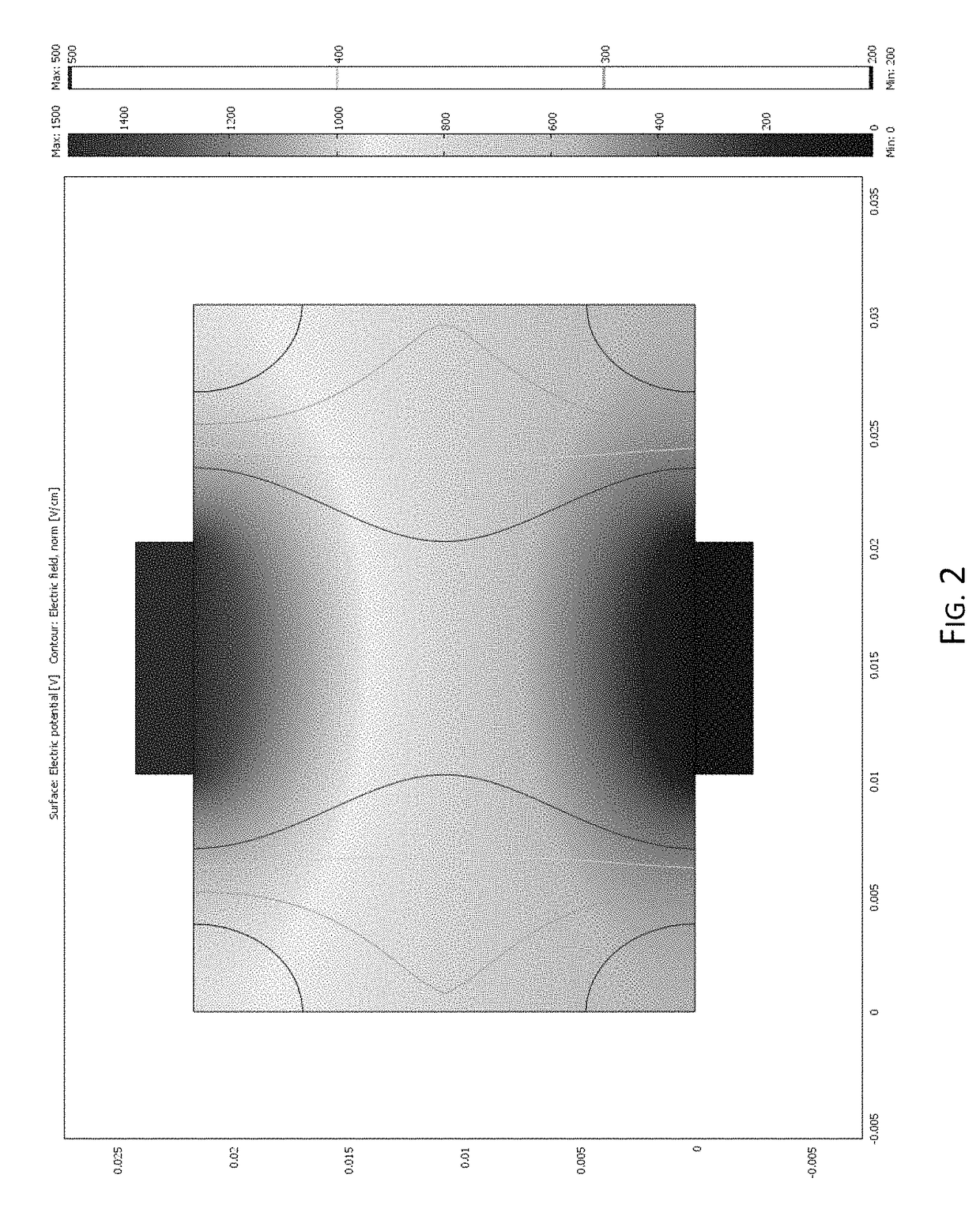
[0109]Preliminary multiphysics modeling was conducted to establish the feasibility of using the vascular system as a pathway for electrical pulse delivery. The electric field distribution, which is the key factor for the development of pores (reversible or irreversible) in the cell membrane, was modeled using a finite element method. Three liver tissue models were developed representing the liver structure with different levels of detail. The Comsol Multiphysics was used to solve for the electric potential (φ) which obeys the Laplace Equation:

∇·(σ∇φ)=0

[0110]where σ is the electrical conductivity.

[0111]Model 1:

[0112]Initially, the liver was modeled as a 2 cm thick homogenous structure with 1 cm diameter plate electrodes on either side of the tissue. This model was used to correlate experimental lesions to electric field intensities. A single 10 microsecond 1500 V / cm pulse was delivered to the top electrode while the bottom electrode was grounded. As shown in FIG. 2, the r...

example ii

Active Perfusion

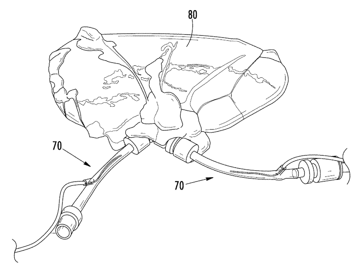
[0142]Young mixed breed pigs were sacrificed by way of barbiturate overdose. Livers were harvested and placed on ice within 15 minutes of death. Vascular anastomosis with the perfusion system was created by inserting Luer lock syringe connections into the portal vein, hepatic artery, and major hepatic vein, which were then secured with zip ties. The livers were flushed with lactated Ringer's solution (LRS) to remove blood / clots before placement on the perfusion system.

[0143]The VasoWave™ Perfusion System (Smart Perfusion, Denver, N.C.) was used to perfuse the livers for 4 and 24 hours. This system produces a cardioemulating pulse wave to generate physiological systolic and diastolic pressures and flow rates within the organ. The system is capable of controlling the oxygen content of the perfusate above and below physiological norms. A perfusate, consisting of modified LRS, was delivered to the portal vein and hepatic artery and recycled back into the system through t...

example iii

Administered IRE with Active Perfusion

[0168]A normal kidney was procured, through standard and approved means, from a pig after death. When obtained, the renal artery and vein of the kidney were isolated and connected with fittings to allow anastomosis to a cardiovascular emulation system (CVES) (VasoWave 2.3™, Smart Perfusion LLC, Denver, N.C.). The organ was connected to the CVES and the kidney was flushed with 1.5 liters of Krebs-Hensleit / Glutathione physiologic solution over a period of 5 minutes.

[0169]The kidney was then anastomosed on the arterial and vascular connectors to the CVES and perfusion was started (arterial systolic pressure: 110 mm Hg, diastolic pressure 60 mm Hg, with a physiologic pulse train delivered at 60 beats per minute). Perfusion was performed at 25° C. The renal vein was left disconnected but the end of the tubing was elevated to generate a low backpressure (this approximated 1 mm Hg). Perfusion was stabilized over a 15 minute interval. The Krebs solution...


PUM

PropertyMeasurementUnit
thicknessaaaaaaaaaa
distancesaaaaaaaaaa
thicknessaaaaaaaaaa

Abstract

The present invention relates to the field of medical treatment of diseases and disorders, as well as the field of biomedical engineering. Embodiments of the invention relate to the delivery of Irreversible Electroporation (IRE) through the vasculature of organs to treat tumors embedded deep within the tissue or organ, or to decellularize organs to produce a scaffold from existing animal tissue with the existing vasculature intact. In particular, methods of administering non-thermal irreversible electroporation (IRE) in vivo are provided for the treatment of tumors located in vascularized tissues and organs. Embodiments of the invention further provide scaffolds and tissues from natural sources created using IRE ex vivo to remove cellular debris, maximize recellularization potential, and minimize foreign body immune response. The engineered tissues can be used in methods of treating subjects, such as those in need of tissue replacement or augmentation.

Description

CROSS-REFERENCE TO RELATED APPLICATIONS[0001]The present application is a National Stage application under 35 USC §371 of Application No. PCT / US11 / 62067, filed Nov. 23, 2011, which application is a Continuation-In-Part (CIP) application of U.S. patent application Ser. No. 12 / 491,151, filed Jun. 24, 2009, which relies on and claims the benefit of the filing date of U.S. Provisional Patent Application Nos. 61 / 075,216, filed Jun. 24, 2008; 61 / 125,840, filed Apr. 29, 2008; 61 / 171,564, filed Apr. 22, 2009; and 61 / 167,997, filed Apr. 9, 2009, and the present application is also a CIP application of U.S. patent application Ser. No. 12 / 432,295, filed Apr. 29, 2009, which relies on and claims the benefit of the filing date of U.S. Provisional Patent Application No. 61 / 125,840, filed Apr. 29, 2008; and the present application relies on and claims the benefit of the filing date of U.S. Provisional Patent Application No. 61 / 416,534, filed Nov. 23, 2010. The entire disclosures of all of these pa...

Claims


Application Information

Patent Timeline
Patent Type & Authority Patents(United States)
IPC IPC(8): A61B18/14A61K48/00A61M5/00A61B18/00A61N1/32
CPCA61B18/14A61K48/0075A61M5/00A61N1/327A61B2018/00613A61B2018/1472A61B2218/002A61B18/1492
Inventor SANO, MICHAEL B.DAVALOS, RAFAEL V.ROBERTSON, JOHN L.GARCIA, PAULO A.NEAL, II, ROBERT E.
Owner VIRGINIA TECH INTPROP INC
Why Eureka
Try Eureka