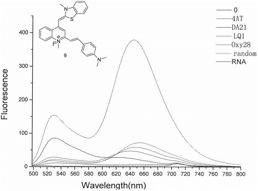
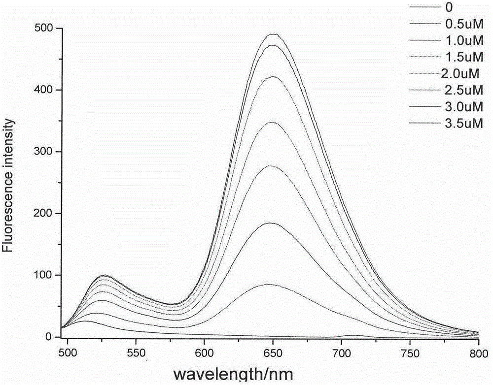
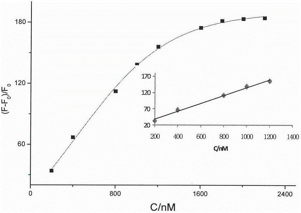
Thiazole orange phenylethylene compound serving as G-quadruplex nucleic acid fluorescent probe

A technology of fluorescent probes and quadruplexes, applied in fluorescence/phosphorescence, luminescent materials, organic chemistry, etc., can solve the problems of poor selectivity limiting the application of TO, failure to recognize the secondary structure of G-quadruplexes, etc., and achieve a good cell membrane Permeability, good photostability, enhanced fluorescence effect

Active Publication Date: 2016-11-23
GUANGDONG UNIV OF TECH
View PDF3 Cites 20 Cited by
  • Summary
  • Abstract
  • Description
  • Claims
  • Application Information

AI Technical Summary

Problems solved by technology

However, TO cannot effectively recognize the G-quadruplex secondary structure from other

Method used


Image

  • Thiazole orange phenylethylene compound serving as G-quadruplex nucleic acid fluorescent probe
  • Thiazole orange phenylethylene compound serving as G-quadruplex nucleic acid fluorescent probe
  • Thiazole orange phenylethylene compound serving as G-quadruplex nucleic acid fluorescent probe

Examples


Embodiment 1

[0035] Embodiment one: the synthesis of compound 2

[0036] Weigh 0.2g (1.1236mmol) of 4-chloro-quinaldine into a 25ml round bottom flask, add methyl iodide and sulfolane as a solvent, heat the mixture to 40-60°C, react for 18 hours, cool, and add anhydrous diethyl ether After shaking, suction filtration, the solid was washed several times, weighed after vacuum drying, and thin layer chromatography showed that there were no by-products, and 0.345g of pure product 2 was obtained, with a yield of 95.8%: 1 H NMR (400MHz, DMSO) δ8.56(d, J=8.4Hz, 1H), 8.46(d, J=8.3Hz, 1H), 8.22(t, J=8.1Hz, 1H), 8.01(t, J =7.9Hz, 1H), 7.55(s, J=7.4Hz, 1H), 4.20(s, 3H), 3.74(s, 1H), 2.68(s, 3H).

Embodiment 2

[0037] Embodiment two: the synthesis of compound 4

[0038] Weigh 0.25g (1.68mmol) of 2-methyl-benzothiazole into a 25ml round bottom flask, add methyl iodide and solvent absolute ethanol, react at 80°C for 15 hours, and cool the reacted solution to room temperature , then add the mixed solution of absolute ethanol and chloroform, shake and filter with suction, and wash the precipitate with a small amount of reagent, and obtain 0.448g of white powdery solid after vacuum drying, the yield is 91.7%: 1H NMR (400MHz, DMSO) δ8.44(d, J=8.1Hz, 1H), 8.30(d, J=8.4Hz, 1H), 7.90(t, J=7.8Hz, 1H), 7.81(t, J =7.7Hz, 1H), 4.20(s, 3H), 3.54(s, 1H), 3.17(s, 3H).

Embodiment 3

[0039] Embodiment three: the synthesis of compound 5

[0040] Weigh 0.50 g each of compounds 2 and 3, add them to a round-bottomed flask containing 10 ml of methanol, stir at room temperature, add a small amount of 0.5 mol / L sodium bicarbonate aqueous solution, and stir for about 1 hour. Add 4ml of saturated KI solution to the reacted solution, stir for about 5 minutes, then filter with suction, then wash with water and acetone to finally obtain a brick-red solid, which is dried, filtered, concentrated, and purified by column chromatography to obtain 0.98g of compound 4. The rate is 81.7%: 1 H NMR (400MHz, DMSO) δ8.77(d, J=8.3Hz, 1H), 8.18(d, J=8.7Hz, 1H), 8.02-7.96(m, 2H), 7.74(d, J=8.2Hz , 2H), 7.59(t, J=7.7Hz, 1H), 7.39(t, J=7.5Hz, 1H), 7.34(s, 1H), 6.85(s, 1H), 4.07(s, 3H), 3.98 (s,3H), 2.87(s,3H).


PUM


Abstract

The invention discloses a fluorescent probe, a preparation method thereof and an application of the fluorescent probe for detecting a nucleic acid G-quadruplex structure. The probe is provided with a structure in a general formula (I) is simple and stable in structure and easy to prepare. The probe can be used for specific detection of a nucleic acid G-quadruplex secondary structure, and the G-quadruplex secondary structure in solution can be rapidly detected by a fluorescence spectrometer or direct visual inspection under irradiation of a fluorescent lamp. The probe can be used for detecting the nucleic acid G-quadruplex structure in agarose gel or polyacrylamide gel and can also be used for detecting, marking or displaying existence or distribution of the G-quadruplex structure in living cells. Fluorescent materials have efficient and specific recognition capability for the nucleic acid G-quadruplex structure, have the advantages of excellent cell membrane permeability, low photo-induced toxicity, biological toxicity and photo-bleaching performance and the like, and overcome the shortcomings of high cost, high equipment requirement, complicated technical operation and the like in other detection methods.

Description

technical field [0001] The invention relates to a fluorescent probe and its preparation method, as well as its use in detecting the secondary structure of nucleic acid G-quadruplex in aqueous solution, in gel and in cells. Background technique [0002] Nucleic acid is not only the basic component of all biological cells, but also plays a dominant role in the growth, development, reproduction, inheritance and variation of organisms and other major life phenomena. Nucleic acid macromolecules are divided into two categories: deoxyribonucleic acid (DNA) and ribonucleic acid (RNA), which play a role in storing and transmitting genetic information in the replication and synthesis of proteins. [0003] G-quadruplex (G-quadruplex) is a special nucleic acid secondary structure. Many guanine-rich regions in the human genome have the ability to form this structure, including the guanine repeat at the end of the telomeric region, and the promoter regions of various genes, such as c-kit...

Claims


Application Information

Patent Timeline
IPC IPC(8): C09K11/06C07D417/06C07D417/14C12Q1/68G01N21/64
Inventor 卢宇靖邓强张焜方岩雄胡冬萍王郑亚杜志云黄宝华陈俊禧黄飞鸿
Owner GUANGDONG UNIV OF TECH
Why Eureka
Try Eureka