
Transparentizing reagent and application thereof in biological tissue material optical imaging as well as living body skin tissue transparentizing imaging method

A reagent and skin technology, applied in the field of clearing reagents, can solve the problems of low scale processing efficiency, destruction of fine tissue structure, increased damage to cell membranes, etc., and achieve the effect of good transparency rate and transparency effect, reducing tissue scattering, and improving imaging depth.

Active Publication Date: 2019-12-20
SHENZHEN TECH UNIV
View PDF6 Cites 4 Cited by
  • Summary
  • Abstract
  • Description
  • Claims
  • Application Information

AI Technical Summary

Problems solved by technology

Among them, SeeDB and FRUIT are gentle and simple, and the fluorescent protein signal is well preserved, and the tissue shape is well preserved, but the processing efficiency is low and the transparency achieved is not high
The process of Scale, CUBIC and ScaleS will lead to a large expansion of the organization, and the processing efficiency of Scale is low
CUBIC has high treatment efficiency and high transparency, but it utilizes a water-based solution containing high concentration (15%) of Triton X-100 to treat tissues. Although high concentration surfactant can accelerate the permeability of cell membrane, it also Substantially increased damage to cell membranes, which leads to disruption of tissue fine structure and incompatibility with cell membrane staining

Method used


Image

  • Transparentizing reagent and application thereof in biological tissue material optical imaging as well as living body skin tissue transparentizing imaging method
  • Transparentizing reagent and application thereof in biological tissue material optical imaging as well as living body skin tissue transparentizing imaging method
  • Transparentizing reagent and application thereof in biological tissue material optical imaging as well as living body skin tissue transparentizing imaging method

Examples


Embodiment 29

[0075] Embodiment 29, a kind of living body skin tissue transparent imaging method:

[0076] A. After anesthetizing the mouse, scrape off the ear hair with a razor blade and clean it up.

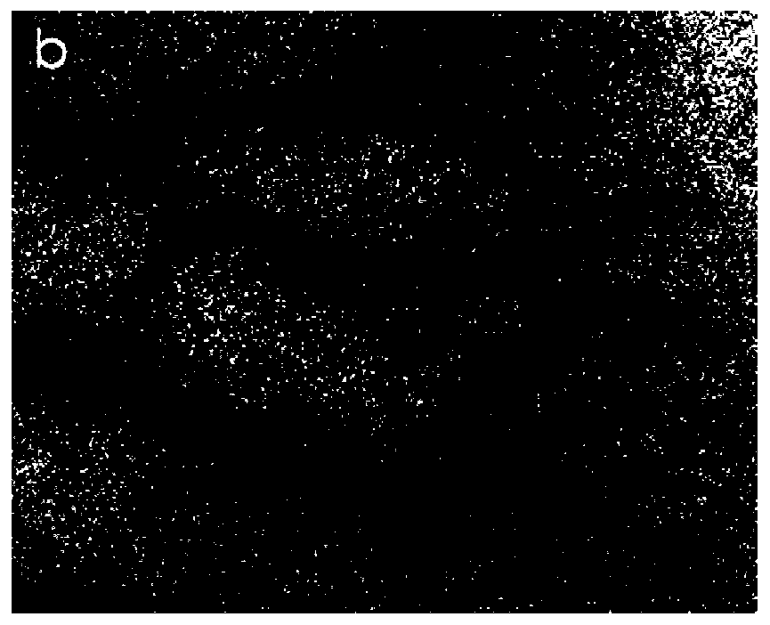
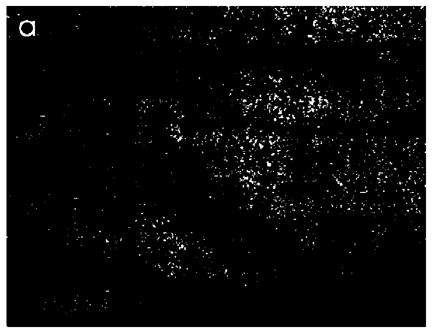
[0077] B. Fix the mouse with a mouse fixer, stick the mouse ear on the fixer with double-sided tape, and place it at the focal plane of the optical imaging system, fix the dried mouse ear at the imaging position, and image the mouse in two experiments get as Figure 1a , 2a The optical speckle image shown.

[0078] C, drip any clearing agent in embodiment 1-28 to the mouse ear that is fixed on the surface of the fixture, realize the light clearing of skin tissue after 5-30min (especially can use the clearing agent of embodiment 15-28 , to accelerate the transparentization process), it can be used for imaging in an optical microscopic imaging system. In this embodiment, the optically transparentized biological tissue is imaged with a laser speckle imaging system; The internal refractive in...

Embodiment 30

[0083] Embodiment 30. Imaging method for transparent skin tissue in vitro:

[0084] After the mouse was killed under anesthesia, its skin tissue (such as: back skin, mouse ear) was removed with a razor blade, and the hair was shaved off and cleaned up. Soak the isolated mouse ear tissue in the clearing reagent of Example 6 (the main component of which is mannose). After 10-30min, the optical transparency of the isolated tissue of the mouse ear was realized, and the camera was used to take pictures to obtain the following: image 3 As shown in the bright field image, it can be seen from the figure that compared with the clearing effects of PBS, fructose, glycerin, PEG 400, etc. in the prior art, the clearing agent using mannose in the present invention has a good clearing effect, and the 30min After that, the background grid lines behind the mouse ears can be clearly seen, but the complete background grid lines behind the mouse ears cannot be observed by using PBS, fructose, g...

Embodiment 31

[0088] Example 31, application in optical imaging of brain tissue.

[0089]For the fixation and separation of brain tissue, the embodiment adopts mouse brain, and its fixation and separation process is as follows: 20mL needle tubes are filled with paraformaldehyde and PBS (phosphate buffer saline) respectively, and the air bubbles are exhausted. The mice were anesthetized with chloral hydrate at a mass concentration of 10%, and after the mice were completely anesthetized, the mice were fixed with the ventral surface upward. Open the mouse chest cavity to fully expose the heart and liver, and expose the heart as much as possible. Find the apex of the mouse heart, pre-clamp the apex of the heart with hemostatic forceps, hold the needle and insert it from the apex slightly to the right, stop when there is a breakthrough feeling, cut the right atrial appendage, inject PBS, the red blood flows out from the right atrium and rushes out of the whole body, time For about 2-3 minutes, ...


PUM


Abstract

The invention discloses a transparentizing reagent and application thereof in biological tissue material optical imaging as well as a living body skin tissue transparentizing imaging method. The transparentizing reagent is mannose or a mixture formed by dissoving mannose and 1,3-dimethyl-2-imidazolinone in a solvent. The transparentizing reagent can be applied to the biological tissue material optical imaging. The living body skin tissue transparentizing imaging method comprises the following steps: removing hairs on the surface, cleaning and drying; fixing and placing a skin tissue on a focalplane of an optical imaging system; dropwise adding the transparentizing reagent to the skin tissue at an imaging position, and standing for 5-30 minutes; imaging; and after the imaging is finished,wiping the transparentizing reagent, and repeatedly scrubbing and cleaning the surface of the skin tissue with normal saline or phosphate buffer solution, so that the skin tissue restores the originalstate. The reagent disclosed by the invention can not produce obvious tissue expansion or tissue shrinkage when in use, a detailed structure of a biological material can be completely stored, transparentizing time is short, transparentizing capability is high, and the reagent can be applied to the biological tissue material optical imaging.

Description

technical field [0001] The invention belongs to the technical field of biological materials, and in particular relates to a clearing reagent for biological tissue and the application and application method of the clearing reagent in optical imaging of biological tissue materials. Background technique [0002] The skin has many important functions such as tissue nutrient supply, thermoregulation and metabolism. Among them, ensuring tissue nutrition is the most important function, and impaired skin function or insufficient nutrient supply often lead to certain diseases. Indeed, these injuries are often reflected in vascular diseases such as diabetes, hypertension, venous insufficiency, etc. Therefore, it is very important to obtain information about skin microcirculation. Non-invasive optical technology has shown great potential for monitoring skin microcirculation, by immersing skin tissue in some chemical reagents with high refractive index and high permeability to reduce ...

Claims


Application Information

Patent Timeline
Patent Type & Authority Applications(China)
IPC IPC(8): G01N21/84G01N21/17G01N21/59G01N21/64G01N1/28G01N1/30A61K49/00
CPCG01N21/84G01N21/1702G01N21/59G01N21/6458G01N1/286G01N1/30A61K49/00
Inventor 陈玲玲刘丽娜李仁剑李贵叶
Owner SHENZHEN TECH UNIV
Learn More