
Endostatin-like angiogenesis inhibition

an angiogenesis inhibitor and endostatin technology, applied in the field of cancer treatment, can solve the problems of difficult administration, degraded by the body, extremely expensive production, and doubtful whether enough protein therapeutics could be produced to treat all the required cancer patients, and achieve the effect of enhancing emission and inhibiting quenching

Inactive Publication Date: 2005-01-20
MINERVA BIOTECH
View PDF5 Cites 4 Cited by
  • Summary
  • Abstract
  • Description
  • Claims
  • Application Information

AI Technical Summary

Benefits of technology

[0036] Certain embodiments of the invention make use of self-assembled monolayers (SAMs) on surfaces, such as surfaces of colloid particles, and articles such as colloid particles having surfaces coated with SAMs. In one set of preferred embodiments, SAMs formed completely of synthetic molecules completely cover a surface or a region of a surface, e.g. completely cover the surface of a colloid particle. “Synthetic molecule”, in this context, means a molecule that is not naturally occurring, rather, one synthesized under the direction of human or human-created or human-directed control. “Completely cover” in this context, means that there is no portion of the surface or region that directly contacts a protein, antibody, or other species that prevents complete, direct coverage with the SAM. I.e. in preferred embodiments the surface or region includes, across its entirety, a SAM consisting completely of non-naturally-occurring molecules (i.e. synthetic molecules). The SAM can be made up completely of SAM-forming species that form close-packed SAMs at surfaces, or these species in combination with molecular wires or other species able to promote electronic communication through the SAM (including defect-promoting species able to participate in a SAM), or other species able to participate in a SAM, and any combination of these. Preferably, all of the species that participate in the SAM include a functionality that binds, optionally covalently, to the surface, such as a thiol which will bind to a gold surface covalently. A self-assembled monolayer on a surface, in accordance with the invention, can be comprised of a mixture of species (e.g. thiol species when gold is the surface) that can present (expose) essentially any chemical or biological functionality. For example, they can include tri-ethylene glycol-terminated species (e.g. tri-ethylene glycol-terminated thiols) to resist non-specific adsorption, and other species (e.g. thiols) terminating in a binding partner of an affinity tag, e.g. terminating in a chelate that can coordinate a metal such as nitrilotriacetic acid which, when in complex with nickel atoms, captures a metal binding tagged-species such as a histidine-tagged binding species. The present invention provides a method for rigorously controlling the concentration of essentially any chemical or biological species presented on a colloid surface or any other surface. Without this rigorous control over peptide density on each colloid particle, co-immobilized peptides would readily aggregate with each other to form micro-hydrophobic-domains that would catalyze colloid-colloid aggregation in the absence of aggregate-forming species present in a sample. This is an advantage of the present invention, over existing colloid agglutination assays. In many embodiments of the invention the self-assembled monolayer is formed on gold colloid particles.
[0037] A drug candidate may he studied for competition with the analyte for binding of one of the species, or binding with one site on the analyte. In this case, the analyte may be provided as a known species. Presence of the drug candidate will thus inhibit immobilization of the first and second colloid particles relative to each other, and thus will inhibit quenching. Alternative embodiments involve enhancing emission or shifting the wavelength of emission or absorption of a first molecule, by a second molecule on a second colloid particle.
[0038] This colloid / colloid aggregation technique can be used to identify the binding partners of drugs or proteins of interest. This can be accomplished by attaching the drug or protein to one set of colloids and possible binding partners to other sets of colloids and assaying for a binding interaction between the two sets of colloids. Once a biological target of a drug or protein has been identified, candidate drugs can be added to the assay in the presence of the colloid-attached binding partners to disrupt binding of the drug or protein to the cognate ligand, allowing identification of synthetic mimics of the drug or protein on the first set of colloids. This technique is very useful in identifying the biological target of orphan drugs or uncharacterized proteins for diagnostic or drug-screening purposes. This technique will also allow identification of synthetic replacements or “mimics” of currently used drugs that are expensive or difficult to produce.
[0039] In one embodiment, an angiogenesis inhibitor is attached to one set of colloids (via an affinity tag linkage, chemical coupling, or nonspecific adsorption), and its biological target is attached to another set of colloids. For the unique case of an angiogenesis inhibitor that has two or more ligand-binding sites, such as endostatin, the ligand may be attached to one set of colloids and the angiogenesis inhibitor may be added in solution. Drug candidates are added and assayed for their ability to disrupt the binding interaction. Any drug that inhibits the interaction is then attached to a third set of colloids and assayed for binding to the angiogenesis inhibitor and the biological target of the angiogenesis inhibitor. A drug that binds to the biological target of the angiogenesis inhibitor and inhibits binding of the angiogenesis inhibitor to its target can be deemed a “mimic” of the angiogenesis inhibitor, and may be used as a replacement drug. This assay may be used to screen for mimics of virtually any drug. It is of specific interest for drug screening for synthetic replacements of angiogenesis inhibitors, which are both costly and difficult to produce. The assay can be used to identify synthetic replacements for endostatin, through disruption of the endostatin-vitronectin or endostatin-RGD-peptide interactions; angiostatin, through disruption of the angiostatin-ATP-synthase or angiostatin-vitronectin interaction; or TNP-470 through disruption of the TNP-470-methionine-aminopeptidase interaction. As in other colloid / colloid assays, color change, fluorescence quenching, or other emissive molecule enhancement or suppression and the like can be indications of a result. Study of RGD / endostatin interaction is described in examples 1 and 2 below.
[0040] This colloid / colloid aggregation technique also can be used for discovery of angiogenesis inhibitors or ligands involved in angiogenesis pathways. In one assay, suspected angiogenesis inhibitors or proteins can be immobilized relative to (e.g., fastened to) a first colloid particle. Second colloid particles can be immobilized with respect to molecules that have been implicated in angiogenesis and / or metastasis, such as basement membrane proteins, integrins, or adhesion molecules. If a particular angiogenesis inhibitor binds to the basement membrane protein, integrin, or adhesion molecule immobilized on the second set of colloids, then the two sets of colloids will become immobilized with respect to each other and the binding interaction will become detectable by methods of the invention such as color change, precipitation, etc. Once an angiogenesis inhibitor is identified by this method, candidate drugs for disruption of the binding can be screened. If the drugs disrupt interactions, then colloid particles will not immobilize relative to each other or will do so to a lesser degree. This assay can be used with known angiogenesis inhibitors to identify or verify the biological targets of the angiogenesis inhibitors. Drug candidates can then be added to the assay to identify other drugs that act on the same biological target.
[0041] Another embodiment in which colloid particles can be immobilized relative to each other in such assays involves colloids each being immobilized with respect to a common surface. The common surface can be a surface of another colloid particle presenting binding partners of species on the first colloid particles. The common surface can also be the surface of an article such as a membrane such as a nitrocellulose membrane, a chip surface, a surface of an article derivatized with a SAM, or the like. In preferred embodiments, the surface to which the colloid particles can bind includes binding sites at a high enough density so that if binding occurs (between species on the common surface and species on the colloid particles), the colloid particles will be brought into close enough proximity that detection (via color change characteristic of aggregation, quenching of fluorescence, or other property described herein) can occur.

Problems solved by technology

A drawback of using angiostatin and endostatin as cancer therapeutics is that they are proteins, which are hard to administer, easily degraded by the body and extremely expensive to produce.
In fact it is questionable whether enough of these protein therapeutics could be produced to treat all the required cancer patients.
For several reasons, it has been difficult to identify new angiogenesis inhibitors.
First, the biological process of vascularization is not yet well understood.
These assays are expensive, time-consuming and not compatible with high throughput.
The complex tertiary structure of proteins and antibodies makes it difficult or impossible to optimize them.

Method used


Image

  • Endostatin-like angiogenesis inhibition
  • Endostatin-like angiogenesis inhibition
  • Endostatin-like angiogenesis inhibition

Examples


example 1

Detection of the Biotin-Streptavidin Interaction Using Gold Colloids

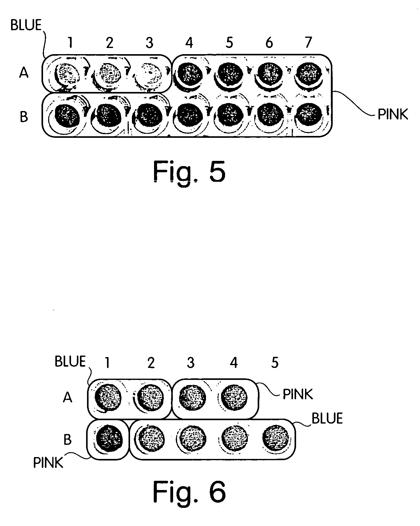
[0053] Gold colloids were prepared using a mixture of 10 μM biotin thiol, 10 μM NTA thiol, and 580 μM C11 thiol. Control colloids were prepared using 20 μM NTA thiol and 580 μM C11 thiol for a total thiol concentration of 600 μM. After deposition, the colloids were heat cycled in 400 μM EG3 thiol, and charged with nickel sulfate. A streptavidin stock solution (1 mg / mL) was prepared in 10 mM sodium phosphate buffer, 100 mM sodium chloride, pH 7.4 (buffer) To detect the biotin-streptavidin interaction, 60 μL buffer, 10 μL streptavidin, and 30 μL colloids, were mixed in a well of a 96-well plate. The plate was incubated at room temperature and observed for color change. At the three highest concentrations of streptavidin (0.1 mg / mL, 0.01 mg / mL, 0.001 mg / mL) the colloids presenting biotin turned blue (wells A1, A2, A3, FIG. 5). At lower concentrations of streptavidin, the wells containing biotin colloids remained pink ...

example 2

The Angiogenesis Inhibitor, Endostatin Specifically Binds to a His-Tagged GRGDS Motif Peptide (HHHHHHSSSSGSSSSGSSSSGGRGDSGRGDS), but Angiostatin Does Not

[0055] 200 μL NTA-Ni agarose (Qiagen) were washed 2× with 100 μL ddH2O, then with wash buffer A, containing 50 mM NaH2PO4, 300 mM NaCl, and 10 mM imidazole at pH 8.0.

[0056] A synthetic peptide, was dissolved in DMSO then diluted in phosphate buffer to a final concentration of 1 mM. 100 μL of this peptide solution was incubated with the NTA-Ni resin for 20 minutes at room temperature to allow binding of the histidine-tagged peptide to the NTA-Ni resin. The resin was then pelleted and the supernatant was removed. The resin was washed in buffer A. The peptide bound resin was then divided into two aliquots. A first aliquot was mixed with 100 μL human recombinant endostatin (0.1 mg / mL in 10 mM sodium phosphate buffer, 100 mM sodium chloride, pH 7.4, diluted from stock endostatin, Calbiochem 324746). A second aliquot was mixed with 100 ...

example 3

Drug Screen for Synthetic Mimics of Endostatin

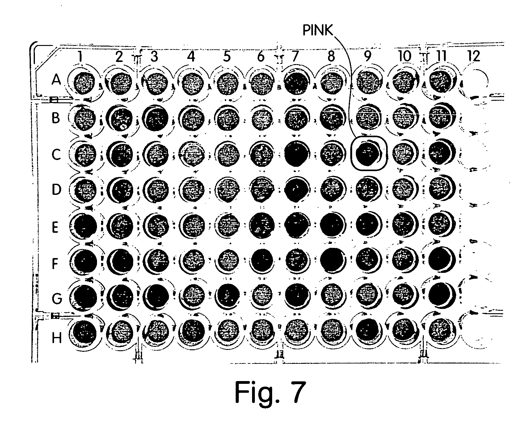
[0058] 40 μM NTA colloids presenting a His-tagged peptide containing a tandem repeat GRGDS motif (Sequence ID No. 1; Table 1) were prepared by incubating 2.1 mL colloids with 210 μl 100 μM His-RGD for ten minutes pelleting the colloids to remove excess unbound peptide, and resuspending the colloids in 10 mM sodium phosphate buffer (pH 7.4). Negative control colloids were prepared by substituting an irrelevant His-tagged FLR peptide (Sequence ID No. 2, see Table 1). 25 μl GRGDS-colloids (or random peptide-colloid for negative controls) was added to each well of a 96-well plate along with 65 μl sodium phosphate buffer per well. DMSO was added in place of a drug to the positive and negative controls. 5 μl of 0.1 mg / ml endostatin (Calbiochem) was added to each well. The plate was incubated in room temperature and observed for color change. After about 20 minutes, the positive controls changed color from pink to blue as the endostatin bound ...


PUM


Abstract

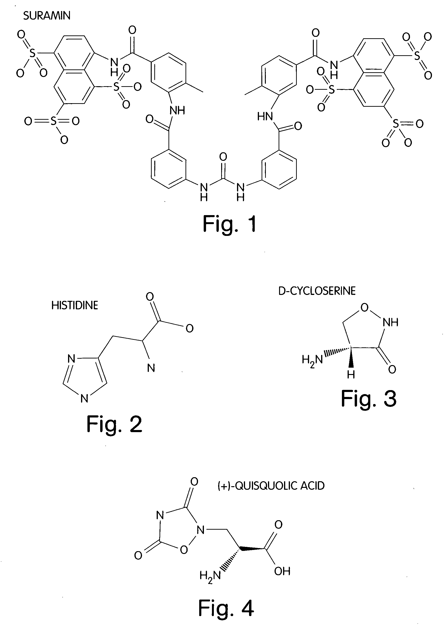
A treatment for cancer is provided. The treatment may include administering a therapeutic amount of L-histidine, D-cycloserine, quisqualic acid or suramin or analogs thereof.

Description

RELATED APPLICATIONS [0001] This application is a continuation of pending U.S. patent application Ser. No. 10 / 003,681, filed Nov. 15, 2001; and claims the benefit of priority to U.S. provisional patent application Ser. No. 60 / 248,865, filed Nov. 15, 2000; and U.S. provisional patent application Ser. No. 60 / 277,922, filed Mar. 22, 2001. Each of these applications is incorporated by reference herein.FIELD OF THE INVENTION [0002] The invention relates to treatments for cancer, and particularly to treatments using angiogenesis inhibitors. BACKGROUND OF THE INVENTION [0003] Angiogenesis is the name given to the in vivo process of new blood vessel formation. It is widely believed that cancer may be effectively treated by reducing or eliminating the supply of blood to a tumor. Angiogenesis inhibitors are a class of compounds that somehow act to interrupt the process of new blood vessel formation. Because adults do not, in general, require much new blood vessel formation, it is thought that...

Claims


Application Information

Patent Timeline
IPC IPC(8): A61K31/185A61K31/255A61K31/365A61K31/4015A61K31/415A61K31/4174A61K31/42A61K31/4245A61P35/00
CPCA61K31/185A61K31/255A61K31/365A61K31/4015B82Y30/00A61K31/4174A61K31/42A61K31/4245A61K31/415A61P35/00
Inventor SHENDELMAN, R. SHOSHANA BAMDADBAMDAD, CYNTHIA C.
Owner MINERVA BIOTECH
Learn More