Plastic microfluidic chip and methods for isolation of nucleic acids from biological samples

Inactive Publication Date: 2007-01-18
TRUSTEES OF BOSTON UNIV
View PDF9 Cites 99 Cited by
  • Summary
  • Abstract
  • Description
  • Claims
  • Application Information

AI Technical Summary

Benefits of technology

[0016] The present invention is directed to methods of manufacture of microfluidic chips such as plastic microfluidic chips, which has channels packed with polymer-embedded particles and uses thereof. The chip of the present invention is designed for application of an untreated biological sample on the chip thus allowing isolation, purification and detection of biomolecules, such as nucleic acids or proteins or peptides in one step. The invention also p

Problems solved by technology

Silicon and glass fabrication can be very expensive, while PDMS lacks dimensional stability and has limited shelf-life.
These limitations necessitate the use of alternative materials to make disposable, point-of-care devices, for example, for diagnostic applications.
However, this device is not suitable for purification and isolation of nucleic acids.
These methods are very expensive.
However, the sol-gel chemistry involves high temperatures and is not suitable for in situ applications of the polymeric devices.
Such methods are difficult to implement in other than full diagnostic laboratory settings.
This prevents them from being used for, for example critical bacterial strain detection when analyzing causative agents for infections.
Problems also exist with conventional immunoassays.
They often require long assay times; require difficult fluid handling techniques and use of relatively large quantity of sample material and reagents.
These problems again prevent these assays from becoming a point-of-care diagnostic technique.
Gett

Method used


Image

  • Plastic microfluidic chip and methods for isolation of nucleic acids from biological samples
  • Plastic microfluidic chip and methods for isolation of nucleic acids from biological samples
  • Plastic microfluidic chip and methods for isolation of nucleic acids from biological samples

Examples


Example

Example 1

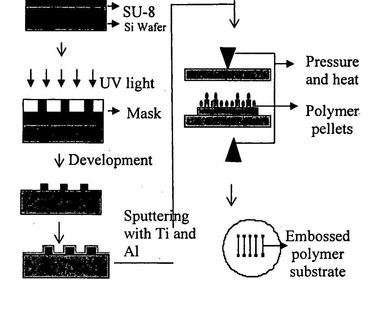
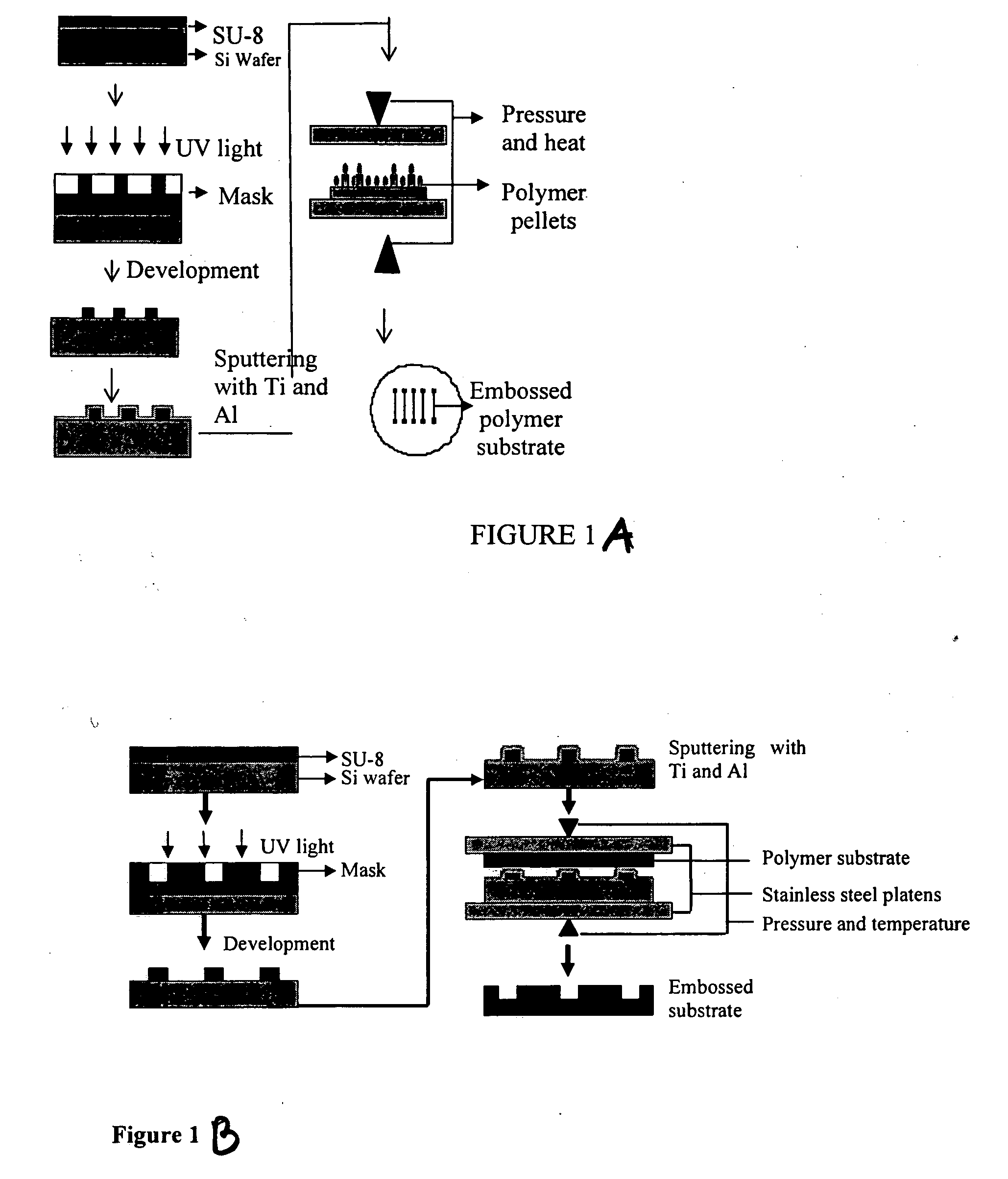
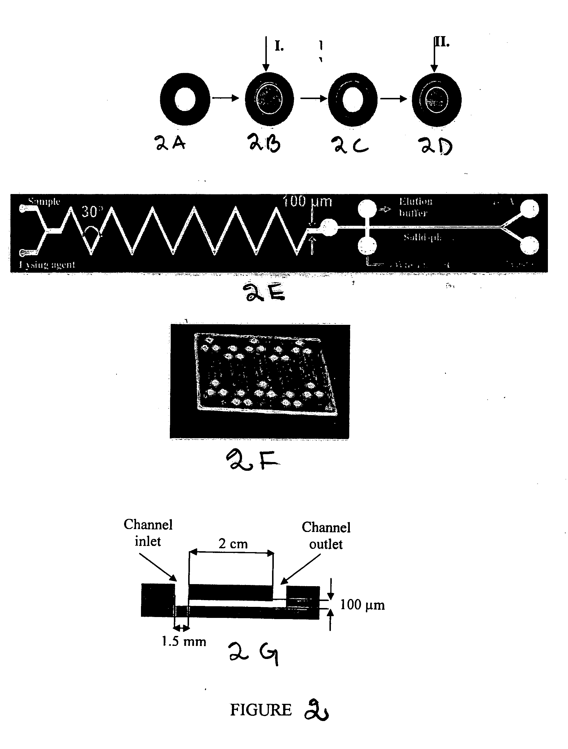
[0146] Device fabrication: The microfluidic channels were fabricated by hot embossing with an SU-8 master. Channels of 100 μm and 165 μm depths were fabricated by this method. The widths of the channels varied from 50 μm to 250 μm and wells of 1.5 mm diameter were drilled at the end of the channels for sample introduction and collection. The SU-8 masters were fabricated on piranha-cleaned silicon wafers by spinning SU-8 50 photoepoxy (Microchem, Newton, Mass.) at a thickness of 100 μm and 165 μm onto the wafers. After pre-baking the wafers for 30 min at 95° C., the pattern was transferred through a mask by contact lithography. This was followed by development with SU-8 developer (Microchem) and post-baking the wafers for 1.5 h at 175° C. After this fabrication process, the SU-8 molds exhibited glass-like mechanical properties and had the negative pattern of the channels. The wafers were then sputter coated with 500 Å of Ti for adhesion, followed by 1000 Å of Al.

[0147] The...

Example

Example 2

Immunoassay Methods Using the Microfluidic Device of the Invention

[0154] This example describes the development of fluorescence and chemiluminescence based microfluidic immunoassay techniques on a thermoplastic microchip. The immunoassays were applied to determine femtomolar concentrations of C-reactive protein (CRP), a classic inflammation marker associated with cardiovascular diseases. Because of the very high sensitivity of the described immunoassay techniques, they are suitable for developing saliva-based diagnostic tests. The microfluidic chips were fabricated in cyclic polyolefin by hot-embossing techniques. The surface of the microchannels were modified by immobilizing protein A to increase the sensitivity of the immunoassays, since protein A has high affinity for immunoglobulin G (IgG) of most species. Concentrations of CRP were determined on-chip by both fluorescence and chemiluminescence based detection methods. A heterogeneous., sandwich immunoassay scheme was ...

Example

Example 3

Purification of Biomolecules

[0176] The DNA extraction efficiency of the monolith / silica columns was tested through spectroscopic measurement of absorption at 260 nm. The extraction procedure itself consisted of load, wash and elution steps. The loading solution consisted of 0.5 μg / mL of λ DNA in chaotropic buffer containing GuSCN (guanidium thiocyanate) and 3% BSA (Bovine Serum Albumin). 3% BSA was added to confirm that the separation column was able to separate nucleic acids in the presence of proteins. The microchannels were conditioned with the loading buffer (without DNA) for 5 min before the subsequent extraction in the channel. Then 75 μL of the loading solution was passed through the microchannel at a flow rate of 300 μL / hour with a KDS100 syringe pump (manufactured by KD Scientific, Holliston, Mass.). The syringe was connected to the microchip using 360-μm-i.d. PEEK tubing and NANOPORT™ fittings. 75 μL of the wash buffer consisting of 70% ethanol was then passed t...


PUM

PropertyMeasurementUnit
Lengthaaaaaaaaaa
Diameteraaaaaaaaaa
Diameteraaaaaaaaaa

Abstract

The present invention is directed to methods of manufacture of microfluidic chip such as a plastic microfluidic chips, which has channels packed with polymer-embedded particles and uses thereof. The chip of the present invention is designed for application of an untreated biological sample on the chip thus allowing isolation, purification and detection of biomolecules, such as nucleic acids, proteins or peptides in one step. The invention also provides a microfluidic chip for combined isolation, purification and detection of biomolecules thus providing a complete Lab-on-a-Chip analysis system for biomolecules such as nucleic acids and proteins. The chips of the invention can be adapted to perform highly specific immunoassays and diagnostic test, for example, for diagnosis of infectious agents, such as bacteria, viruses or parasites.

Description

CROSS-REFERENCE TO RELATED APPLICATIONS [0001] This application claims benefit of U.S. provisional applications Ser. No. 60 / 674,833, filed Apr. 26, 2005, and 60 / 760,691, filed Jan. 20, 2006, the contents of which are herein incorporated by reference in its entirety.BACKGROUND OF THE INVENTION [0002] 1. Field of the Invention [0003] The present invention relates to a device and methods for their manufacture as well as isolation, purification and detection of biological molecules, such as nucleic acids and proteins. Specifically, the invention relates to the preparation of microfluidic chips that comprise polymer-embedded particles and methods for solid-phase isolation, purification and detection, of biological molecules using such microfluidic chips. Immunoassays and diagnostic tests, for example for detecting microorganisms, such as bacteria, using the device are also provided. [0004] 2. Description of the Related Art [0005] The extraction and detection of biomolecules, such as nucl...

Claims


Application Information

Patent Timeline
IPC IPC(8): C12Q1/68C12P19/34C12M3/00
CPCB01J20/28026B01J20/28042B01L3/5023B01L3/502707B01L2200/0631B01L2200/10G01N1/405B81B2201/051B81B2203/0338B81C1/00206B81C2201/0197C12Q1/6813B01L2200/12
Inventor KLAPPERICH, CATHERINE M.BHATTACHARYYA, ARPITA
Owner TRUSTEES OF BOSTON UNIV
Why Eureka
Try Eureka