Compositions and Methods for Making and Using Laminin Nanofibers

a technology of laminin nanofibers and compositions, applied in the field of compositions, methods, and apparatuses for preparing and using electrospun laminin, can solve the problems of mesh loss, large percentage of porosity and surface roughness, and inability to manufacture feature sizes on the nanometer scale with traditional printing and etching techniques, etc., to achieve novel biomimetic effects, improve the effect of tensile strength and long shelf li

Inactive Publication Date: 2010-05-13
UNIV OF VIRGINIA ALUMNI PATENTS FOUND
View PDF9 Cites 26 Cited by
  • Summary
  • Abstract
  • Description
  • Claims
  • Application Information

AI Technical Summary

Benefits of technology

[0020]The nanofiber meshes prepared by the methods of the invention should have a very long shelf life stored with desiccation. They have far greater tensile strength than matrigel gels. The nanoscale fibers are more similar to the fibers seen by cells encountering laminin in real basement membranes, thus they may be expected to demonstrate novel biomimetic effects. The materials fabricated by this process may, for example, be used as an anhydrous coating of scaffold biomaterials for tissue engineering, as well as substrate for ex vivo cultivation of both specialized tissue cells and stem cells. The latter could be a tremendous aid to basic science research as differentiation and phenotype expression of cells on biomimetic laminin scaffolds may be more representative of in vivo behavior.
[0021]Due to the sensitivity of laminin nanofibers, glutaraldehyde crosslinking may destroy the bioactivity of the laminin protein. The present invention provides compositions and methods for electrospun laminin which does not have to be crosslinked. In one aspect, the solvent HFP is used and laminin activity remains, and no cross-linking is required.

Problems solved by technology

Previous efforts to manufacture feature sizes on the nanometer scale have been unsuccessful with traditional printing and etching techniques [11].
However, previous studies demonstrated that the bioactive properties of laminin are fragile and often destroyed by processing methods required to form laminin substrates for in vitro cell culture studies including lyophilization and exposure to ultraviolet light [16].
Other groups have faced this challenge when electrospinning interstitial collagens, and one might expect to encounter similar obstacles with laminin.
While glutaraldehyde crosslinking does add some structural stability to the nanofiber matrices, the meshes lose a large percentage of their porosity and surface roughness.
In addition, glutaraldehyde is cytotoxic, and may be difficult to entirely remove after crosslinking treatment [18].

Method used


Image

  • Compositions and Methods for Making and Using Laminin Nanofibers
  • Compositions and Methods for Making and Using Laminin Nanofibers
  • Compositions and Methods for Making and Using Laminin Nanofibers

Examples


embodiments

[0159]The present invention provides compositions and methods for mimicking three dimensional scaffolding as found in vivo to better mimic how cells grow and differentiate. Cell proliferation and differentiation are regulated by unique spatial interactions between cells. Spatial cues in conjunction with the topologically distinct location of specific attachment molecules, and the release of specific humoral factors, such as growth and differentiation factors, function as signals to the cell to proliferate, differentiate, migrate, remain in a resting state, or initiate apoptosis. The capacity of the cell to respond to these signaling triggers is dependent on the availability of specific cell surface and intracellular receptors. The signal transduction pathways that are stimulated by these molecules depend on the organization and structure of the cell cytoskeleton whose architecture is a function of multipoint cell surface interactions with these signaling molecules, surrounding cells...

example 1

Materials and Methods

[0257]The solvent, 1,1,1,3,3,3-hexafluoro-2-propanol (HFP) was purchased from Sigma (St Louis, Mo.). All cell culture reagents were purchased from Fisher Scientific (Pittsburgh, Pa.).

[0258]Laminin Isolation

[0259]Laminin I was purified from the EHS tumor according to previously established methods. The final laminin solution was subjected to 2 rounds of precipitation with 45% ammonium sulfate to remove most growth factors present. Purity of laminin was evaluated by SDS-PAGE and Western analysis with affinity purified antibodies to type IV collagen, entactin / nidogen and perlecan, the major contaminants of such preparations. Purity was determined to be greater than 99% laminin (w / v). Laminin was stored at −80° C.

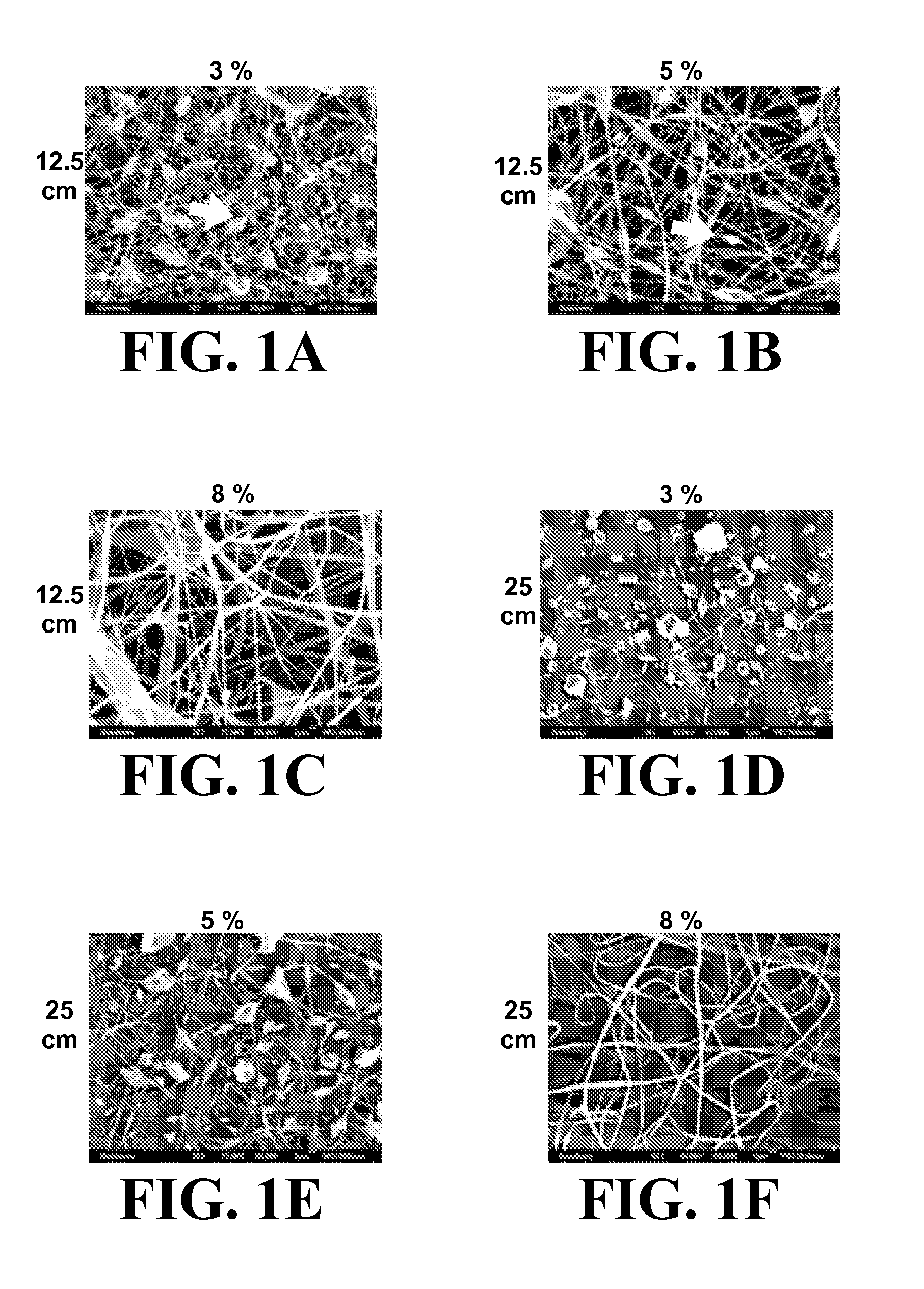
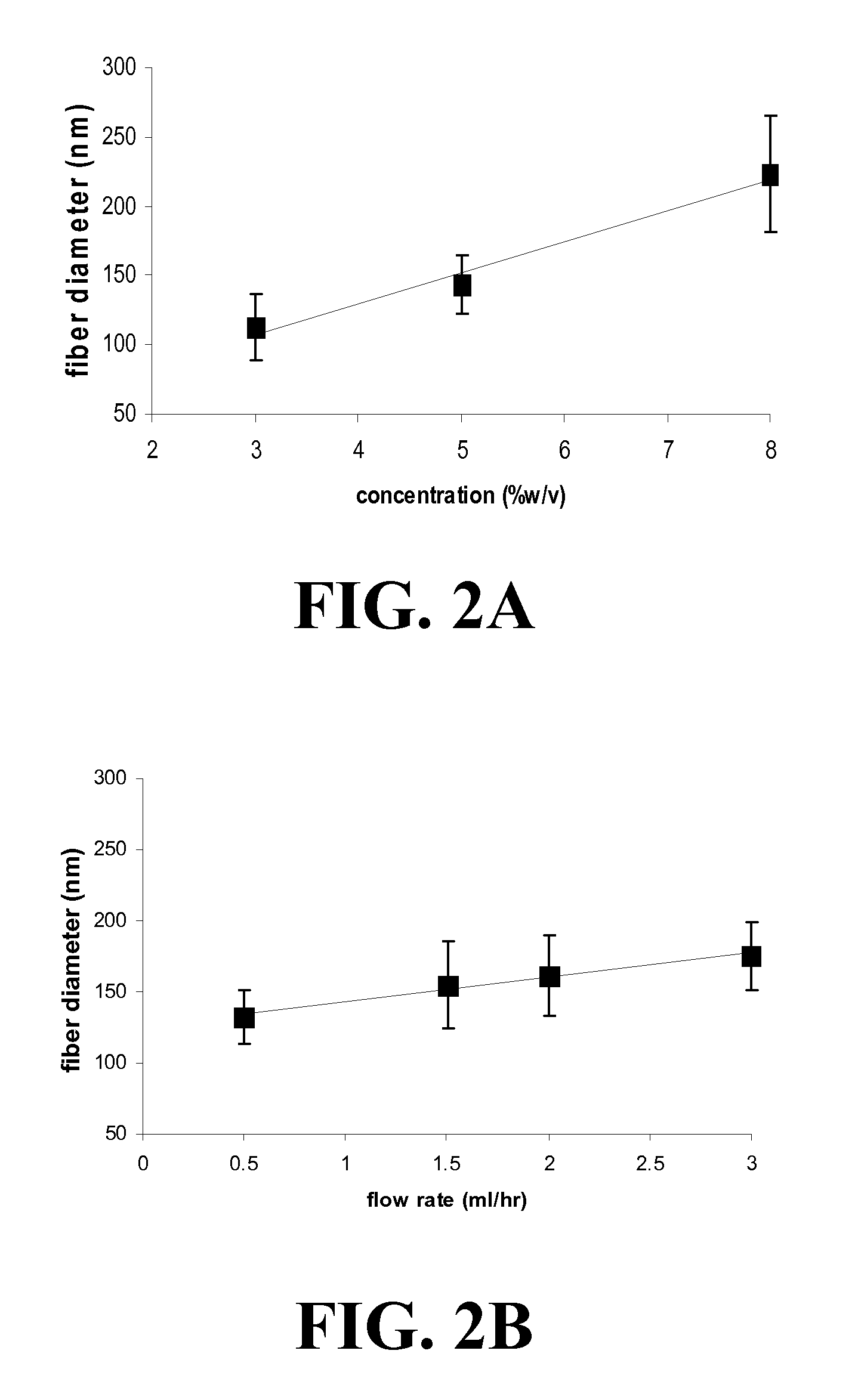
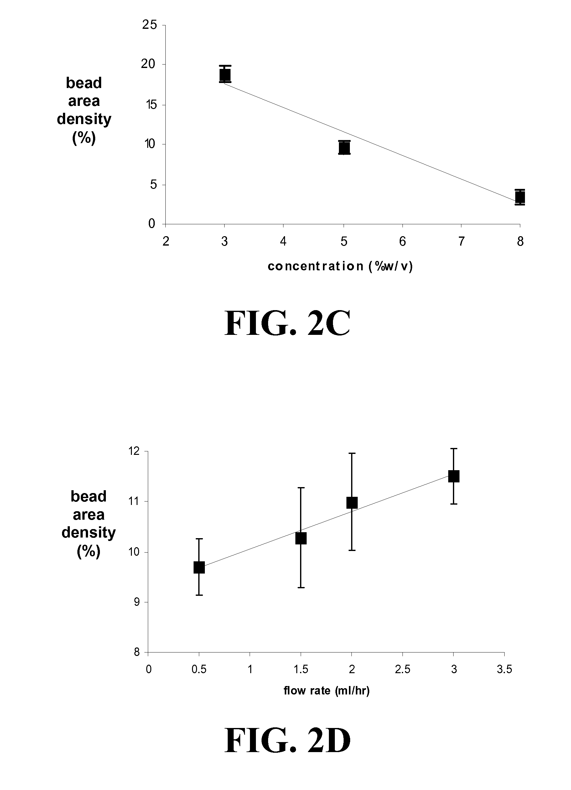
[0260]Laminin Electrospinning

[0261]For the parametric study, a series of process parameters was chosen within ranges shown to be successful in creating submicron or nanoscale fibers of other ECM proteins such as collagens [13] and fibrinogen. Laminin was di...

example 2

Laminin Nanofiber Mesh Substrates for Stem Cell Growth and Differentiation

[0295]Methods—Embryonic Stem Cell Culture: D3 and ES-E14TG2a murine embryonic stem cells were cultured on STO or CF1 mouse embryonic fibroblast feeder layers, fed daily and sub-cultured every 2 or 3 days. The media used was DMEM+15% ES-qualified FBS supplemented with L-glutamine, non essential amino acids, pyruvate, 2-mercaptoethanol, and leukemia inhibitory factor (Chemicon). All tissue culture reagents were from GIBCO except as noted.

[0296]Fabricated meshes of laminin I nanofibers (LNFs) with fiber size (10-150 nM dia.), geometry, and porosity of authentic basement membranes were fabricated using electrospinning methods. Unlike previously described NFs synthesized from protein polymers, meshes of LNFs retain their structural features when wetted and do not require fixation by chemical cross-linking, which often destroys biological activity. Embryonic stem cells (ESCs) and multipotent stem cells from adipose ...


PUM

PropertyMeasurementUnit
flow rateaaaaaaaaaa
flow rateaaaaaaaaaa
flow rateaaaaaaaaaa

Abstract

The present invention encompasses methodologies and parameters for the formation of nanofibrous (to microfibrous) laminin via electrospinning. The present application discloses conditions and appropriate parameters to synthesize laminin fibers from a diameter of about 10 nM to a diameter of over 1,000 nM via electrospinning.

Description

CROSS REFERENCE TO RELATED APPLICATIONS[0001]This application is entitled to priority pursuant to 35 U.S.C. §119(e) to U.S. provisional patent application No. 60 / 927,583, filed on May 4, 2007, the entirety of which is incorporated by reference herein.STATEMENT REGARDING FEDERALLY SPONSORED RESEARCH OR DEVELOPMENT[0002]This invention was supported in part by Grant No. DE-010369-08 awarded by the National Institutes of Health and Grant No. 736002 awarded by the National Science Foundation. The United States Government therefore has certain rights in the invention.FIELD OF THE INVENTION[0003]The present invention relates to compositions, methods, and apparatuses for preparing and using electrospun laminin.BACKGROUND[0004]Laminins are a family of large extracellular matrix (ECM) proteins found primarily in basement membranes associated with all epithelial, endothelial, muscle, fat and Schwann cells. The laminins serve critical functions in cell attachment, growth, migration, and differe...

Claims


Application Information

Patent Timeline
Patent Type & Authority Applications(United States)
IPC IPC(8): C12N11/02C07K1/02C12M1/00
CPCC12N5/0068D01F4/00D01D5/0038C12N2533/52
Inventor OGLE, ROY CLINTONBOTCHWEY, III, EDWARD A.
Owner UNIV OF VIRGINIA ALUMNI PATENTS FOUND
Why Eureka
Try Eureka