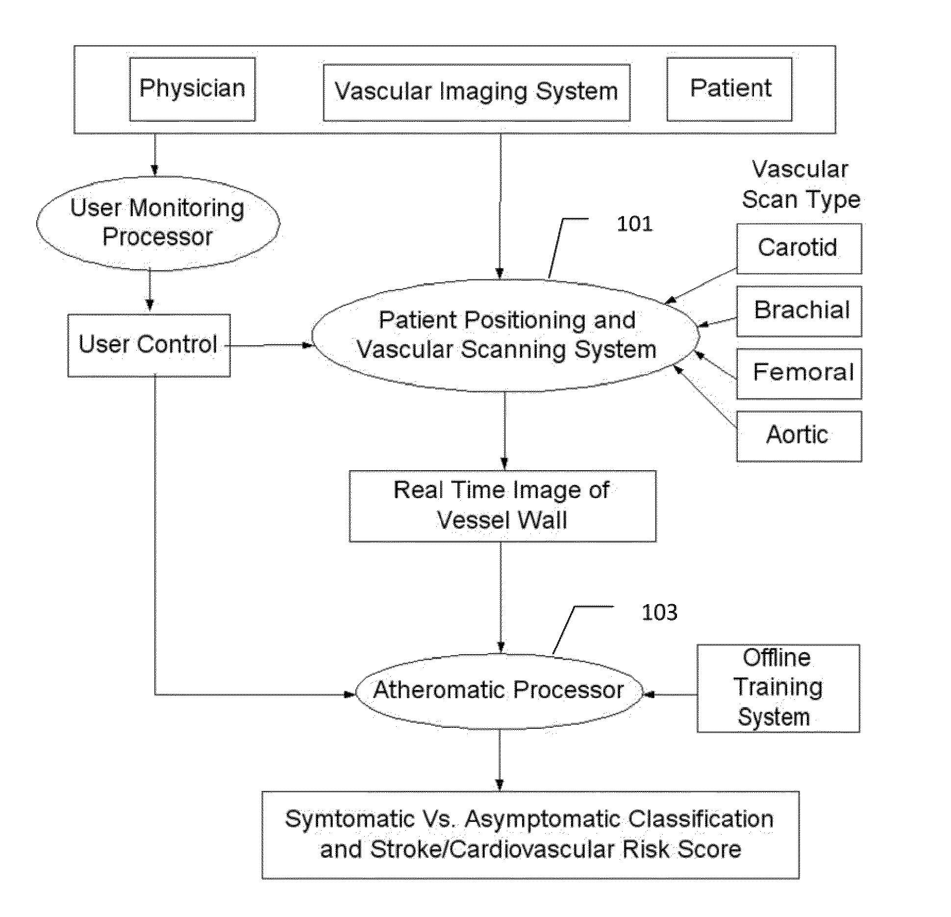
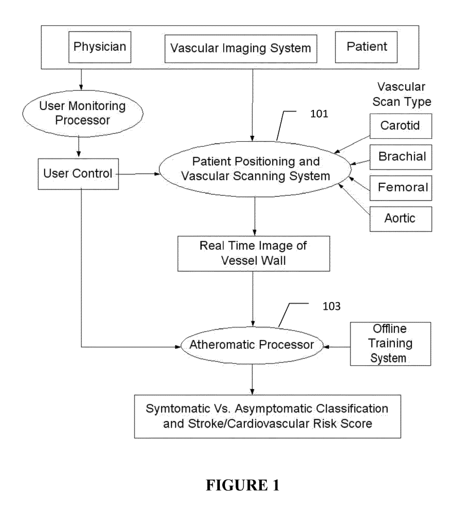
[0008]The on-line system consists of Atherosclerotic Wall Region estimation using AtheroEdge™ (for longitudinal Ultrasound) or Athero-CTView™ (for CT) or Athero-MRView (for MR) and extendable to 3D carotid Ultrasound or 3D IVUS. This
grayscale Wall Region is then fed to a
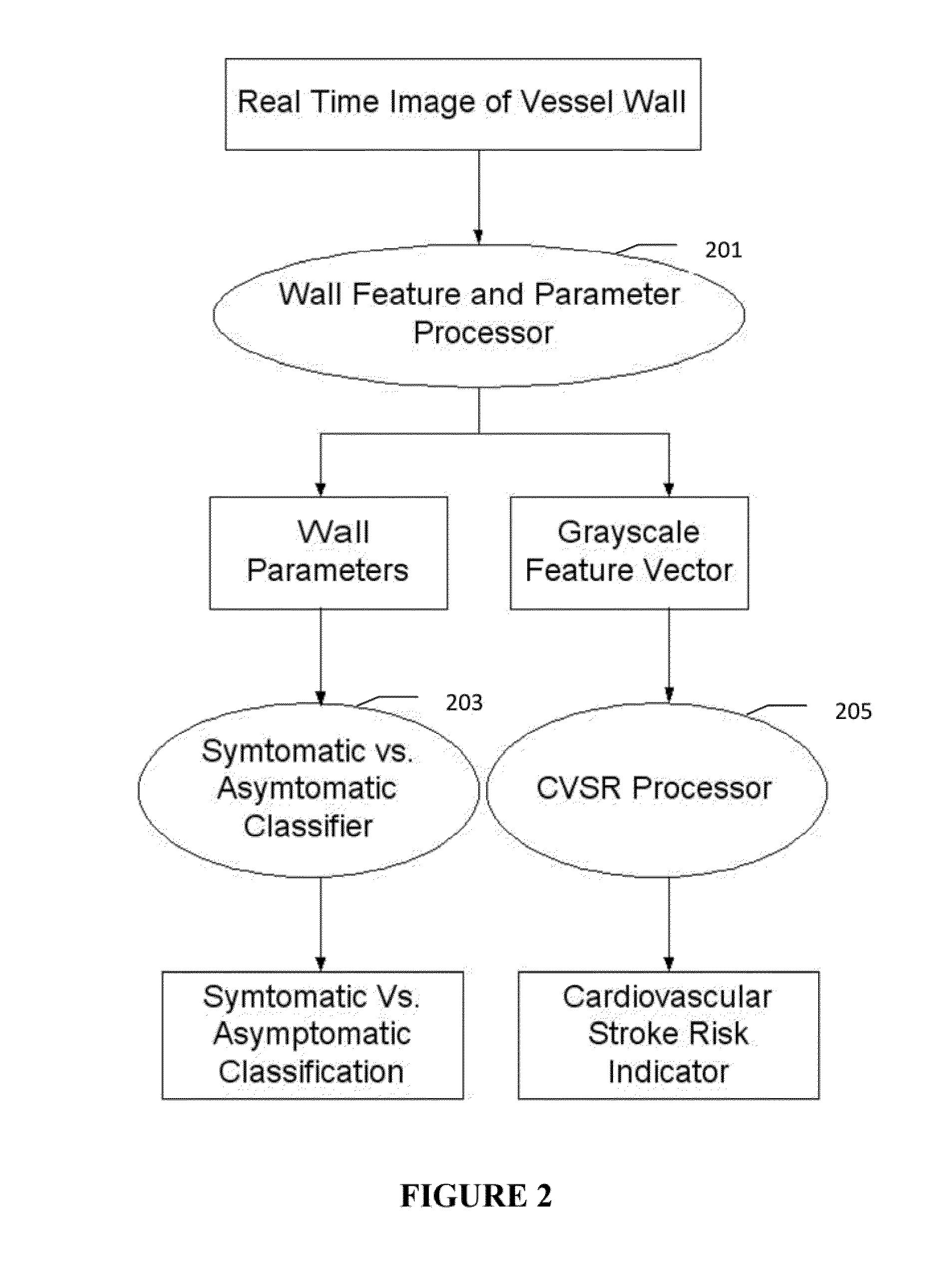
feature extraction processor which computes: (a) Higher Order Spectra-based features; (b) Discrete
Wavelet Tansform (DWT)-based features; (c) Texture-based features and (d) Wall Variability. The output of the Feature Processor is fed to the Classifier which is trained off-line from the
Database of similar Atherosclerotic Wall Region images. The off-line Classifier is trained from the significant features from (a) Higher Order Spectra; (b) Discrete
Wavelet Tansform (DWT); (c) Texture and (d) Wall Variability, selected using t-test. Symptomatic
ground truth information about the training patients is drawn from
cross modality imaging such as CT or MR or longitudinal Ultrasound or 3D ultrasound or 3D IVUS in the form of 0 or 1 (1 for symptomatic).
Support Vector Machine (SVM) or similar classifier (such as KNN, PNN or
Decision Tree or
Adaboost) supervised classifier of varying kernel functions is used off-line for training. The obtained training parameters are then used to evaluate the
test set. One can then achieve high accuracy with the
radial basis function kernel and the one with a
polynomial kernel of order two. The system also yields the risk score value on the basis of the wall features. The proposed technique demonstrates that plaque classification between symptomatic vs. asymptomatic could be achieved with a completely automated CAD tool with a significant accuracy. Performance of the system can be evaluated by computing the accuracy, sensitivity, specificity, and Positive
Predictive Value (PPV). Hence, such a tool would prove to be a valuable inclusion in the current treatment planning protocol by vascular surgeons.
[0021](11) A
data mining on-line system for Symptomatic vs.
Asymptomatic classification of the patient image and Cardiovascular risk score estimation, where the training-based system computes features in the AWR and the training
ground truth information can be taken from the cross-modality CT or MR or 3D ultrasound or longitudinal ultrasound itself, where the on-line features are computed using a non-linear behaviour of a carotid
stenosis and cerebrovascular
disease. The non-linear behaviour uses Higher Order Spectra for
feature extraction such as
Bispectrum.
Higher order statistics denote
higher order moments (order greater than two) and non-linear combinations of
higher order moments, called the higher order cumulants. The
Ultrasound image is subjected to
Radon Transform for computation of Phase Entropy. Also the on-line feature computed is Normalized Bispectral Entropy and Normalized Squared Bispectral Entropy. This on-line feature then combines with other features such as from
Discrete Wavelet Transform (DWT), Texture, Wall Variability to improve the robustness of the Symptomatic vs.
Asymptomatic classification of the patient image and cardiovascular risk score estimation.
[0033](ii) Validation Embedded Segmentation of Vascular IMT estimation: Here the recognition of
artery has been validated by the anatomic information during the segmentation process. Since lumen is the anatomic information which is blood carrier to brain and is next to the far
adventitia borders, which needs to be located, therefore, this patent application uses the anatomic information (lumen) to ensure that the far
adventitia borders are robustly computed and do not penetrate the lumen region or near wall region, while estimating the far
adventitia walls. This adds robustness to our automated recognition and Atherosclerotic Wall Region estimation process.
[0034](iii) Faster than the conventional
processing: Since the recognition is strategized at coarse level down sampled twice from its original size of the image, it is therefore
processing ¼th the number of pixels for automated recognition of the
media layer. This improves the speed of the system for computation of Atherosclerotic Wall Region.
[0036](v) Guiding Method for the Calibration
System: Since the recognition is followed by the calibration (segmentation) process, the calibration system becomes very robust since the calibration processing is done in the
region of interest determined by the automated
recognition system. Thus the calibration system adds the value determined by the automated
recognition system for
vascular ultrasound such as IMT measurement for carotid, femoral, aortic and brachial. Such a combination where the calibration system is guided by the automated recognition system for Atherosclerotic Wall Region estimation and helps in
mass processing of huge
database processing.
[0043]Extracting LIMA borders in presence of
Calcium Shadow:
Calcium is an important component of the
media layer. It is not exactly known how the
calcium is formed, but it is said that
calcium accumulates in the plaques. During the beginning of Atherosclerosis
disease, the
arterial wall creates a
chemical signal that causes a certain type of WBC (white blood cells) such as monocytes and T cells that attaches the
arterial wall. These cells then move into the wall of the
artery. These T cells or monocyles are then transformed into foam cells, which collect
cholesterol and other fatty materials and trigger the growth of the
muscle cells (which are smooth in nature) in the artery. Over time, it is these fat-laden foam cells that accumulate into plaque covered with a fibrous cap. Over time, the calcium accumulates in the plaque. Often times, the calcium is seen in the near wall (proximal wall) of the carotid artery or aortic arteries. This causes the shadow cone formation in the distal wall (far wall). As a result the LI boundaries (for Atherosclerotic Wall Region computation) are over computed from its actual layer. The shadow causes the LI lining over the actual LI boundary. As a result, the LI-MA distances are over computed in the
shadow zone. Because of this, the Atherosclerotic Wall Region formation is over computed in these cases. This application particularly takes care of Atherosclerotic Wall Region computation during the shadow cone formation. We will see how the actual LI boundaries are recovered if calcium is present causing the shadow cone. As a result, the Atherosclerotic Wall Region computation has the following advantages when using shadow cones (a) Accurate Atherosclerotic Wall Region computation in real time when the calcium is present in the proximal wall (near wall) causing the shadow cone formation; (b) The system allows computing the Atherosclerotic Wall Region in both cases: (a) when calcium is present and when calcium is not present.

