
Plasma modified medical devices and methods

a technology of medical devices and plasma, applied in chemical vapor deposition coatings, magnetic recording, record information storage, etc., can solve the problems of affecting the safety of patients, so as to increase the apoptosis of smooth muscle cells, and increase the apoptosis of coronary artery endothelial cells

Inactive Publication Date: 2013-02-21
CHEN MENG
View PDF9 Cites 34 Cited by
  • Summary
  • Abstract
  • Description
  • Claims
  • Application Information

AI Technical Summary

Benefits of technology

The present invention provides a novel coating for metallic biomaterials, such as coronary stents, which is resistant to blood clots and restenosis. The coating is deposited using a low temperature plasma process and is thin enough to allow for stent expansion without cracking. The coated surfaces exhibit non-clot formation, increased attachment of coronary artery endothelial cells, and reduced intimal hyperplasia. The coating can also be used for other implantable medical devices.

Problems solved by technology

Coronary heart disease (CHD) caused by atherosclerosis, the narrowing of the coronary arteries due to fatty build up of plaque, remains a major healthcare problem in the US.
However, concerns were raised in recent years about the safety of DES due to a reportedly small but significantly increased risk of blood clots in the stent within 1 year after stenting (i.e. stent thrombosis).
While not frequent, late stent thrombosis is a life-threatening problem.
Each year about 600,000 Americans are getting DES [Stent facts. http: / / americanheart.mediaroom.com / ], which means that even a small increased risk could result in thousands of heart attacks and deaths.
Furthermore, the requirement for prolonged, aggressive anti-thrombotic therapy after placement of a DES (usually aspirin and clopidogrel) can introduce major complications into the management of patients who require surgical procedures (which necessitate temporary discontinuation of anti-thrombotic drugs) within the first year after PCI.
Restenosis following angioplasty, however, is a major clinical problem since the biological response to this vessel damage is stimulation of accelerated growth of arterial smooth muscle cells.
Biomater Sci Polym Ed, 21(4): 529-552, 2010], but further investigation for clinical use is needed, and recent morphology studies of biodegradable coatings have shown cracks in the coatings after stent expansion [Basalus et al.
However, no systematical study for stent application has been reported.
J Biomed Mater Res (Appl Biomater), 53: 244-251, 2000], but the polyurethane used as a sealant for the radioactive agent on the stent surface remains problematic in causing chronic inflammation.
In summary, none of those aforementioned approaches addresses both issues of late thrombosis and in-stent restenosis with one specific coating.
However, in large animal studies with this patented plasma technology, certain, often large variations have been observed on the patency of plasma treated stents after implantation, which is believed to be due to the potential instability of surface bioactivity generated by the single-step NH3 / O2 plasma surface treatment on bare stainless steel surfaces.
As discussed above, the currently available coronary stents and the methods under development for improved biocompatibility of stents have the following crucial problems: 1) existing stent procedures with BMS still experience a high incidence of restenosis; 2) although DES, in comparison with BMS, have been much more widely used due to their better ability in controlling restenosis carry the risk of developing late stent thrombosis, which is associated with a clinically significant risk of mortality; and 3) most existing coating processes investigated have the major limitation of being incapable of preventing restenosis and thrombosis at the same time.

Method used


Image

  • Plasma modified medical devices and methods
  • Plasma modified medical devices and methods
  • Plasma modified medical devices and methods

Examples


example 1

Preparation of Stents

[0095]Stainless steel coronary artery stents when unexpanded had dimensions of 1.6 mm (diameter)×12 mm (length) with a total exposed wire surface area of 20.66 mm2. The stents were cleaned with a 2% (v / v) Detergent 8 solution for 30 min at 50° C. in an ultrasonic bath. The stents were then sonicated in distilled water for 30 min at 50° C. Stents were given a final rinse with distilled water and dried in an oven at 50° C. for 30 min.

[0096]The stents were then threaded through an electrically conductive metal wire that had been attached to aluminum panels with a surface area 15.3 cm×7.6 cm, using silver epoxy. For DC treatment groups, we used an oxygen pretreatment step (1 sccm oxygen, 50 mTorr, 20 W DC, 2 min) followed by TMS plasma polymer deposition (1 sccm TMS, 50 mTorr, 5 W DC, 15 s) and a 2:1 ammonia / oxygen plasma surface modification treatment for 2 min at 50 mTorr and 5 W DC. For RF treatment groups, we used an oxygen pretreatment step (1 sccm oxygen, 50 m...

example 2

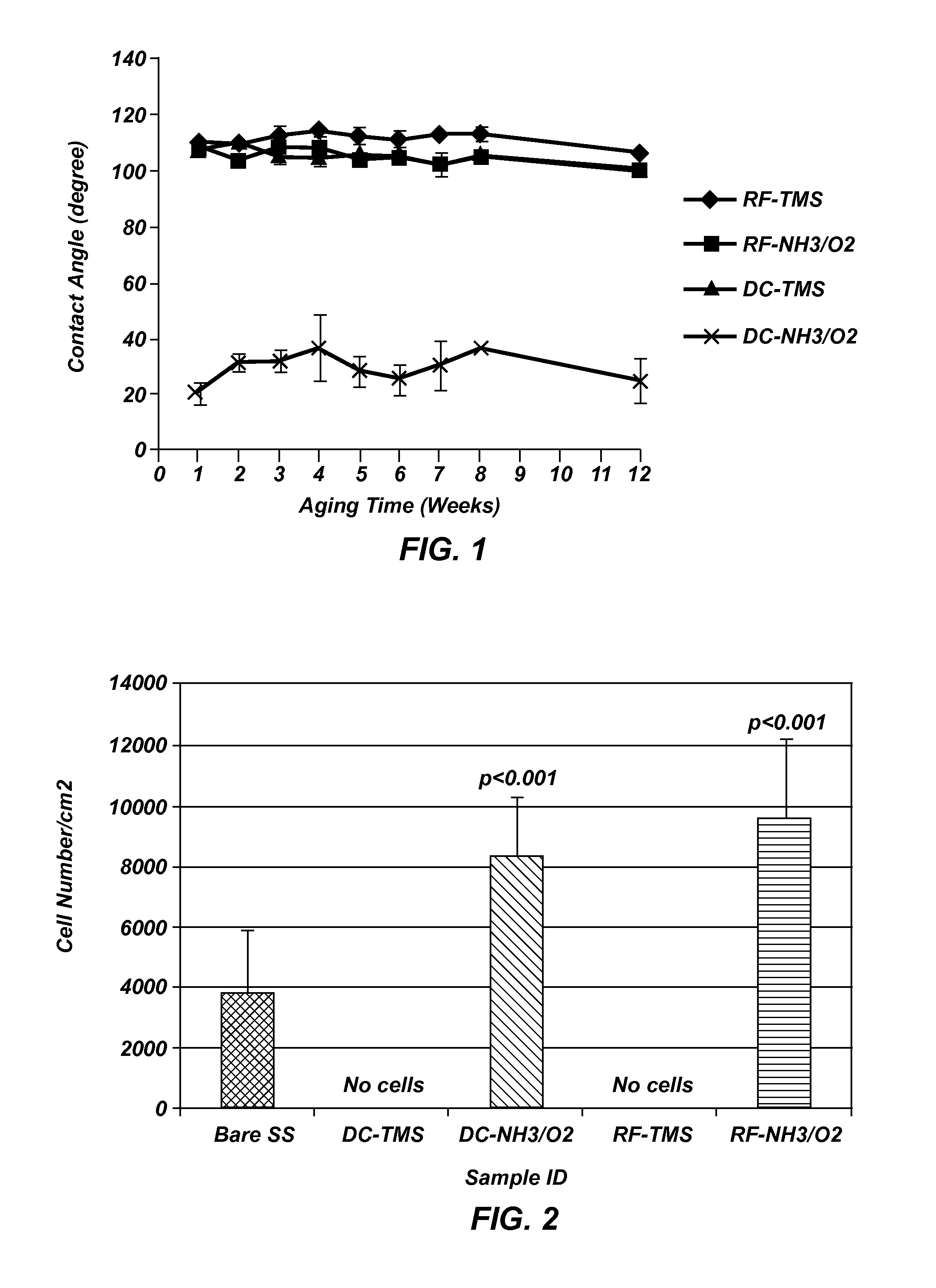
Water Contact Angle of Plasma Coated Stainless Steel Wafers

[0097]Measurements were taken on plasma coated wafers for up to 12 weeks following the plasma coating to evaluate the long teen stability. The results indicated the plasma coated surfaces tend to stabilize at about two weeks after plasma processing, and the wafers coated with TMS followed by NH3 / O2 plasma treatment using DC plasma (FIG. 1) remained very hydrophilic at 12 weeks after the plasma coating process as compared to uncoated controls, indicating long-lasting surface bioactivity generated by the plasma coating process.

example 3

Plasma Coating Adhesion to Substrate Surface and Coating Integrity

[0098]A cross-hatch was made using a razor blade on plasma coated stainless steel wafers followed by a Scotch® tape pull test. Visual inspection showed that there was no coating coming off the cross-hatched or its surrounding area, indicative of strong adhesion to the underlying surface, which warrants the coating integrity when flexed during stent crimping, navigation and expansion in clinical application.

[0099]Stainless steel stents of generic design in the dimension of Ø1.6 mm×12 mm (diameter×length) before dilation were used for the coating cracking test. After plasma coatings, the stents were imaged using an optical microscope at 20× and 50× magnifications. Following imaging, the samples were expanded with a balloon catheter (monorail™ Maverick PTCA Dilatation Catheter, Boston Scientific, Natick Mass.) and inflated to 3.0 mm in diameter; the stents were then visualized again via optical microscopy and Scanning El...


PUM

PropertyMeasurementUnit
temperatureaaaaaaaaaa
thicknessaaaaaaaaaa
contact angleaaaaaaaaaa

Abstract

Coatings, devices and methods are provided, wherein the contacting surface of a medical device with at least one contacting surface for contacting a bodily fluid or tissue, wherein long-lasting and durable bioactive agents or functional groups are deposited on the contacting surface through a unique two-step plasma coating process with deposition of a thin layer of plasma coating using a silicon-containing monomer in the first step and plasma surface modification using a mixture of nitrogen-containing molecules and oxygen-containing molecules in the second step. The two-step plasma coating process enables the implantable medical device to prevent both restenosis and thrombosis under clinical conditions. The invention also relates to surface treatment of metallic and polymeric biomaterials used for making of medical devices with significantly improved clinical performance and durability.

Description

CROSS-REFERENCE TO RELATED APPLICATIONS[0001]This application claims the benefit of U.S. Provisional Application No. 61 / 524,434, filed Aug. 17, 2011, which is hereby incorporated by reference herein in its entirety, including but not limited to those portions that specifically appear hereinafter, the incorporation by reference being made with the following exception: In the event that any portion of the above-referenced provisional application is inconsistent with this application, this application supercedes said above-referenced provisional application.STATEMENT REGARDING FEDERALLY SPONSORED RESEARCH AND DEVELOPMENT[0002]This invention was made with government support under grant No. 1R44HL097485-01A2 from the National Institutes of Health. The government has certain rights in the invention.FIELD OF THE INVENTION[0003]This invention relates to applications and methods for glow discharge plasma coatings for medical devices with improved long-term biocompatibility for clinical pract...

Claims


Application Information

Patent Timeline
Patent Type & Authority Applications(United States)
IPC IPC(8): A61F2/06B32B3/00C23C16/50A61L27/34B32B15/04B82Y5/00B82Y40/00
CPCA61L27/34Y10T428/265B82Y40/00A61L27/50A61L31/10A61L31/14A61L2400/18B82Y5/00C23C16/56C23C16/30B05D1/62B05D3/148C08L83/04A61L31/022A61L31/16A61L33/0094A61L33/068A61L2300/204A61L2300/412A61L2300/416A61L2300/42A61L2420/02B05D5/00Y10T428/31678
Inventor CHEN, MENG
Owner CHEN MENG
Learn More