Macroporous bioengineered scaffolds for cell transplantation

Inactive Publication Date: 2013-04-11
CONVERGE BIOTECH INC +1
View PDF5 Cites 5 Cited by
  • Summary
  • Abstract
  • Description
  • Claims
  • Application Information

AI Technical Summary

Benefits of technology

[0004]To address these issues, we have designed and developed a highly porous organosilicone (polydimethylsiloxane, PDMS) scaffold capable of providing structural support and adjustable spatial distribution to cells without hindering nutrient delivery. The scaffold provides a superior, more physiological environment for the cells, leading to improved viability and function. Furthermore, the scaffold material itself can be modified to provide sustained delivery of biologically active agents that improve the engraftment, survival, function and long-term viability of the cells. Such agents include, but are not limited to, oxygen generating, releasing or transport-enhancing agents, growth factors or growth-stimulating factors, anti-inflammatory compounds and immunosuppressive agents. Table 1 (below) indicates certain advantages of the scaffold and select reasons for their importance.
[0005]The implanted cells may be, e.g., insulin-producing cells. Structural support to insulin-producing cells in the form of a scaffold is critical to reducing pelleting and agglutination of the cells, which results in decreased avai

Problems solved by technology

Structural support to insulin-producing cells in the form of a scaffold is critical to reducing pelleting and agglut

Method used


Image

  • Macroporous bioengineered scaffolds for cell transplantation
  • Macroporous bioengineered scaffolds for cell transplantation
  • Macroporous bioengineered scaffolds for cell transplantation

Examples


example 1

Fabrication of Macroporous Silicone Scaffolds

[0079]Macroporous silicone scaffolds were fabricated using the solvent casting and particulate leaching technique (SCPL). The silicone polymer was prepared by mixing PDMS monomer with platinum catalyst, 4:1 v / v. The silicone molds were created by combining varying ratios (50-90% v / v) of sodium chloride crystals (Mallinckrodt Baker, N.J.) (250 to 425 μm diameter) and silicone polymer solution. The salt / silicone mixture was loaded into prefabricated, stainless steel molds (10 mm diameter, 2 mm height), pressurized to 1500 psi and incubated at 37° C. for 48 hrs to complete silicone cross-linking. The NaCl was then leached out from the scaffolds for at least 72 hrs. Pore size and degree of porosity were individually controlled by varying the particle size and polymer to particle ratio, respectively. For enhanced cell adhesion, the scaffold surface was modified by incubating overnight with fibronectin at 250 μg / mL.

example 2

Characterization of Macroporous Silicone Scaffolds

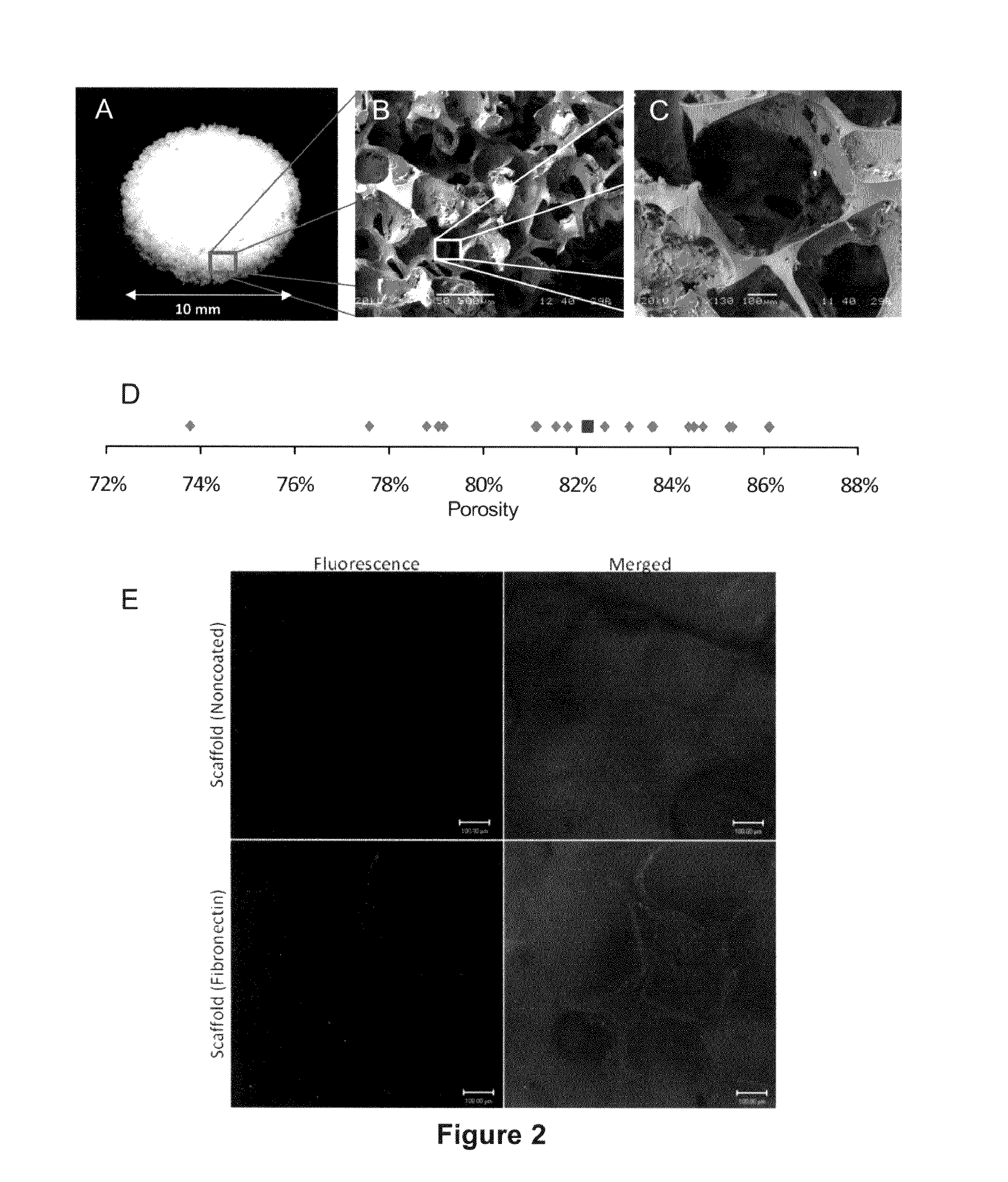
[0080]The macroporous structure of the scaffold was visualized photographically (FIG. 2A) and by scanning electron microscopy (SEM) (FIG. 2B, C). As seen in the SEM images, the scaffold is highly porous and the pore size is representative of the salt crystal diameter. Moreover, the pores are interconnected and tortuous. Final porosity was determined using gross measurements and weights (dry and wet), and calculated with the following formula:

porosity=mw / ρwmw / ρw+msilicone / ρsilicone

Porosity of the scaffolds manufactured with 90% w / v sodium choloride crystals was determined to be 85%±5% w / v (FIG. 2D).

[0081]The protein-modified scaffold surface was stained with anti-fibronectin-biotin primary and streptavidin-FITC secondary antibodies, and visualized through confocal imaging. The fluorescence imaging demonstrated a homogenously modified scaffold surface with protein coating (FIG. 2E).

[0082]We performed in vivo studies to assess biocompat...

example 3

Islet Viability and Function in Macroporous Silicone Scaffolds

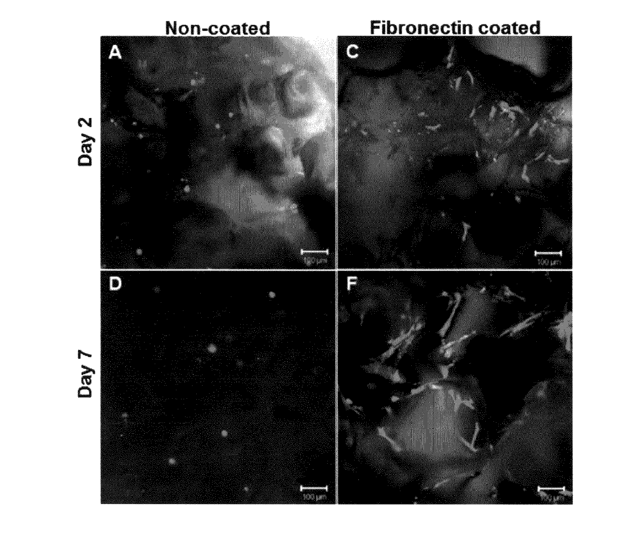
[0085]Pancreatic islets from male Lewis rats, non-human primate (NHP) baboons, and human sources were used in experiments to measure islet viability and function in macroporous silicone scaffolds. Islets were loaded into the scaffolds at the desired islet equivalent (IEQ) density by suspending them in a small volume, pipetting them onto the scaffolds and applying a light pressure gradient to distribute the islets into micro-sized pores. Fibrin glue was added to select groups to evaluate islet retention. The islets were cultured in tissue culture dishes at 20% oxygen for up to 24 hours. Two-dimensional cultures were used as controls.

[0086]Scaffolds seeded with rat, non-human primate and human islets were inspected for islet spatial distribution and viability by fluorescent live / dead dye staining (calcein AM and EthD-1) and confocal microscopy (FIG. 9). The adherence of the rat and non-human primate islets, and the viabilit...


PUM

PropertyMeasurementUnit
Pore sizeaaaaaaaaaa
Porosityaaaaaaaaaa
Pressureaaaaaaaaaa

Abstract

The present invention provides highly porous, biocompatible and biostable scaffold constructs for improving overall cell engraftment, survival, function and long-term viability. These scaffolds can provide mechanical protection to implanted cells, afford retrievability from a subject, and allow for both intra-device vascularization and a means to spatially distribute the cells within the device. The scaffold surface or material may be modified with one or more different adhesion proteins and optionally other biological factors for enhanced cell adherence and viability. Further, the scaffold surface or material may be modified with one or more agents with slow/sustained release characteristics to aid engraftment, survival, function or long-term viability. Implanted cells of the invention may be insulin-producing cells such as islets.

Description

BACKGROUND[0001]Cell replacement therapy is a promising potential treatment option for a wide variety of diseases. Many clinical conditions and disease states result from the lack of factor(s) produced by living cells or tissues, including, for example, diabetes, in which insulin production is inadequate; Parkinson's disease, in which dopamine production is decreased; and anemia, in which erythropoietin is deficient. Such conditions or diseases may be treated by cell / tissue implants that produce the missing or deficient factor(s).[0002]However, many challenges remain in the field of cell replacement therapy. The viability and functionality of transplanted cells is compromised by, for example, lack of mechanical protection, lack of necessary factors / nutrients (e.g., due to inadequate vascularization or inability of the vascular system to reach parts of the transplant), and inflammatory responses. Thus, there is a need for methods and devices that optimize the viability and functional...

Claims


Application Information

Patent Timeline
IPC IPC(8): A61L27/18A61L27/26
CPCA61K9/0024A61K35/12A61L27/18A61L27/3804A61L27/54A61L27/26A61L2300/414C12N5/0676C12N2533/30A61L27/56C08L83/04
Inventor ANDERSON, CHERYL STABLERPEDRAZA, EILEENFRAKER, CHRISTOPHER A.BUCHWALD, PETERKENYON, NORMA SUEINVERARDI, LUCAPILEGGI, ANTONELLOLATTA, PAULHUBBELL, JEFFREYWEAVER, JESSICARICORDI, CAMILLO D.
Owner CONVERGE BIOTECH INC
Why Eureka
Try Eureka