

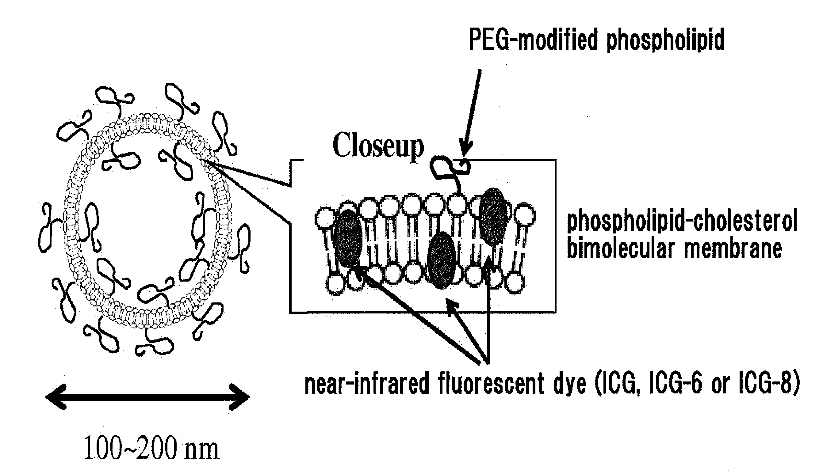
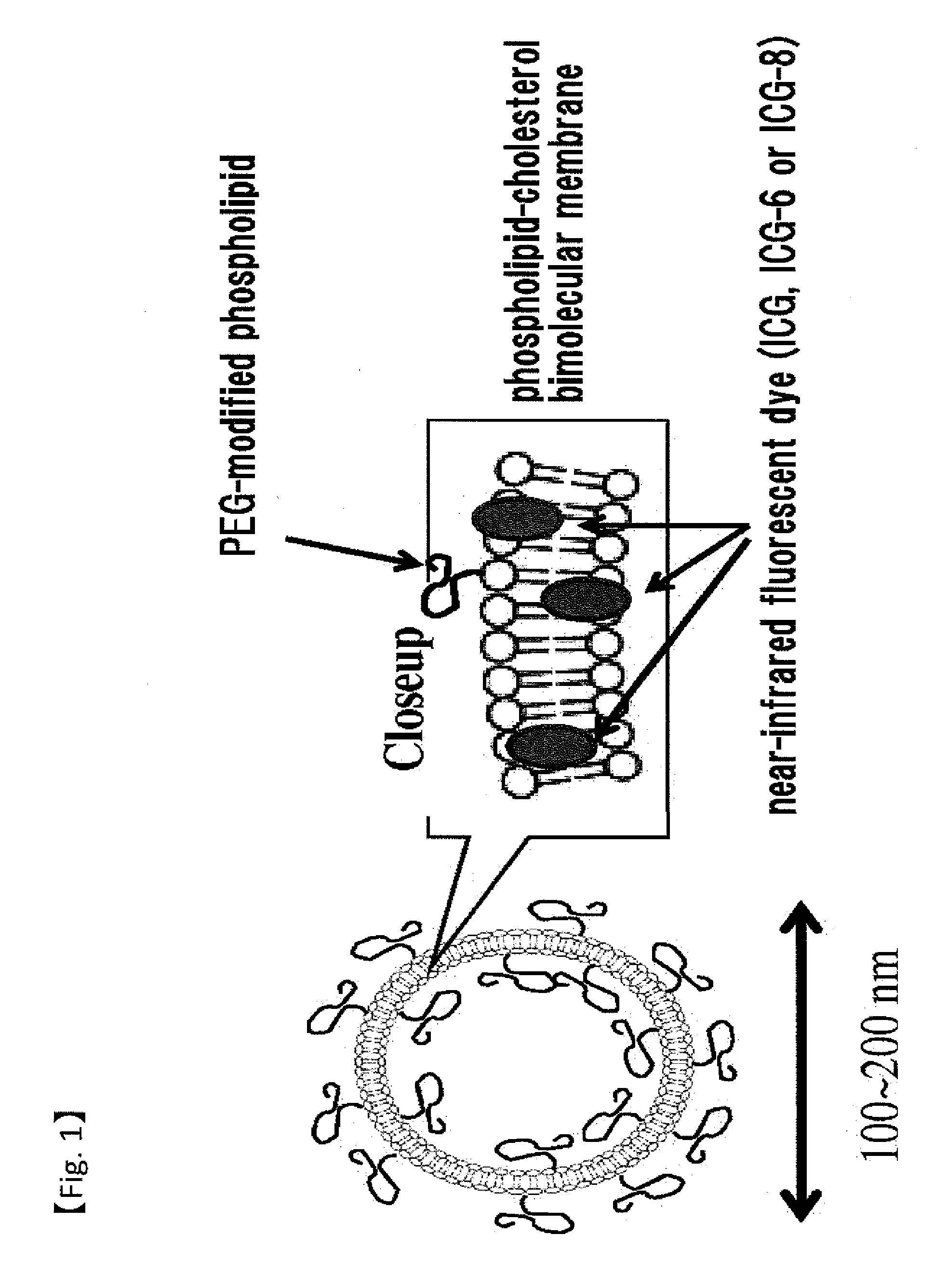
Fluorescent Probe for Imaging Lymph Nodes
a fluorescent probe and lymph node technology, applied in the direction of ultrasonic/sonic/infrasonic diagnostics, methine/polymethine dyes, diagnostic recording/meauring, etc., can solve the problem of difficult to distinguish between stained lymph ducts and native venous vessels having blue or green color, and the detection accuracy is significantly reduced, so as to achieve high fluorescence intensity, increase fluorescence intensity, and high affinity for lipids
- Summary
- Abstract
- Description
- Claims
- Application Information
AI Technical Summary
Benefits of technology
Problems solved by technology
Method used
Image

Examples
example 1
Synthesis of Fluorescent Dyes ICG-6 and ICG-8
[0059]Fluorescent dyes ICG-6 and ICG-8 of the present invention were synthesized according to the scheme indicated below.
2,3,3-trimethyl-1-(sulfobutyl)-4,5-benzoindolium inner salt (1)
[0060]2,3,3-trimethyl-4,5-benzo-3H-indole (3.1 g, 15 mmol) and 1,4-butanesultone (2.1 g, 15 mmol) were mixed in a 25 ml four-mouth flask and filled with nitrogen. After stirring for 4 hours at 80° C., the mixture was allowed to stand to cool to room temperature. Acetone was added and stirred briefly, then the resulting crystals were filtered, washed with 10 ml of acetone, and dried to obtain gray crystals (1.17 g, 23%).
2,3,3-trimethyl-1-(2-carboxyethyl)-4,5-benzoindolium bromide (2)
[0061]A mixture of 2,3,3-trimethyl-4,5-benzo-3H-indole (2.3 g, 11 mmol), 3-bromo-1-propionic acid (1.5 g, 9.8 mmol) and acetonitrile (10 ml) was stirred for 16 hours at 65° C., allowed to stand to cool to room temperature, and poured into ethyl acetate (50 ml). The crystals were f...
example 2
Synthesis of Fluorescent Dye ICG-9
[0068]Fluorescent dye ICG-9 of the present invention was synthesized according to the scheme indicated below.
p-(di-n-hexylamino)phenol (4)
[0069]A mixture of p-aminophenol (0.55 g, 5.0 mmol), 1-iodohexane (2.5 g, 11.8 mmol), potassium carbonate (0.70 g, 5.1 mmol) and dimethylformamide (10 ml) in a 25 ml four-mouth flask was stirred for 3 hours at 75° C. Potassium carbonate (0.70 g, 5.1 mmol) was added and stirred for 6 hours. The mixture was allowed to stand to cool to room temperature, poured into water and extracted with ethyl acetate. The organic layer was washed with water (3 times) and saturated salt solution, dryed over anhydrous sodium sulfate and concentrated under reduced pressure to obtain a crude product (1.3 g). The crude product was purified by silica gel column chromatography (toluene / ethyl acetate=25 / 1 to 15 / 1) to obtain the titled compound as an oil (0.81 g, 58%).
3-chloro-2,4-trimethylene glutaconaldehyde dianil hydrochloride (5)
[0070...
example 2-2
Synthesis of Additional Compounds
[0074]ICG-8-C4:
4(2-((1E,3E,5E,7E)-7-(1,1-dimethyl-3-butyl-1H-benzo[e]indol-2(3H)-ylidene)hepta-1,3,5-trienyl)-1,1-dimethyl-1H-benzo[e]indolium-3-yl)butane-1-sulfonic acid (ICG-8-C4)
[0075]1-butyl-2,3,3-trimethyl-4,5-benzo[e]indolium iodide (10.7 g, 62%) was obtained in the same manner as Example 1 with replacing 1-iodooctadecane (16.8 g, 44 mmol) (used for the synthesis of 1-octadecyl-2,3,3-trimethyl-4,5-benzo[e]indolium iodide (4) in Example 1) with 1-iodobutane (12.1 g, 66 mmol). The titled compound (1.35 g, 44%) was obtained in the same manner as the synthesis of ICG-8. The primary molecular ion peak as determined by LC-MS was m / z=673 (positive). The maximum absorbance wavelength and molar extinction coefficient in ethanol were λmax=787 nm and c=2.52×10⁵, respectively.
[0076]ICG-8-C6
4(2-((1E,3E,5E,7E)-7-(1,1-dimethyl-3-hexyl-1H-benzo[e]indol-2(3H)-ylidene)hepta-1,3,5-trienyl)-1,1-dimethyl-1H-benzo[e]indolium-3-yl)butane-1-sulfonic acid (ICG-8-C6)
[00...
PUM
| Property | Measurement | Unit |
|---|---|---|
| particle diameter | aaaaa | aaaaa |
| particle diameter | aaaaa | aaaaa |
| particle diameter | aaaaa | aaaaa |
Abstract
Description
Claims
Application Information