

Protein nanofibers from self-assembling pentamers
a technology of nanofibers and pentamers, applied in the field of nanofibers, can solve the problems of increasing the difficulty of manufacturing materials with the same level of structural and molecular specificity on various length scales, and the difficulty of effective production of engineered materials with the desired dimensions and properties, and achieve the effect of preventing oxidation
- Summary
- Abstract
- Description
- Claims
- Application Information
AI Technical Summary
Benefits of technology
Problems solved by technology
Method used
Image

Examples
example 1
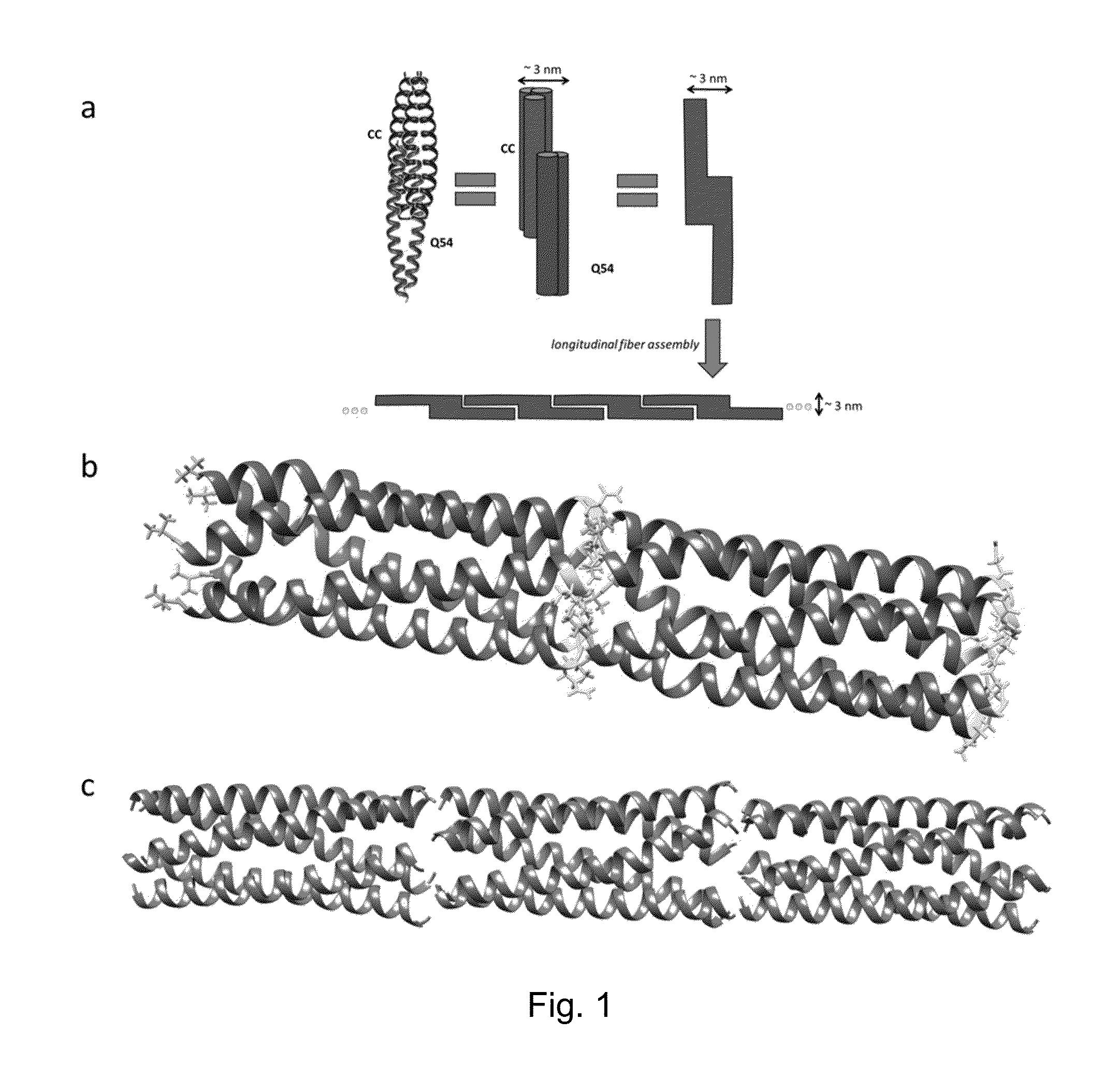
[0098]This example describes characterization of proteins CC and Q54. The proteins were characterized for assembly and structure using transmission electron microscopy (TEM), atomic force microscopy (AFM), scanning electron microscopy (SEM), fluorescence and confocal microscopy, zeta potential, dynamic light scattering, and circular dichroism. Circular dichroism curves show that the combination of CC and Q54 results in a very α-helical protein that is comprised of the assembly of both complementary proteins. The protein fibers have an average diameter on the order of 80 nm extending for several μm in length. Fibrils composing the fibers have a regular width of approximately 3 nm. In addition, fibers generated by CC and Q54 have been shown to be able to bind small molecules. The fluorescent molecule curcumin was bound to these protein assemblies and studied under fluorescence and confocal microscopes. Curcumin is known to bind within the hydrophobic pore formed by the homopentamer of...
example 2
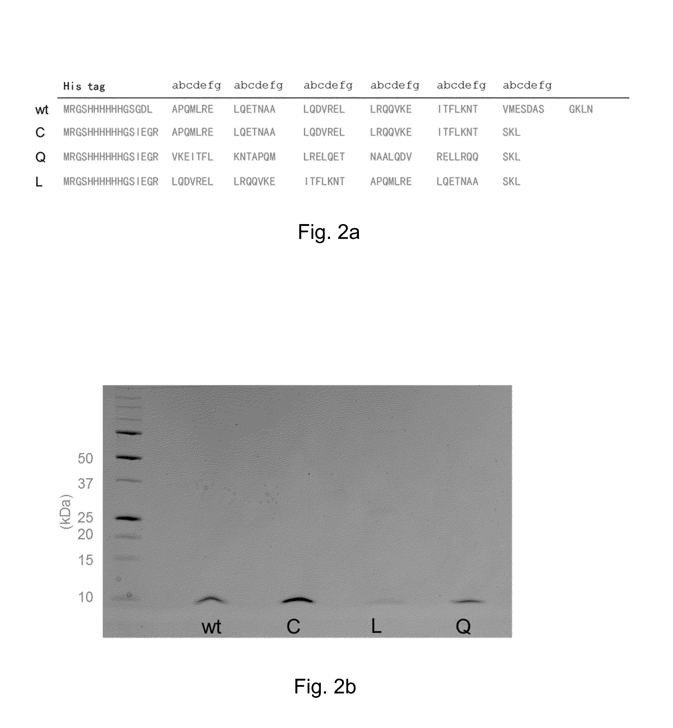
[0108]In this example, we describe the self-assembly of a novel protein (Q) designed by swapping regions of wt (COMPcc), and compare it to wt as well as a negative control swap protein, called L. Q assembled into robust nanofibers with unprecedented diameters up to 560 nm under pH 4. In the presence of the small molecule curcumin, the Q fibers further assembled into microfibers with diameter of 16 μm, akin to natural keratin and spider silk fibers measuring tens of micrometers in diameter, providing the first example of an engineered protein microfiber.
Materials
[0109]Sodium phosphate (monobasic and dibasic) and nickel-nitrilotriacetic acid resins were purchased from Sigma-Aldrich. Ampicillin, isopropyl-β-D-thiogalactopyranoside (IPTG), tryptone, urea, tris-HCl, and sodium chloride were obtained from Fisher Scientific. Yeast extract, methanol, and curcumin were purchased from Acros Organics and BCA kit was obtained from Pierce. Imidazole was purchased from Alfa Aesar and copper grids...
example 3
[0141]This example describes further characterization of the protein fibers of this disclosure. As discussed above, we observed that nanoscale Q fibers aggregate to form mesoscale protein fibers upon addition of curcumin. In addition, we examined the ability of C, Q, and wt COMPccs to bind with two fatty acid chains: myristic acid and palmitic acid. Protein fibers formed in the presence of these fatty acids are shown in scanning electron micrographs in FIGS. 27a-27c and FIGS. 28a-28c. Samples were prepared by mixing fatty acid solution in a 1:1 molar ratio with COMP solution. The resulting solution was allowed to stand for 6 hours. Samples were prepared by depositing the fatty acid and COMP solution onto TEM grids that are supported on an SEM stub with carbon tape. The solution was allowed to dry, and was washed 3-5 times with water. The sample was allowed to dry between each wash. The images were taken on an SEM under high vacuum. Buffer conditions were pH 4 in 50 mM phosphate buff...
PUM
| Property | Measurement | Unit |
|---|---|---|
| Diameter | aaaaa | aaaaa |
| Diameter | aaaaa | aaaaa |
| Diameter | aaaaa | aaaaa |
Abstract
Description
Claims
Application Information