Si-o-n-p related fabrication methods, surface treatments and uses thereof

Active Publication Date: 2017-12-07
TEXAS A&M UNIVERSITY +1
View PDF0 Cites 0 Cited by
  • Summary
  • Abstract
  • Description
  • Claims
  • Application Information

AI Technical Summary

Benefits of technology

The present invention provides new chemistry for implant coatings that can increase the usability of metal dental and orthopedic implants for patients with disorders involving soluble bone formation. These coatings use a Si—O—N—P elemental chemistry to promote rapid formation of bone in both healthy and disordered bone. In one aspect, these coatings can also be used for implants to promote rapid regeneration of bone for healthy patients suffering from bone fractures. Additionally, the patent describes a treated surface that enhances neovascularization, which may provide an increase in vasculogenesis and angiogenesis compared to a surface not treated with a composition comprising the phosphorous-doped silicon oxynitride.

Problems solved by technology

When fractures occur, the site is structurally unstable and hypoxic due to severe bone loss and ischemia.
However, methods and / or materials that provide for the effective and therapeutic control and / or prevention of destructive high ROS levels through enhancing the generation of anti-oxidants remain lacking in the medical arts.
Though process seems ideal in theory in the sense that it appears to mimic the natural bone remodeling process, in practice it is difficult to control the rate of dissolution of the biomaterial to match the rate of replacement of the host bone tissue.
These types of biomaterials are therefore rarely used in load-bearing tissue applications.
Bioactive biomaterials form a strong chemical bond with local bone host tissue through a series of chemical reactions at the bioceramics-bone interface that results in the formation of a carbonated apatite layer at the interface surface.2 This strong interface makes them ideal for use in load bearing bone implant applications such as hip stems and dental screws; however, most bioactive biomaterials used in bone tissue applications—particularly silica-based bioactive glasses—do not have the necessary mechanical strength required to replace load bearing bone.
Bioinert metals like titanium have traditionally been used in load bearing bone implant applications, but they do not bond to local bone tissue and have a finite fatigue life in vivo.
These Ti-bioglass systems generally suffer from the fact that while the bioglass-bone forms a strong interface, the bioglass-Ti interface is much weaker.
Current structural materials used to support large missing bone volume do not provide a mechanism whereby deleteriously high levels of ROS can be controlled.
Nor do the metal surfaces of these implants foster the formation of an apatite surface layer, an important aspect of the bone formation process.
Thus, while metal implants (e.g., titanium [Ti]) are useful as structural materials for bone implant because of their strength, they suffer the disadvantage of long healing times15.
However, while these materials do provide for the formation of a surface hydroxyapatite layer, they unfortunately fail to provide stable, strong bone attachment because of the large coefficient of thermal expansion mismatch with the underlying metal.
This thermal expansion mismatch problem results in resorption and delamination of the bone from the metal surface of the implant, and ultimately failure of the implant attachment16.
In addition, these coatings fail to provide a solution to the damaging effects of high ROS levels, so common to the physiological environment of an implant site in vivo.
Synthesis routes for synthetic hydroxyapatite overlay cause inhomogeneity in the coating and have thermal expansion mismatch with the underlying Ti, leading to delamination and instability of the coating1.
Bioactive glass coatings, which are modified from commercially available bioglass with added MgO for improved thermal expansion matching, have shown a drawback to down-regulate important osteogenic markers associated with bone formation due to Mg2+ release2.
However these coating are limited in that they can only be applied by enameling, which has a large coating to interface thickness ratio (1000:1), leaving them more susceptible to delamination15.
Bioactive glasses, however, cannot be used in load-bearing osseoapplications.

Method used


Image

  • Si-o-n-p related fabrication methods, surface treatments and uses thereof
  • Si-o-n-p related fabrication methods, surface treatments and uses thereof
  • Si-o-n-p related fabrication methods, surface treatments and uses thereof

Examples


example 2

cterization

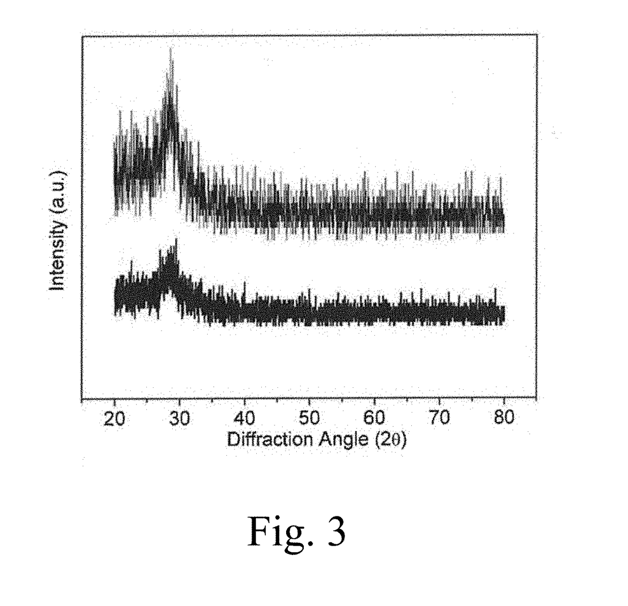
[0140]X-ray diffraction was used to investigate the film for the formation of any crystalline phases. A Bruker D8 Advance diffractometer was used to collect Bragg-Brentano scans using Cu kα radiation (λ=1.5418 Å) at room temperature. Data was recorded over the 2θ range of 20-80° with a 0.02° step size and a dwell time of 1 second.

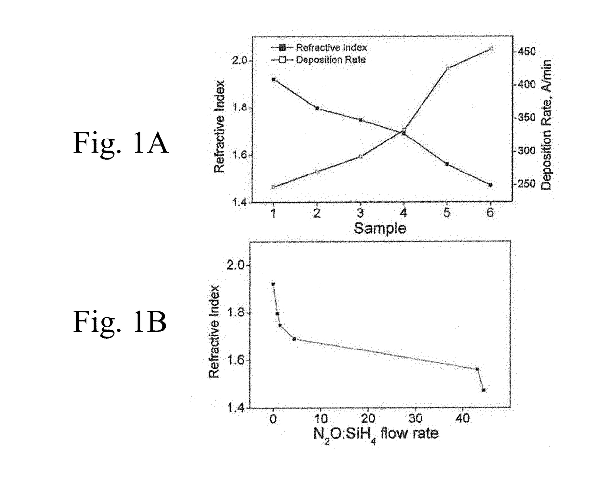
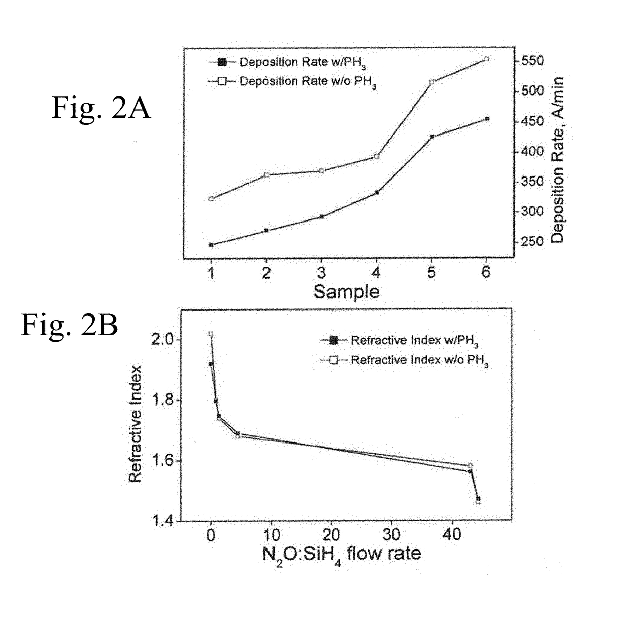
[0141]X-ray diffraction was used to confirm the amorphous nature of the coatings. Though X-ray spectra do not show two broad 2theta peaks typical to amorphous silicon oxynitrides117: one around 20-30° and one around 60-80°; it is assumed that the SiO2 sub-layer dominates the spectra resulting in one amorphous silica peak. FIG. 3 shows representative spectra from the highest N content Sample 1 and the highest O content sample 6. Other samples show the same general spectra, and spectra are too noisy to provide a basis for comparison and establish a general trend in the data.

[0142]XRD spectra confirm that the coatings formed were amorphous.

example 3

DS Evaluation

[0143]Surface morphology, film composition, and film thickness were investigated using scanning electron microscopy (Hitachi S-3000N Variable Pressure SEM) equipped with an energy dispersive X-ray spectrometry system (EDAX). SEM images were taken with an acceleration voltage of 20 keV and scans for EDX mapping and compositional studies were taken at 12 keV to prevent interference from sub-coating layers. EDAX software was used to quantify spectral mapping data from 50 μm×50 μm areas. Four regions of interest (ROIs) corresponding to Si, O, N, and P Kα lines were defined and compositional information was calculated using EDAX's proprietary ZAF method.

[0144]Coating thickness and surface morphology was confirmed by SEM imaging and found to be in agreement with ellipsometry measurements. EDS mapping showed no major carbon or other contaminants and a uniform elemental distribution normal to the wafer surface. A ZAF quantification method was used to determine the atomic % of e...

example 4

nergy Evaluation

[0148]A sessile drop technique was used to determine contact angles of distilled, deionized water and diiodomethane (>99%, Sigma-Aldrich, St. Louis, Mo., USA) on the wafer coating surface. For each liquid, 9 repeat drops were measured at 25° C. and samples were tested in triplicate. The surface tension of the testing fluids including the polar and dispersive components was taken from literature values at the same experimental conditions and can be seen in Table 3. Surface energy (γLV) and its corresponding dispersive (γd) and polar (γp) component values for probing liquids113,114.

TABLE 3Probing fluidγLV (mJ m−2)γd (mJ m−2)γp (mJ m−2)Water72.821.851.0Diiodomethane50.850.40.4

[0149]The hydrophilicity of the surface was inferred from the contact angle with water and the surface energy values of the samples were obtained by relating the measured probing liquid contact angles (θ) and relating them to solid surface energy dispersive and polar components by the Owens-Wendt-K...


PUM

PropertyMeasurementUnit
Fractionaaaaaaaaaa
Thicknessaaaaaaaaaa
Thicknessaaaaaaaaaa

Abstract

Disclosed are compositions, methods and processes for fabricating and using a device or other implement including a surface or surfaces having a nanoscale or microscale layer or coating of Si—O—N—P. These coatings and / or layers may be continuous, on the surface or discontinuous (e.g., patterned, grooved), and may be provided on silica surfaces, metal (e.g., titanium), ceramic, and combination / hybrid materials. Methods of producing an implantable device, such as a load-bearing or non-load-bearing device, such as a bone or other structural implant device (load-bearing), are also presented. Craniofacial, osteogenic and disordered bone regeneration (osteoporosis) uses and applications of devices that include at least one surface that is treated to include a nanoscale or microscale layer or coating of Si—O—N—P are also provided. Methods of using the treated and / or coated devices to enhance enhanced vascularization and healing at a treated surface of a device in vivo, is also presented.

Description

CROSS-REFERENCE TO RELATED APPLICATIONS[0001]The present application claims priority to U.S. Provisional application Ser. No. 62 / 305,956, filed Mar. 9, 2016. The contents of the provisional application are specifically incorporated herein by reference in its entirety.FEDERAL GRANT SUPPORT[0002]The work was supported by a grant from National Institutes of Health 1R03DE023872-02. The United States federal government owns rights in the present invention.FIELD OF THE INVENTION[0003]The present invention relates to the field of surface treatments and / or surface thin-film / nano coatings for medical devices, dental implants and semi-conductor, solar cell, and microelectronic devices. The invention also relates to methods for enhancing osteogenesis, bone generation (formation), fracture healing and wound healing. The invention also relates to the field of medical device surface design and chemical deposition methods.SUBMISSION OF SEQUENCE LISTING[0004]The contents of the electronic submissio...

Claims


Application Information

Patent Timeline
IPC IPC(8): A61L27/32A61C8/02C09D1/00C23C16/22C04B35/597A61F2/30A61L27/10A61L27/38A61L27/54C23C16/50A61C8/00
CPCA61L27/32A61F2002/3093A61L27/10A61L27/38C09D1/00C04B35/597C23C16/50C23C16/22A61C8/0006A61C8/0015A61F2/30771A61L2400/12A61L2420/02A61L2430/02A61L2400/18A61L2430/12A61L2420/06A61L2300/412A61L2300/606A61L2300/112C04B2235/42A61F2002/30981A61F2002/3084A61F2310/00796A61F2310/00874A61F2310/0061A61L27/54C04B35/5154C23C16/30C09D7/60A61F2/3094
Inventor VARANASI, VENUASWATH, PRANESHKRAMER, PHILIPVELTEN, MEGENILYAS, AZHAR
Owner TEXAS A&M UNIVERSITY
Why Eureka
Try Eureka